CORD BLOOD – Constituents of Cord Blood, Purpose, Cord Blood Collection, Cord Blood Banking, Advantages and Disadvantages of Cord Blood
Umbilical
Cord Blood (Cord Blood) is the blood which found in the placenta and in the
attached umbilical cord after childbirth.
Cord blood
contains stem cells which are used to treat hematopoietic and genetic
disorders. There are several advantages to use cord blood as a main source of
stem cells because these cord blood is less likely to induce immunological
reaction during transplantation.
Constituents of Cord Blood
Cord blood
contains more number of granulocyte-macrophage (CFU-GM), erythroid (CFU-E), and
multipotential (CFU-GEMM) progenitor stem cells. It is composed of substance
found in whole blood such as red blood cells, white blood cells and plasma.
Along with whole blood and progenitor stem cells, cord blood contain high number of natural killer cells,
low level of T-cells and higher level of immature T-cells.
Stem cells
are found in cord blood mainly hematopoietic stem cells which is used to for
stem cell transplantation during bone marrow transplant. Some non-hematopoietic
stem cells is also found, which contain mesenchymal stem cells.
Haematopoietic
stem cells (HSCs) are normally found in the bone marrow. Haematopoietic stem
cells can make any type of blood cells (red cells, white cells and platelets). These
stem cells are needed for blood production. HSCs are used in bone marrow
transplants to treat blood diseases.
Purpose
Cord blood is used as a source of
stem cells.
Cord blood can be preserved and used
in the same individual when stem cell need arises.
Cord blood is used for allogenic
transplant and autologous transplant.
Cord blood is multipotent stem cells,
so it can be used for reconstitution and repaired damaged tissues.
Cord blood contains T-cell which is
immunologically less active than adult marrow or peripheral blood.
Cord blood is used for stem cell
replenishment in hematological malignancies such as acute leukemia, chronic
myeloid leukemia and myelodysplastic syndrome.
Cord blood is also used in
non-malignant conditions such as thalassemias, aplastic anemia, hemoglobinopathies
and immunodeficiency.
CORD BLOOD COLLECTION
After child
birth, physician clamps the umbilical cord in two regions, about 10 inches
apart and cut the cord, separating mother from baby. After that doctor collect
blood (about 40 ml of blood) from cord using needle. Blood is collected in
sealed bag and sent to cord blood bank for storage. The cord blood is
cryopreserved in the viable state for many years. These cord blood will be used
for the same individual who is need for future use.
CORD BLOOD BANKING
Cord blood
is the collected of stem cells from the umbilical cord and placenta either in
utero before the delivery of the placenta or ex-utero after its delivery. These
cord blood is stored either in private or public cord banks. In Public Cord
Banking, Donor can donate cord blood for free for future research as well as for
future need for same individual.
In private
cord blood banking, individual cord blood is stored for certain period of time
for their personal or family purposes. For storage, private cord blood banking
will charge for the individual, but it’s expensive.
ADVANTAGES OF CORD BLOOD
Availability of cord blood
Lower incidence of graft versus host
disease (GVHD)
Transplantation success rate is high
for two different antigens.
DISADVANTAGES OF CORD BLOOD
High infection rates compared to
mature stem cells from bone marrow or peripheral blood (adult donors)
Multipotent cells but not pluripotent
cells.
CORD BLOOD – Constituents of Cord Blood, Purpose, Cord Blood Collection, Cord Blood Banking, Advantages and Disadvantages of Cord Blood
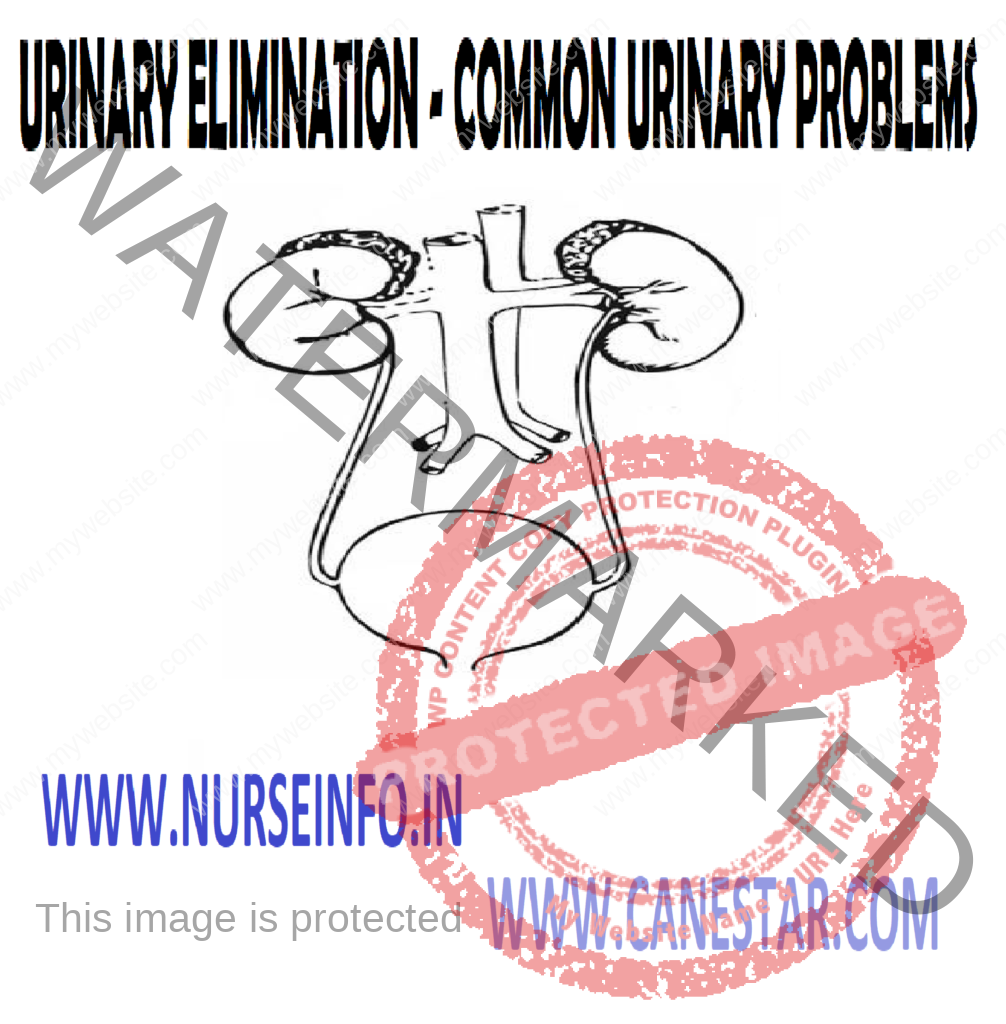
There are various urinary diseases and conditions that can affect the urinary system, which includes the kidneys, bladder, ureters, and urethra. Here are some common urinary diseases:
Urinary Tract Infections (UTIs): UTIs are infections that can occur in any part of the urinary system, including the bladder, urethra, and kidneys. They are often caused by bacteria, and common symptoms include pain or burning during urination, frequent urination, and cloudy or strong-smelling urine.
Kidney Stones: Kidney stones are hard deposits that form in the kidneys and can cause severe pain when passing through the urinary tract. They may result from the accumulation of minerals and salts in the urine.
Interstitial Cystitis (IC): Also known as painful bladder syndrome, IC is a chronic condition characterized by bladder pain, urinary urgency, and frequency. The cause of interstitial cystitis is not well understood.
Bladder Infections: Infections of the bladder, also known as cystitis, can cause symptoms such as frequent urination, urgency, and pain or discomfort in the lower abdomen.
Urinary Incontinence: This is a condition characterized by the loss of bladder control, leading to involuntary urine leakage. It can be caused by various factors, including age, childbirth, and neurological disorders.
Prostatitis: Inflammation of the prostate gland, known as prostatitis, can cause urinary symptoms such as pain or discomfort during urination, increased frequency, and urgency.
Urinary Infections in Children: Children can also experience urinary tract infections, which may present with symptoms like fever, abdominal pain, and changes in urinary habits.
Polycystic Kidney Disease (PKD): PKD is a genetic disorder that causes the growth of fluid-filled cysts in the kidneys, leading to kidney enlargement and potential kidney function decline.
Renal Failure: Chronic kidney disease (CKD) or acute kidney injury (AKI) can result in the gradual or sudden loss of kidney function, affecting the body’s ability to filter waste and maintain fluid and electrolyte balance.
Urinary
elimination, a natural process in which the body excretes waste products and
materials those exceeded bodily needs, usually is taken for granted. When the
urinary system fails to function properly, virtually organ systems can be
affected. Persons with alternations in urinary elimination may also suffer
emotionally from body image changes. The proper functioning of the urinary
system is vital to the body’s physical well being, to life itself, and a
person’s general sense of well bring.
Nursing
therapies promote or minimize factors that influence urinary elimination. Each
client has a different pattern of elimination. The nurse must assess this pattern
and design therapies to promote normal urinary elimination when necessary. The
nurse uses devices such as a condom or an indwelling catheter to assist the
client with urinary elimination. The nurse assisting a client with urination or
intervening to resolve health related to urinary needs may have specialized
abilities
DEFINITION
Urinary
elimination is defined as expulsion of waste products from the body through the
urinary system.
Elimination
from the urinary tract helps to remove the waste products from body. It is
essential to the body’s physical well-being
PHYSIOLOGY
Urinary
elimination depends on the function of the kidneys, ureters, bladder, and
urethra. Kidneys remove waste from the blood to form urine. ureters transport
urine from the kidneys to the bladder. The bladder holds urine until the urge
to urinate develops
Growth and
development of individual: it influences urination. Usually infants or children
with 6 to 8 kg excrete 400 to 500 ml per day and child cannot withhold urination.
The adult normally voids 1500 to 1600 ml per day and has normal urine color;
also has control over urination. Aging impairs urination, e.g. elder adults
Food and
fluid: foods high in water content increased urine production. Certain foods
affect the color and odor of urine. Certain fluid needed to urinate develops.
Urine leaves the body through the urethra. All organs of the urinary system must
be intact and functional for successful removal of urinary wastes
The process
of emptying the bladder is known as micturition or voiding or urination. The
bladder normally holds as much as 600 ml of urine. However, the desire to
urinate can be sensed when the bladder contains only a small amount of urine
(150 to 200 ml in adults and 50 to 200 ml in a child). As the volume increases,
the bladder walls stretch, sending sensory impulses to micturition center in
the sacral spinal cord. Parasympathetic impulses from the micturition center
stimulate the detrusor muscle to contract rhythmically. The internal sphincter
also relaxes so that urine may enter the urethra, although voiding does not yet
occur. As the bladder contracts, nerve impulses travel up the spinal cord to
the midbrain and cerebral cortex. A person is thus conscious of the need to urinate.
If the person chooses not to void, the external urinary sphincter remains
contracted, and the micturition reflex is inhibited. However, when a person is
ready to void, the external sphincter relaxes, the micturition reflex
stimulates the detrusor muscle to contract and urination occurs. The act of
micturition normally is painless
Factors Influencing
Developmental Considerations: infants are born without voluntary control of urination and with the little ability to concentrate urine. Older children and adults have general control of urination voluntarily. Physiological may affect urination
Lifestyle: many individual’s families and sociocultural variables influence a person’s normal voiding habits. For some individuals voiding is a very personal and private act
Fluid and food intake: the healthy body maintains a sensitive balance between the amount of fluid ingested and the amount of fluid eliminated. When fluid intake increases, the output also increases
Environment: during summer, due to excessive perspiration urine output is less. During winter, due to lack of perspiration, urine output is more
Psychological factors: stress can also interfere with the ability to relax external urethral sphincter as a result, emptying the bladder completely becomes difficult or impossible
Medication: Many medications interfere with the normal urination process and may cause retention. Diuretics, e.g. frusemide, increase urine formation by preventing the reabsorption of water and electrolytes from the tubules of the kidney into the bloodstream
Muscle tone and activity: People who exercise regularly will have good muscle tone increased body metabolism and good urine production
Pathological conditions: endocrine disorders such as diabetes insipidus increase urine formation. Diseases of the kidney themselves can reduce kidney function and perhaps eventually result in renal failure
Surgical and diagnostic procedure: surgery on structures adjacent to the urinary tract can also voiding because of swelling in the lower abdomen and often necessitates the use of retention catheter for a short time
DIAGNOSTIC EXAMINATION
Diagnostic
examination of the urinary system can also influence micturition, for example,
intravenous pyelogram
Conditions Which Alter Urinary Elimination
The most
common conditions which alter urine elimination encountered by the nurse,
involve disturbance in the act of micturition. These disturbances result from impaired
bladder function, obstruction to urine outflow, or inability or voluntary
control of micturition. The common renal conditions causing alternation in
urinary elimination are as follows:
Prerenal Conditions
Decreased intravascular volume,
dehydration, hemorrhage, and burns shock
Altered peripheral vascular
resistance; sepsis, anaphylactic shock and reactions
Ureteral, bladder or urethral obstructions,
due to calculi, blood clot, tumors, and strictures
Prostatic hypertrophy
Neurogenic bladder
Pelvic tumor
Retroperitoneal fibrosis
ROLE OF NURSE
The role and
responsibilities of nurse, when managing the urinary elimination in their clients
include the following:
Taking nursing history pertaining to
client with partial emphasis on urinary elimination
Conducting or assessing physical
assessment of kidneys, bladder, urethral orifice, skin integrity and hydration
and urine
In addition, carrying out the
following assessment measures like measuring urine output, collecting urine
specimens, determining the presence of abnormal constituents, assisting with
diagnostic procedure
COMMON URINARY DISEASES
Anuria: technically, no urine is voided for 24-hour-urine output is less than 100 ml
Dysuria: difficulty in voiding, may or may not be associated with pain, a feeling of warm local irritation occurring during voiding is called “burning”
Frequency: increased incidence of voiding
Glycosuria: presence of sugar in the urine. it may be due to an unusually large intake of sugar or to marked emotional disturbance and is temporary
Hematuria: presence of blood in the urine
Incontinence: inability to voluntarily control the discharge of urine
Nocturia: frequency of urination during the nights
Oliguria: scanty or greatly diminished amount of urine voided in a given time (24 hours urine output is 100-400 ml)
Orthostatic albuminuria: presence of albumin in urine that is voided after periods of standing, walking or running. It is the phenomenon of circulatory systems
Pneumaturia: passage of urine containing gas
Polyuria: excessive output of urine (diuresis)
Proteinuria: presence of protein, usually albumin, in the urine
Pyuria: pus in the urine. Urine appears cloudy.
Enuresis: it is defined as repeated involuntary urination in children beyond 4 to 5 years of age, when voluntary bladder control is normally acquired
Enuresis can
be nocturnal (night time) and diurnal (day time) or both
Urinary incontinence: it is the ability to control passage of urine to continence may be caused by stress. Neurological impairment and injury to urethral sphincter
Urinary retention: it is the accumulation of urine in the bladder associated with inability of the bladder to empty itself
Urinary catheterization is a medical procedure in which a thin, flexible tube called a catheter is inserted into the urinary bladder through the urethra to drain urine. This procedure may be necessary for various reasons, and it can be performed in different settings, such as hospitals, clinics, or even at home under certain circumstances.
Here are some common reasons for urinary catheterization:
Urinary Retention: When a person is unable to empty their bladder naturally, either due to a medical condition, surgery, or other factors, a catheter may be inserted to allow for the drainage of urine.
Surgery: Catheterization is often performed before, during, or after certain surgical procedures, especially those involving the genitourinary system or pelvic area.
Monitoring Urine Output: In critically ill patients or those undergoing surgery, healthcare providers may use catheters to closely monitor urine output and assess kidney function.
Urinary Incontinence: In some cases, especially in individuals with severe urinary incontinence, catheters may be used as a means of managing and collecting urine.
There are different types of urinary catheters, and the choice depends on the specific needs of the patient and the medical situation. The main types include:
Indwelling Catheters (Foley Catheters): These are left in place for an extended period and have a balloon at the tip to hold them in the bladder. They are often used for patients who are unable to void on their own.
Intermittent Catheters: These are inserted and removed several times a day to empty the bladder. They are commonly used for short-term purposes or in cases where regular emptying is needed.
External Catheters (Condom Catheters): These are used in males and are attached externally to the penis to collect urine. They are typically used for managing urinary incontinence.
Urinary
catheterization is the introduction of a tube (a catheter) through the urethra
into the urinary bladder to drain the bladder
Urinary
catheterization is an aseptic method of introducing the catheter into the
urinary bladder through the external urethra for withdrawal of urine
Purpose
To obtain a clear specimen for
diagnostic purpose
To relieve distension of bladder
caused by retention of urine
To determine whether the failure to
void is due to retention or suppression
To determine the amount of residual
urine present in the bladder
To empty the bladder prior to
surgery, bladder, irrigation or before instillation of a drug
To avoid soiling and infection of the
wound following operations on the genital region
To manage incontinency, when all
other measures to prevent skin breakdown have failed
To provide for intermittent or
continuous bladder drainage and irrigation
To prevent urine from passing over a
wound, e.g. after repair of the perineum
Principle Involved
Pathogenic organisms are transmitted
from the source to a new host directly on by contaminated articles
Urinary bladder is a sterile cavity
and the urinary meatus act as a portal of entry for pathogenic organisms
Cleaning an area minimize the spread
of organisms
A break in the integrity of the skin
and mucus membrane provides ready entrance for microorganism
Lubrication reduces friction
Through knowledge of anatomy and
physiology of the genitourinary system facilitates catheterization of the
urinary bladder
Systematic ways of doing saves times,
energy and material
Unfamiliar situation produce anxiety
General Instruction
Apply all the nursing measures to
induce urination before the catheterization of the bladder
Observe strict aseptic techniques to
prevent the urinary tract infection
Catheterization should be done slowly
and never use force
Always catheterize in a good light
Clean the perineum from the pubis
downwards to the anal region
Use one cotton ball for one swabbing
Do not touch the portion of the
catheter that is going into the urinary tract
Lubricate the catheter well before
introducing into the urinary tract
Keep the patient relaxed by providing
privacy and adequate explanations
Preliminary Assessment
Check
Doctors order for any specific
precautions
Identify the purpose of catheterization
Level of consciousness
Any contraindications
General condition of the patient
Mental status to follow instructions
Articles available in the unit
Preparation of Patient and Environment
Explain the sequence of the procedure
Arrange the articles at the bed side
locker
Provide privacy
Position the patient in dorsal
recumbent
Place the Mackintosh and towel under
the buttocks
Provide adequate light by placing
extra spotlight
Types of Urinary Catheters
Round-ended catheter
Double lumen catheter
Triple lumen catheter
Tirmann catheter
Whistle-tipped catheter
Equipment
A sterile
tray containing:
Catheter of correct size
Small bowl containing an antiseptic
Cotton swabs
Pair of gloves
Thumb forceps and artery forceps-one
each
Sterile kidney tray – 1 prefilled
syringe with sterile water
Sterile towel, sterile drainage
tubing and collection bag
Test tube or specimen bottle
Small cup containing lubricant
A clean tray
containing
Mackintosh and towel
Flashlight or spotlight
Bath blanket
Kidney tray
Adhesive tape and scissors
Bed pan to empty the urine from the
kidney tray
Measuring jar
Urobag or collection bag
Procedure
Scrub hands as for a surgical
procedures
Lift the draping sheet back towards
abdomen
Open the sterile tray with aseptic
techniques
Place the sterile towel and the slit
in position
Place the sterile kidney tray on the
sterile towel in front of the patient
Lubricate the catheter and place it
in the sterile tray ready for insertion
Clean the perineum with the cotton
balls dipped in the antiseptic lotion using the forceps
Discard the swab in the paper bag and
discard the forceps in an unutterable kidney tray
Pick up the catheter with the gloved
hand, holding it about 7.5 cm from the tip and place the distal end in the
sterile kidney tray
Gently insert the catheter about 5 to
7.5 cm (female) the urine will flow into the kidney tray
Collect the urine specimen if
required. Attach the drainage tubing if an indwelling catheter is put in
Clean the Perineum in Female Patients
Clean only in one direction
Use one swab for one swabbing
Clean labia majora on both sides
Clean the inside of the labia majora
on both sides
Clean the labia minora on the both
sides
Clean the vulva
Cleaning the Perineum for Male Patients
Retract the foreskin during the
cleaning process
Draw the penis upward and forward at
90 degree angle to the patients leg in order to straighten the urethra before
the catheter is introduced
Foreskin is replaced as quickly as
possible after the insertion of the catheter
After Care
Wash and dry the perineum
Remove the drapes, replace the
garments and bed covers
Place the patients comfortably
Take all the articles to the utility
room, clean it and replace it
Send specimen to the laboratory
immediately
Wash hands
Record the procedure in the nurse’s
record sheet
Types of catheterization
Intermittent catheterization
Short-term indwelling catheterization
Long-term indwelling catheterization
CATHETERIZATION OF THE URINARY BLADDER – Purpose, Principle, Instruction, Preliminary Assessment, Preparation of Patient and Environment, Types of Urinary Catheters, Equipment, Procedure, After care, Types of Catheterization
Urinals in hospitals serve several purposes, contributing to the overall functionality and hygiene of healthcare facilities. Here are some ways in which urinals are used in hospitals:
Patient Care and Convenience: Hospitals often use urinals as a convenient way for patients to urinate while in bed or when mobility is limited. This is particularly important for patients recovering from surgery, those with medical conditions affecting mobility, or those who are bedridden.
Postoperative Recovery: After certain surgeries or medical procedures, patients may have difficulty getting to a bathroom. Portable urinals can be used to collect urine, providing a more comfortable and convenient option for patients during the recovery period.
Bedside Care: Healthcare professionals use urinals as part of bedside care, assisting patients who may have difficulty reaching the bathroom independently. This is common in situations where patients are weak, recovering from surgery, or have mobility limitations.
Incontinence Management: Urinals are utilized in hospitals to manage urinary incontinence. Patients who may have difficulty controlling their bladder function can use urinals as a means of maintaining hygiene and dignity.
Patient Monitoring: Monitoring urine output is an essential aspect of patient care. Urinals can be used to collect urine for measurement, helping healthcare providers assess kidney function, fluid balance, and other aspects of a patient’s health.
Emergency Situations: In emergency situations, such as when a patient cannot be moved quickly to a bathroom, urinals provide a practical solution for immediate waste elimination.
Preventing Falls and Injuries: For patients at risk of falls or injuries during bathroom trips, using urinals in the bedside setting can reduce the likelihood of accidents and improve overall patient safety.
Urinal is
used for male patients to void the urine the nurse should insist the bedridden
to void into a urinal (a plastic or metal receptacle for urine) in bed
In case of
female patients nurse should provide bed-pan for bedridden to collect the urine
Purposes
To promote comfort
To assist the void
To prevent bed wetting
To maintain the urinary output record
To minimize the physical strain
Factors Influences
Normal urinary elimination habits
Nature of disease condition
Environment (privacy)
The amount of intake (food and
fluids)
Availability of equipment and
personal
Preliminary Assessment
Check
Doctors order for specific
precautions such as movements and position
Level of consciousness
Self-care ability of the patient
Frequency of urination
Articles available in the unit
Preparation of the Patient and Environment
Provide adequate privacy
Arrange the article (urinal) ready at
bed side
Place the Mackintosh and draw sheet
Place the patient in proper body
alignment
Equipment
Clean urinal
Disposable gloves
Clean linen if required
Hand washing basin, mug and water
Soap with soap dish
Measuring jar
Procedure
Wash hands thoroughly
If the patient is conscious, allow
him to place or else position penis into urinal
Prevent soiling of urine on bed or
patients body
Remain with helpless patient; get
assistance from relatives if needed
Remove urinal after patient has
voided
Measure and empty the urine in sluice
room
After Care
Assist the patient to wash perineal
area and hands
Place the patient in a proper body
alignment
Change the bed linen, if required
Replace the articles used after
cleaning
Wash hands
Record the procedure in nurse’s
record sheet and the amount in intake output chart
USE OF URINAL – Purpose, Factors, Preliminary Assessment, Preparation of patient and environment, Equipment, Procedure, After care
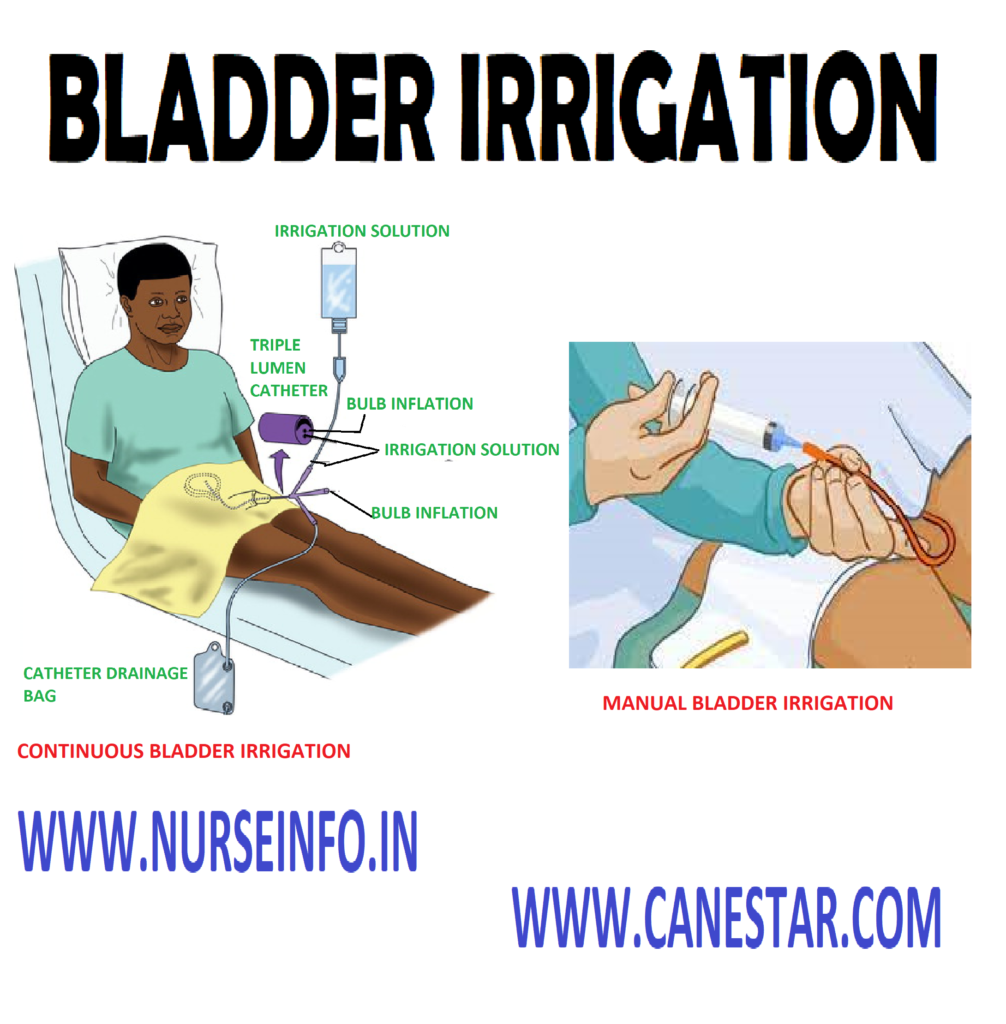
Bladder irrigation is a medical procedure commonly used to flush out the bladder with a sterile solution. This procedure is typically performed for therapeutic or diagnostic purposes and is carried out by healthcare professionals.
Here are some common reasons for bladder irrigation:
Hematuria (Blood in the Urine): Bladder irrigation may be used to treat or prevent blood clot formation in the bladder in cases of significant hematuria. The irrigation helps remove clots and blood from the bladder, reducing the risk of obstruction.
Postoperative Care: After certain urological surgeries, such as transurethral resection of the prostate (TURP) or bladder tumor removal, bladder irrigation may be performed to prevent blood clot formation and ensure clear urine drainage.
Infection Control: Bladder irrigation with an antimicrobial solution may be used to manage or prevent urinary tract infections (UTIs) in some cases.
Chemotherapy or Immunotherapy: Bladder irrigation may be employed in the treatment of bladder cancer. Medications, such as chemotherapy or immunotherapy agents, can be instilled into the bladder and then drained out to target cancer cells.
Diagnostic Procedures: Bladder irrigation may be part of diagnostic procedures, such as cystoscopy, to provide clear visualization of the bladder lining.
The procedure involves the insertion of a catheter into the bladder through the urethra. A sterile irrigation solution is then introduced into the bladder, and the fluid is allowed to drain out, carrying away debris, blood, or other substances. The process is typically repeated until the drained fluid is clear.
Bladder irrigation requires careful monitoring to avoid complications, and it should only be performed by trained healthcare professionals. Patients undergoing bladder irrigation may experience temporary discomfort, urgency, or increased frequency of urination.
Bladder
irrigation or wash is defined as washing of the urinary bladder by directly a
stream of solution into the bladder through the urinary meatus by means of a
catheter tubing and funnel
Purpose
To cleanse the bladder from
decomposed urine bacteria, excess mucus and pus
To medicate the lining of the bladder
of antiseptic irrigation
To prepare the bladder for surgery as
a preoperative measure
To promote healing
To relieve congestion and pain in
case of inflammatory conditions of cystitis
To arrest bleeding and prevent
clothing of blood
Solution Used
Normal saline 0.9%
Boric acid solution 2%
Sterile water
Acetic acid 1:4000 to treat
pseudomonas infection
Sodium nitrate 1:8000 to prevent clot
formation
KMO4 1:5000 – 1:10,000
Acriflavin 1:10,000
Silver nitrate 1:5,000
Mercury compounds in low
concentration
General Instructions
The temperature of the solution
needed for cleaning purpose body temperature in enough
The temperature of the solution
needed for therapeutic purposes ranging from 100-110 degree F
The maximum amount of solution used
for cleaning is 2 pints and also depends on the patient’s condition
Methods of Administration
Funnel and tubing method (open
method)
Irrigation can, rubber tubing and Y
connection
Asepto syringe (open method)
Preliminary Assessment
Check
Doctors order for specific
precautions and instructions
General condition of the patient
Diagnosis of the patient
Self care ability of the patient
Mental status to follow instructions
Articles available in the unit
Preparation of the Patient and Environment
Explain the sequence of the procedure
Arrange the articles at the bed side
Provide privacy
Place the patient in comfortable
position
Place the Mackintosh under the
buttocks
Equipment
Sterile Catheterization Pack
A sterile
tray containing:
Funnel, tubing 3 feet long which fits
the connection screw clip and glass connection
A small mug or pint measures to pour
solution
A sterile pint jug with required
solution
Solution thermometer kept in
antiseptic solution in a bottle if available
Medication if ordered
Bucket for emptying the return flow
Litmus paper
Procedure
Wash hands thoroughly
Wear gloves and empty the bladder
keeping outlet of catheter uncontaminated
After urine withdrawal, attach glass,
connection, tubing and funnel to the catheter
Place bucket or kidney tray
conveniently near the meatus
Hold the funnel lowered with one hand
and with other hand pour 75-100 ml of solution along sides of the funnel
Raise the tube and keep the funnel 30
cm above bed level
Never allow the funnel to be empty,
lower the funnel and slowly invert in over the bucket
Repeat procedure until the return
flow is clear
At the end of the procedure, clamp
tubing disconnects glass connection, tubing and funnel, gently remove catheter
and complete
In case of self-retaining catheter
connect it to the drainage bag
After Care
Provide catheter care
Remove the Mackintosh and position
the patient comfortably
Cover the patient with bed sheets
Replace the articles after cleaning
Wash hand thoroughly
Record the procedure and observations
in the nurse’s record sheet
BLADDER IRRIGATION – Purpose, Solution, Instructions, Administration methods, Preliminary Assessments, Preparation of patient and environment, Equipment, Procedure, After care
Sterilization in hospitals is a critical process designed to eliminate or destroy all forms of microbial life, including bacteria, viruses, fungi, and spores, from surfaces, instruments, and equipment. Sterilization is essential to prevent the transmission of infections between patients and to maintain a safe and sanitary healthcare environment.
STERILIZATION – Definition, Purpose, Scientific Principles, Methods of Sterilization
Sterilization
in hospitals is one of the important processes in order for prevention of
hospital acquired infections. Bacterial spores are the most resistant of all
living organisms because of their capacity to withstand external destructive
agents. Although the physical or chemical process by which all pathogenic and
microorganisms, including spores, are destroyed is not absolute, supplies and
equipment are considered sterile when necessary conditions have been met during
a sterilization process
DEFINITION
Sterilization is the process by which
an object becomes free of all the microorganisms. By sterilization, both the
pathogenic and non-pathogenic organisms are destroyed
Sterilization is a process by which
the pathogenic as well as spores and viruses are destroyed
Sterilization is the process by which
an article, a surface or a medium free from all microorganisms, both in
vegetative and sporing states, by removing or killing them
PURPOSE
To render the supplies/articles free
from pathogens
To make complete destruction of
microorganism
To sterilize instruments and
equipments used in the surgical practice
To keep the articles in such a
condition that they are ready for use at any time
For the safety of the patients
SCIENTIFIC PRINCIPLES
Dust and dirt harbors microorganisms
which adversely affect the well-being of patients and retards recovery
Proper care of articles prolong its
life ensures their utility and provides a neat and finished appearance which
promotes a feeling of comfort
Selection of appropriate simple
methods of sterilization saves energy, time and material
Water is a universal solvent and so
produces surface tension
Friction helps in removing dirt and
microorganisms from surface
Unpleasant odor, sight and noise are
disturbing to the patient
METHODS OF STERILIZATION
Natural method of sterilization: this
method is used to sterilize contaminated linen and bedpans. Direct sunlight
will have an effect on acid-fast microorganism. Place the linen or bedpans in
direct sunlight for 6 hours for two consecutive days
Physical method of sterilization:
heat kills all types of bacteria. Boiling is the most commonly used method, but
spore forming bacteria and viruses are not killing by boiling
Chemical method of sterilization: it
is also called as cold sterilization or disinfection by the disinfectants. A
chemical disinfectant is used which acts by coagulating the bacterial protein
or by changing the composition of protein so that is no longer exists in the
same form
Radiation or ultraviolet light
sterilization: this method is expensive. But nowadays, it is used for the
sterilization of plastic items such as disposable saline sets, catheters,
Ryle’s tubes, disposable syringes, etc
Physical methods of sterilization involve the application of heat, radiation, or filtration to eliminate or destroy microorganisms and their spores.
Here are some common physical methods of sterilization:
Autoclaving:
Method: Autoclaving utilizes steam under pressure.
Process: The items to be sterilized are exposed to high-pressure saturated steam at temperatures typically ranging from 121 to 134 degrees Celsius (250 to 273 degrees Fahrenheit).
Application: Autoclaving is effective for sterilizing surgical instruments, laboratory glassware, and other heat-resistant materials.
Dry Heat Sterilization:
Method: Dry heat sterilization involves hot air.
Process: Items are exposed to high temperatures (e.g., 160 to 180 degrees Celsius) for an extended period.
Application: Dry heat is suitable for items that may be damaged by moisture, such as powders, oils, and certain glassware.
Incineration:
Method: Incineration involves burning materials.
Process: The items are exposed to high temperatures until they are completely burned.
Application: Incineration is often used for the disposal of contaminated waste, particularly in laboratories.
Pasteurization:
Method: Pasteurization uses heat.
Process: This method involves heating liquids or food products to a specific temperature for a predetermined time to kill or reduce the number of pathogenic microorganisms.
Application: Commonly used in the food and beverage industry for items like milk and juices.
Radiation Sterilization:
Method: Radiation includes gamma radiation and electron beams.
Process: Items are exposed to ionizing radiation, disrupting the DNA of microorganisms.
Application: Gamma radiation is used for sterilizing medical devices, pharmaceuticals, and certain disposable items.
Filtration:
Method: Filtration uses porous materials.
Process: Liquids or gases are passed through filters with pore sizes small enough to trap microorganisms.
Application: Filtration is common for sterilizing liquids, especially in pharmaceutical and biotechnology industries.
PHYSICAL METHODS OF STERILIZATION
Heat kills
all types of bacteria. Boiling is the most commonly used method in day-to-day
working
Heat is the
safest and most useful agent for sterilization in hospitals. Methods of
applying heat for sterilization are exposure to steam under pressure, boiling,
etc
Boiling (Moist heating): boiling an instrument/article immersed fully in boiling water (100 degree Celcius) for 10 minutes will kill most of the pathogenic organisms
General Instructions
The articles should be clean
The articles should be fully immersed
in water
Close the sterilizer lid tightly
Note the time after the water has
started to boil
Boil it for 7 to 10 minutes
Remove the articles with chattel
forceps
Precautions
Do not pick articles in between, when
the boiling is in process
Do not boil sharp instruments such as
scissors, knives, needles, etc. because boiling blunt them
Advantages
Boiling can be used in the home
environment and other situation
It is one of the economic ways of
sterilizing articles
Disadvantages
Some bacteria and viruses and all spores are resistant to boiling
Boiling method cannot be used for the articles which are destroyed by moisture and heat
PHYSICAL METHODS OF STERILIZATION – Instructions, Precautions, Advantages, Disadvantages
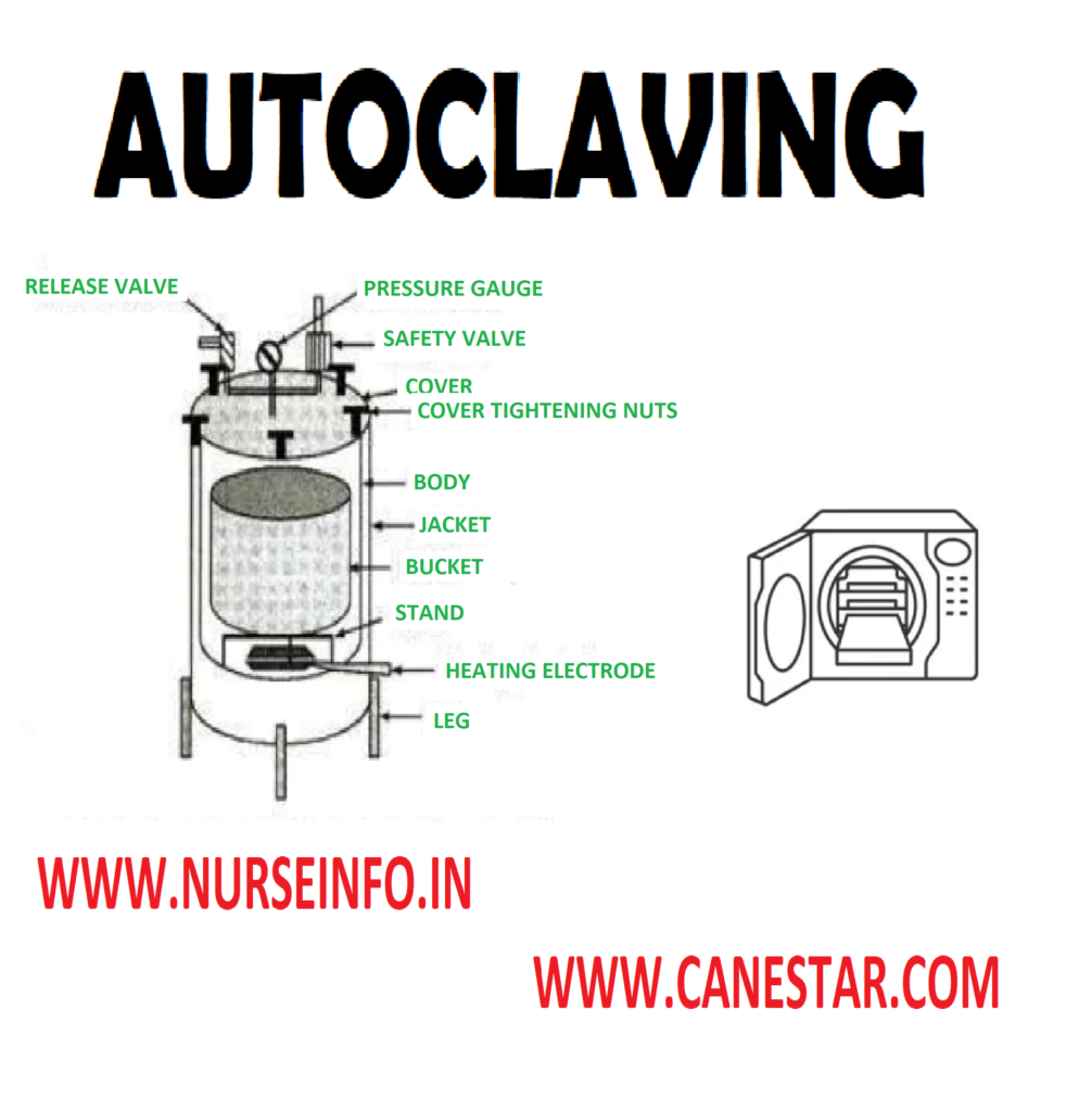
Autoclaving is a widely used and effective method of sterilization that involves the use of steam under pressure to kill or eliminate microorganisms, including bacteria, viruses, and spores.
AUTOCLAVING or AUTOCLAVE
Autoclaving
is the most common method used for sterilizing surgical instruments. It
accomplishes sterilization dependably without damage to most of the instrument
It is the
best, safest and effective method of sterilization. It destroys the spore
forming microorganisms. In this method, high temperature, pressure and humidity
is used to destroy the bacteria
Mechanism of Autoclave
In autoclaving, the sterilization is
done by steam under pressure. In an autoclave water boils and its vapor
pressure equals that of the surrounding atmosphere.
When pressure increases inside a
closed vessel the temperature at which boils also increases. Saturated steam
has better penetrating power
When steam comes into contact with a
cooler surface, it condenses into water and given up its latent heat to that
surface
Temperature: 121 degree celcius
Pressure: 15 pounds per square inch (PSI)
Time: 15-45 minutes
Instruments Used for Sterilization
Surgical instruments
Syringes and needles
Linen including gowns
Masks
Abdominal swabs and dressing
General Instructions
All articles must be clean and dry
The wrapper and container should
allow penetration of the steam into the article
The drum should not be too full nor
the contents arranged too compactly
Cans and jars must be opened and
turned to their sides so that steam can penetrate the contents
The temperature and pressure of the
steam must be 121 degree Celcius and 1.05 kg/cm2. So that it will
kill all types of microorganisms
The destruction of bacteria depends
upon the length of time the articles are exposed to steam under pressure. The
minimum time is 30 minutes
While operating an autoclave, all the
air in the chamber must be driven out and replaced by steam
When the autoclaving is over, wait for half an hour to dry the materials
Autoclave Uses:
Sterilization of Surgical Instruments:
Autoclaves are commonly used in healthcare settings to sterilize surgical instruments, medical devices, and equipment.
Laboratory Sterilization:
Research laboratories use autoclaves for sterilizing glassware, media, and other laboratory supplies.
Pharmaceutical Industry:
Autoclaves are utilized in the pharmaceutical industry to sterilize production equipment and ensure the sterility of pharmaceutical products.
Waste Disposal:
Autoclaving is employed in the disposal of laboratory waste and certain medical waste to render it non-infectious.
Food Industry:
In the food industry, autoclaves may be used for canning and preserving food by eliminating harmful microorganisms.
Autoclaving Tips:
Proper Loading:
Items must be properly arranged to ensure effective steam penetration.
Avoid overloading the autoclave to allow for adequate sterilization.
Use of Indicators:
Chemical and biological indicators are often used to monitor and confirm the effectiveness of the autoclaving process.
Regular Maintenance:
Autoclaves require regular maintenance to ensure proper functioning and calibration.
AUTOCLAVING – Mechanism, Temperature, Pressure, Time, Instructions
A hot air oven is a type of dry heat sterilization device commonly used in laboratories, research facilities, and industries. It operates by using hot air to achieve high temperatures, effectively eliminating microorganisms and achieving sterilization. Here are key features and uses of hot air ovens:
Features:
Design:
Hot air ovens typically consist of an insulated chamber made of metal, with a heating element to generate heat.
Temperature Control:
They are equipped with a thermostat or temperature controller to regulate the internal temperature accurately.
Timer:
Ovens often include a timer to set the duration of the sterilization cycle.
Air Circulation:
Efficient air circulation is important for even heat distribution throughout the chamber, ensuring uniform sterilization.
Ventilation:
Some ovens have ventilation systems to release excess moisture during the sterilization process.
Working Principle:
Heating Element:
The heating element in the oven generates heat.
Thermostat Control:
The thermostat controls the temperature, maintaining it at the desired level for the specified duration.
Sterilization Cycle:
Items to be sterilized are placed inside the oven. The sterilization cycle begins when the set temperature is reached.
Cooling:
After the sterilization process, the oven may include a cooling phase before the items can be safely removed.
HOT AIR OVEN
High
temperature and comparatively long exposure times are required for hot air
oven. Various types of powders, glass materials, etc. are sterilized by this
method
Mechanism of Hot Air Oven
It works on the principle of
sterilization by dry heat
Temperature: 160 degree celcius
Time: one hour
Articles sterilized include
glassware, forceps, scissors, scalpels, syringes, liquid paraffin and dusting
powder
Advantages
All types of microorganisms including
spores are killed by this method
It is safest method of sterilization
Disadvantage
It is costly method of sterilization
General Instructions
Glassware should be perfectly dry before placing in the oven
Oven must be allowed to cool down for 2 hours before door is opened after sterilization
It should not be overloaded
Articles should be arranged in such a manner that free circulation ofd air is possible
Uses:
Laboratory Sterilization:
Hot air ovens are commonly used in laboratories for sterilizing glassware, instruments, and other equipment that can withstand dry heat.
Pharmaceutical Industry:
In pharmaceutical manufacturing, hot air ovens are used for sterilizing equipment and containers used in drug production.
Dental Clinics:
Dental instruments that are heat-resistant can be sterilized in hot air ovens.
Research Facilities:
Research laboratories use hot air ovens for sterilizing media, instruments, and other materials.
Textile Industry:
Some textile materials and products may be subjected to hot air oven treatment for sterilization.
Advantages:
Dry Sterilization:
Hot air ovens provide dry heat sterilization, which is suitable for items sensitive to moisture.
Ease of Use:
They are relatively simple to operate and do not require water or other chemicals.
Uniform Heating:
Efficient air circulation ensures uniform heat distribution, contributing to effective sterilization.
Economical:
Hot air ovens are generally cost-effective compared to other sterilization methods.
HOT AIR OVEN – Mechanism, Temperature, Time, Advantage, Disadvantage, General Instructions
Radiation sterilization is a method that uses ionizing radiation, such as gamma rays or electron beams, to eliminate or reduce the microbial load on various products, surfaces, or materials. This process disrupts the DNA and other cellular components of microorganisms, rendering them unable to reproduce or cause infections. Here are key aspects of radiation sterilization:
Types of Ionizing Radiation:
Gamma Radiation:
Source: Gamma radiation is often emitted from a radioactive isotope, such as cobalt-60.
Penetration: Gamma rays penetrate deep into materials, making them suitable for the sterilization of dense or bulky items.
Application: Commonly used for the sterilization of medical devices, pharmaceuticals, and certain disposable items.
Electron Beam (E-beam) Radiation:
Source: E-beam radiation is generated using an electron accelerator.
Penetration: Electron beams have limited penetration and are suitable for surface sterilization or treating thin materials.
Application: Used for sterilizing medical devices, packaging materials, and some pharmaceutical products.
Radiation Sterilization Process:
Preparation:
Products or items to be sterilized are prepared and packaged appropriately.
Irradiation:
The items are exposed to ionizing radiation in a controlled environment, either by gamma radiation from a gamma source or by passing through an electron beam.
Dose Control:
The dose of radiation is carefully controlled based on the type of material and the required level of sterilization.
Dosimetry:
Dosimeters are used to measure the absorbed dose of radiation, ensuring that the products receive the necessary amount for effective sterilization.
Post-Irradiation Handling:
After irradiation, the products are handled carefully to avoid recontamination.
Advantages of Radiation Sterilization:
Cold Sterilization:
Radiation sterilization is a cold process, meaning it doesn’t involve high temperatures that could damage heat-sensitive materials.
Uniform Sterilization:
Radiation provides uniform sterilization, even in complex or densely packed materials.
No Residue:
Unlike some chemical sterilization methods, radiation leaves no residues on the sterilized items.
Time Efficiency:
The process is relatively quick, allowing for efficient sterilization on a large scale.
Applications:
Medical Devices:
Single-use medical devices, implants, and surgical instruments.
Pharmaceuticals:
Sterilization of pharmaceutical products and ingredients.
Packaging Materials:
Sterilization of packaging materials for medical and pharmaceutical products.
Cosmetics:
Sterilization of cosmetic products to ensure safety and shelf life.
Food Industry:
Used for sterilizing certain food products, spices, and packaging materials.
RADIATION STERILIZATION
Radiation or ultraviolet light sterilization: this method is expensive. But nowadays it is used for the sterilization of plastic items such as disposable saline set, catheter, Ryle’s tubes, etc
Gas sterilization: ethylene oxide gas is employed as a sterilizing agent in especially designed chambers in which temperature and humidity can be controlled and from which air can be evacuated
After
exposure period of 3 to 6 hours is needed. Other gases employed for
sterilization are formaldehyde and betapropiolatone
Articles Sterilized
Surgical instruments with optical
lenses
Tubing and plastic parts of heart
lung machines
Ventilator tubes
Disposable syringe
Pillows and mattresses
Advantages
Exposure to formaldehyde gas under
conditions of controlled humidity, temperature, and the time exposure will
destroy all vegetative forms of bacteria, viruses, and most of the spores
The best results can be obtained with
high concentration of gas humidity above 60% and temperature of not less than
180 degree celcius
Disadvantages
Ethylene oxide has a pungent smell
It is an irritant to eye, mucous
membrane and skin
Radiation Method
There are
two type of radiation are non-ionizing radiation and ionizing radiation
Non-ionizing
radiation methods are infra-red and ultra-violet radiation
Ionizing
radiation methods include X-rays, gamma rays; and cosmic rays are highly lethal
to DNA and other vital cell constituents
Advantages
Instruments like disposable syringe
catheters hypo-dermic needles and sharp instruments that cannot withstand heat,
can be sterilized by this method
Instruments which are covered in
plastic packs or aluminum foils can be sterilized by this method
Disadvantages
Since radiation in a straight line
and do not penetrate only the surface of an object in straight line is irradiated
The bacteria in shadows are
unaffected, so all the surfaces should be exposed to the radiation
RADIATION METHOD OF STERILIZATION – Gas & Radiation sterilization, advantages , disadvantages