
PULMONARY STENOSIS – Etiology, Risk Factors, Signs and Symptoms, Diagnostic Evaluations and Management
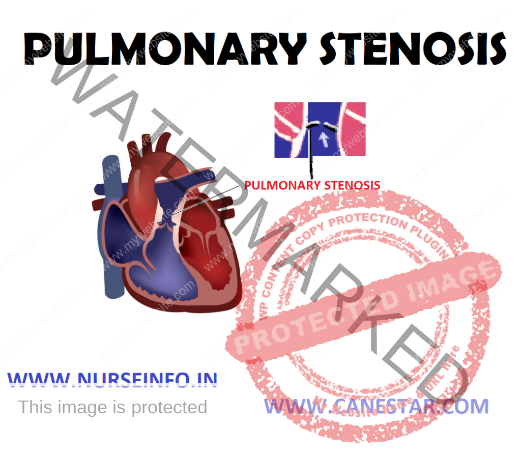
Pulmonary valve stenosis is a condition in which the flow of blood from heart to lungs is slowed by a deformity or near pulmonary valve. In pulmonary valve stenosis, one or more of the leaflets may be defective or too thick, or the leaflets may not separate from each other properly. If this happens, the valve does not open correctly, restricting blood flow.
ETIOLOGY
- Pulmonary valve stenosis usually occurs when the pulmonary valve does not grow properly during fetal development
- Other heart abnormalities also are often present at birth (congenital) in babies who have pulmonary valve stenosis. It is not known what causes the valve to develop abnormally.
Other Contributing Conditions
Sometimes other medical conditions or having an artificial valve can cause the condition in older people.
- Carcinoid syndrome: this syndrome is a combination of signs and symptoms, including flushing of the skin and diarrhea. Carcinoid syndrome results from the release of a chemical, serotonin, from growths called carcinoid tumors located in the digestive system. People with carcinoid syndrome may develop problems with their heart valves from the serotonin.
- Rheumatic fever: this is a complication of an infection caused by Streptococcus bacteria, such as strep throat or scarlet fever. Rheumatic fever may injure the heart valves.
RISK FACTORS
Because most causes of pulmonary valve stenosis develop before birth, there are not many known risk factors. However, certain conditions can increase your risk of developing pulmonary valve stenosis, including:
- Carcinoid syndrome
- Rheumatic fever
- Noonan’s syndrome
SIGNS AND SYMPTOMS
- Pulmonary valve stenosis symptoms vary, depending on the extent to which the valve is obstructed. People with mild pulmonary stenosis will usually not have any symptoms. Those with more significant stenosis often first notice symptoms while exercising.
- Pulmonary valve stenosis signs and symptoms may include:
Heart murmur – an abnormal whooshing sound heard using a stethoscope, caused by turbulent blood flow
Shortness of breath, especially during exertion
Chest pain
Loss of consciousness
Fatigue
Palpitations
DIAGNOSTIC EVALUATIONS
A variety of tests to confirm the diagnosis:
- Electrocardiogram: an electrocardiogram records the electrical activity of heart each time it contracts. During this procedure, patches with wires are placed on chest, wrists and ankles. The electrodes measure electrical activity, which is recorded on paper. This test helps determine if the muscular wall of right ventricle is thickened (ventricular hypertrophy).
- Echocardiography: echocardiography use high-pitched sound waves to produce an image of the heart. Sound waves bounce off the heart and produce moving images that can be viewed on a video screen. This test is useful for checking the structure of the pulmonary valve, the location and severity of the narrowing and the functioning of the right ventricle of the heart.
- Other imaging tests: magnetic resonance imaging and CT scans are sometimes used to confirm the diagnosis of pulmonary valve stenosis.
- Cardiac catheterization: during this procedure, doctor inserts a thin, flexible tube into an artery or vein in groin and weaves it up to the heart or blood vessels. A dye is injected through the catheter to make blood vessels visible on X-ray pictures. Doctors also use cardiac catheterization to measure the blood pressure in the heart chambers and blood vessels.
MANAGEMENT
A physician may prescribe medications that make it easier for blood to flow through the heart’s chambers. Examples of medications prescribed may include:
- Prostaglandins to improve blood flow
- Blood thinners to reduce clotting
- Antiarrhythmic that prevents irregular heart rhythms
SURGICAL MANAGEMENT
- Some cases of pulmonary stenosis are mild and do not require treatment except for routine checkups. However, if the case is more serious, it may need either balloon valvuloplasty or oper-heart surgery
- The decision to perform a balloon valvuloplasty or open-heart surgery depends on the extent to which the pulmonary valve is obstructed. Pulmonary stenosis is classified as mild, moderate or severe, depending on a measurement of the blood pressure difference between the right ventricle and pulmonary artery.
Balloon Valvuloplasty
This technique, which tends to be the first choice for treatment, uses cardiac catheterization to treat pulmonary valve stenosis. During this procedure, doctor threads a small tube through a vein in leg and up to heart. An uninflated balloon is placed through the opening of the narrowed pulmonary valve. Then he inflates the balloon, opening up the narrowed pulmonary valve and increasing the area available for blood flow. The balloon is then removed.
Open-Heart Surgery
- Balloon valvuloplasty cannot be used for cases of pulmonary stenosis that occur above the pulmonary valve (supravalvular) or below the valve (subvalvular). Open-heart surgery is required for these types of stenosis and occasionally for valvular stenosis.
- During this surgery, doctor repairs the pulmonary artery or the valve to allow blood to pass through more easily. In certain cases, doctor may replace the pulmonary valve with an artificial valve.
- Some people with pulmonary stenosis have other congenital heart defects, and these may be repaired at the time of surgery. As with balloon valvuloplasty, there is a slight risk of bleeding, infection or blood clots associated with the surgery.
NURSING MANAGEMENT
Nursing Diagnosis
- Ineffective airway clearance related to bronchoconstriction, increased mucus production, ineffective cough, possible bronchopulmonary infection
- Risk for infection related to compromised pulmonary function, retained secretions and compromised defense mechanism
- Impaired gas exchange related to chronic pulmonary obstruction due to destruction of alveolar capillary membrane
- Imbalanced nutritional status less than body requirements related to increased work of breathing, air swallowing, drug effects with resultant wasting of respiratory and skeletal muscles
- Activity intolerance related to compromised pulmonary function, resulting in shortness of breath and fatigue
- Ineffective coping related to the stress of living with chronic disease, loss of independence, depression, anxiety disorder
Nursing Interventions
- Improving airway clearance:
Eliminate all pulmonary irritants, particularly cigarette smoking
Cessation of smoking usually results in less pulmonary irritation, sputum production, and cough
Keep patient’s room as dust-free as possible
Add moisture to indoor environment
Administer bronchodilators to control bronchospasm and assist with raising sputum
Assess for side effects – tremulousness, tachycardia, cardiac dysrhythmias, central nervous system stimulation, and hypertension
Auscultate the chest after administration of aerosol bronchodilator to assess for improvement of aeration and reduction of adventitious breath sounds.
Observe if patient has reduction in dyspnea
Monitor serum theophylline level as ordered, to ensure therapeutic level and prevent toxicity
Use postural drainage position to aid in clearance of secretions, because mucopurulent secretions are responsible for airway obstruction
Encourage the patient to take high-liquid fluids
Provide inhalation-nebulized water to humidify bronchial tree and liquefy sputum
- Controlling infection:
Recognize early manifestations of respiratory infection – increased dyspnea, fatigue, and change in color, amount and character of sputum, nervousness, irritability, low-grade fever
Obtain sputum for smear and culture
Administer prescribed antimicrobial tree, thus clearing the airway
Advice to live in proper ventilated room
Advice to take medicine regularly as prescribed by doctor
Advice to live in hygienic place, i.e. free from dust, etc
- Improving gas exchange
Watch for restlessness, aggressiveness, anxiety, or confusion; central cyanosis and shortness of breath at rest, which frequently is caused by acute respiratory insufficiency and may signal respiratory failure
Give low-flow oxygen therapy as ordered to correct hypoxemia in a controlled manner and minimize carbon dioxide retention
Be prepared to assist with intubation and mechanical ventilation if acute respiratory failure and rapid carbon dioxide retention occur
Advice to live in ventilated room and contact with fresh air daily
Advice to do breathing exercise as prescribed by doctors
- Improving nutrition:
Take nutritional history, weight, and anthropometric measurements
Encourage frequent small meal if patient is dyspneic, even small increase in abdominal contents may press on diaphragm and impede breathing
Avoid food which causes abdominal discomfort
Give supplemental oxygen therapy while patient is eating to relieve dyspnea, as directed
Monitor body weight regularly
- Increased activity tolerance:
Encourage the patient for daily exercise and discuss about walking, bicycling, swimming and its benefits
Encourage patient to carry out regular exercise program to increase physical endurances
Train patient in energy conservation techniques
Educate about the benefits of exercise to patient as well as to family
- Enhancing coping:
Understand that the constant shortness of breath and fatigue make the patient irritable, anxious and depressed, with feeling of helplessness/hopelessness
Assess the patient for reactive behavior (anger, depression, and acceptance)
Strengthen the patient’s self-image
Allow the patient to express feelings and retain the mechanisms of dental and repression
Support spouse/family members
PULMONARY STENOSIS – Etiology, Risk Factors, Signs and Symptoms, Diagnostic Evaluations and Management
