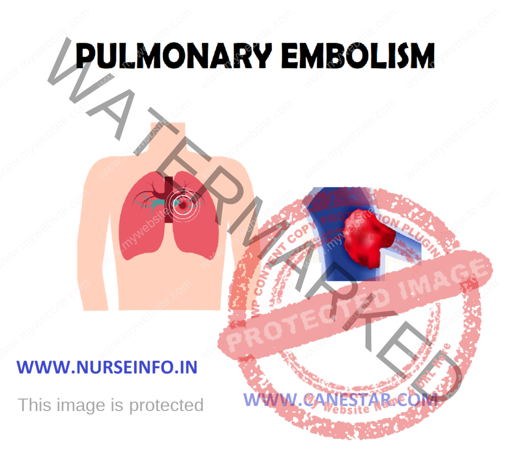
PULMONARY EMBOLISM – Etiology, Pathophysiology, Clinical Manifestation, Diagnostic Evaluation, Management and Complication
- Pulmonary embolism refers to the obstruction of one or more pulmonary arteries, by a thrombus that originates somewhere in the venous system or in the right heart. It may be associated with trauma, surgery, pregnancy CCF, advanced age (above 60 years), and immobility.
- Pulmonary embolism is defined as an obstruction of one or more pulmonary arteries of the lungs which are blocked, or thrombus that originates somewhere in the venous system or right side of the heart
ETIOLOGY
- Venous stasis
- Hypercoagulation
- Damage to the endothelium of blood vessels
The predisposing factors of pulmonary embolism:
- Prolonged immobilization
- Concurrent phlebitis
- Heart failure, strokes
- Malignancy
- Advancing age, estrogen therapy
- Oral contraceptives
- Obesity
PATHOPHYSIOLOGY
Emboli gets dislodged in pulmonary circulation —- decreased perfusion of the lungs —- increased pulmonary embolism resistance —- increased in pulmonary artery and venous pressure —- increase in ventricular overload —- right ventricular hypertrophy —- stroke volume and decreased cardiac output —- cardiogenic shock and circulatory failures —- pulmonary vasoconstriction and bronchospasm —- atelectasis
CLINICAL MANIFESTATION
- Rapid onset of dyspnea at rest, pleuritic chest pain, cough and syncope, delirium, apprehension, tachypnea, diaphoresis, hemoptysis.
- Chest pain with apprehension and a sense of impending doom occurs when most of the pulmonary artery is obstructed
- Tachycardia, rales fever, hypotension, cyanosis, heart gallop, loud pulmonic component of S2.
- Calf and thigh pain, edema, erythema, tenderness or palpable cord
DIAGNOSTIC EVALUATION
- History-taking
- Assess for signs and symptoms; blood study
- Thrombotic imaging; V/Q scan or single photoemission computerized
- Pulmonary angiography
- D-Dimer assay for low intermediate probability of pulmonary embolism
- ABG levels: Decreased PaO2 usually found due to perfusion abnormality of lung
- Chest X-ray – normal or possible wedge-shaped infiltrate
MANAGEMENT
Assessment
- Assess the signs and symptoms of pulmonary embolism
- Assess the vital signs of patient, especially respiration
- Assess the chest pain intensity
- Assess for edema and fever
- Assess the hypotension and cyanosis of the patient
MEDICAL MANAGEMENT
It is focused on anticoagulant to reduce the size of thrombus and maintain the cardiopulmonary stability
- Anticoagulant: it begins with heparin 5000 IU and warfarin, 2.5 mg/day, as maintained dose regulation upon prothrombin time
- Thrombolytic therapy: cytokines resolve the thrombus or emboli quickly and restore normal hemodynamic therapy to reducing pulmonary hypertension improving pulmonary perfusion, oxygenation, and cardiac output.
- Also to improve patient’s respiratory and vascular status:
Oxygenation therapy to correct hypoxia
Venous stasis is reduced by using elastic stoking
Elevating leg for increase venous flow
Hypotension relaxed with fluids
Chest pain and apprehension one treated with analgesics
Surgical Management
When anticoagulation is contraindicated or patient has recurrent embolization or develops serious complication from drug therapy.
- Interruption of vena cava: reduces channel size to prevent lower extremity from reaching lungs. Accompanied by:
Ligation, placation or clipping of the inferior vena cava
Placement of transversely inserted intraluminal filter inferior vena cava to prevent migration of emboli
- Embolectomy
Nursing Diagnosis
- Ineffective breathing pattern related to increase in alveolar dead airspace and possible changes in lung mechanics from embolism
- Ineffective tissue perfusion (pulmonary) related to decreased blood circulation
- Acute pain (pleuritic) related to congestion, possible pleural effusion, lung infraction
- Anxiety related to dyspnea to altered hemodynamic factors and anticoagulant therapy
Nursing Interventions
Improve the Breathing
- Assess for hypoxia, dyspnea, headache, restlessness, apprehension, pallor, cyanosis, behavioral changes
- Monitor vital signs, ECG, oximetry and ABG levels for adequacy of oxygenation
- Monitor patient’s response to IV fluids/vasopressor
- Monitor oxygen therapy – used to relieve hypoxia
- Prepare – patient for assisted ventilation does not respond to supplemental oxygen. Hypoxia is due to abnormalities of V/Q mismatch
Improving Tissue Perfusion
- Closely monitor shock – decreasing BP, tachycardia, cool, clammy skin
- Monitor prescribed medication given to preserve right-sided heart filling pressure and increase BP
- Maintain patient on bed rest during acute phase to reduce oxygen demand and risk of bleeding
- Monitor urinary output hourly because this reduces renal perfusion
- Antiembolism compression stocking should provide a compression of 30-40 mm Hg.
Relieving Pain
- Watch patient for sign of discomfort and pain
- Ascertain if pain worsens with deep breathing and coughing for friction rub
- Give morphine as prescribed and monitor for pain relief
- Monitor signs of hypoxia thoroughly when anxiety, restlessness and agitation of new onset are noted
Relieving Anxiety
- Correct dyspnea and relieve physical discomfort
- Explain diagnostic procedure and the role and correct the misconception
- Listen to the patient’s concern; attentive listing relieves anxiety and reduces emotional distress
- Speak calmly and slowly
COMPLICATIONS
- Bleeding as a result of treatment
- Respiratory failure
- Pulmonary hypertension
Health Education
- Advice patient for possible need to continue taking anticoagulant therapy for 6 weeks up to an indefinite period
- Teaching about sign of bleeding, especially of gum, nose, bruising, blood in urine and stool
- Instruct patient to tell dentist about taking anticoagulant therapy
- Warm against inactivity for prolonged periods or sitting with legs crossed to prevent recurrence
- Warn against support activity that may cause trauma or injury to legs and predispose to thrombus
- Encourage to wear medic alert bracelet, identifying as an anticoagulant user
PREVENTION
Preventing clots in the deep veins in legs (deep vein thrombosis) will help prevent pulmonary embolism. For this reason, most hospitals are aggressive about taking measures to prevent blood clots:
- Anticoagulants: anticoagulants are given to people at risk of clots before and after an operation as well as to people admitted to the hospital with a heart attack, stroke or complications of cancer
- Graduated compression stockings: compression stockings steadily squeeze your legs, helping veins and leg muscles move blood more efficiently. They offer a safe, simple and inexpensive way to keep blood from stagnating after general surgery
- Pneumatic compression: this treatment uses thigh-high or calf-high cuffs that automatically inflate with air and deflate every few minutes to massage and squeeze the veins in legs and improve blood flow
- Physical activity: moving as soon as possible after surgery can help prevent pulmonary embolism and hasten recovery overall. This is one of the main reasons nurse may push to get up, even on your day of surgery, and walk despite pain at the site of your surgical incision.


