
PNEUMOTHORAX – Classification, Etiology, Risk Factors, Pathophysiology, Signs and Symptoms, Diagnostic Evaluations and Management
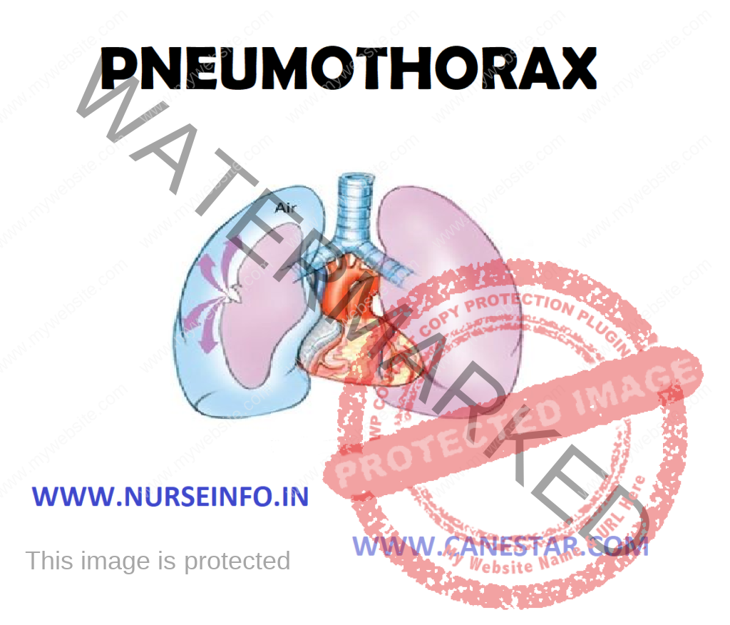
- Air in the pleural space occurring spontaneously or from trauma. Pneumothorax is the presence of air in the pleural space that prohibits complete lung expansion
- Pneumothorax is an abnormal collection of air or gas in the pleural space separating the lung from the chest wall which may interfere with normal breathing
- The lung expansion occurs when the pleural lining of the chest wall and the visceral lining of the lung maintain negative pressure in the pleural space. When the continuity of this system is lost, the lung collapses, resulting in pneumothorax
CLASSIFICATION OF PNEUMOTHORAX
- Spontaneous pneumothorax: sudden onset of air in the pleural space with deflation of the affected lung in the absence of trauma
- Open pneumothorax: implies an opening in the chest wall large enough to allow air to pass freely in and out of thoracic cavity with each attempted respiration
- Tension pneumothorax: build-up of air under pressure in the pleural space resulting in interference with filling of both the heart and lungs
ETIOLOGY
Primary spontaneous: the exact cause of primary spontaneous pneumothorax is unknown
RISK FACTORS
- Male
- Smoking
- Family history
- Secondary spontaneous pneumothorax occurs in the setting of a variety of lung diseases
In children, additional causes include:
- Measles
- Echinococcosis
- Inhalation of a foreign body
- Congenital malformations
- Marfan’s syndrome
- Homocystinuria
- Ehlers-Danlos syndrome
- Alpha 1-antitrypsin deficiency (which leads to emphysema)
PATHOPHYSIOLOGY
- When there is a large hole in the chest wall, the patient will have a steal in the ventilation of other lung
- A portion of the tidal volume will move back and forth through the hole in the chest wall, rather than the trachea as it normally does
- Spontaneous pneumothorax is usually due to rupture of a subpleural bleb
- May occur secondary to chronic respiratory disease or idiopathically
- May occur in healthy people, particularly in thin, white males and those with family history of pneumothorax
SIGNS AND SYMPTOMS
- Hyperresonance (tympany) to percussion on, diminished breath sounds on affected side
- Tracheal deviation away from affected side of tension pneumothorax
- Clinical picture of open or tension pneumothorax is one of air hunger, agitation, hypotension and cyanosis
- Mild to moderate dyspnea and chest discomfort may be present with spontaneous pneumothorax
- Tachypnea
- Asymmetrical chest expansion
DIAGNOSTIC EVALUATION
- Chest X-ray: chest X-ray of left-sided pneumothorax. The left thoracic cavity is partly filled with air occupying the pleural space. The mediastinum is shifted to the opposite side
- Computed tomography: computed tomography can be useful in particular situations. In some lung diseases, especially emphysema, it is possible for abnormal lung areas such as bullae (large air-filled sacs) to have the same appearance as a pneumothorax on chest X-ray, and it may not be safe to apply any treatment before the distinction is made and before the exact location and size of the pneumothorax is determined
- Ultrasound: ultrasound is commonly used in the evaluation of people who have sustained physical trauma. Ultrasound may be more sensitive than chest X-rays in the identification of pneumothorax after blunt trauma to the chest.
MANAGEMENT
Conservative
Oxygen given at a high flow rate may accelerate resorption as much as fourfold
Aspiration
Aspiration is equally effective as the insertion of a chest tube. This involves the administration of local anesthetic and inserting a needle connected to a three-way tap, up to 2.5 liters of air (in adults) are removed. If there has been significant reduction in the size of pneumothorax on subsequent X-ray, the remainder of the treatment can be conservative.
Chest Tube
A chest tube (or intercostals drain) is the most definitive initial treatment of pneumothorax. These are typically inserted in an area under the axilla called the ‘safe triangle’, where damage to internal organs can be avoided. This is delineated by a horizontal line at the level of the nipple and two muscles of the chest wall (latissimus dorsi and pectoralis major). Local anesthetic is applied. Two types of tubes may be used. In spontaneous pneumothorax, small-bore (smaller than 14 F, 4.7 mm diameter) tubes may be inserted by the Seldinger technique, and larger tubes do not have an advantage. In traumatic pneumothorax, larger tubes (28F, 9.3 mm) are used.
Surgical Management
- Pleurodesis and surgery
- Pleurodesis is a procedure that permanently obliterates the pleural space and attaches the lung to the chest wall
- Thoracotomy (surgical opening of the chest), with identification of any source of air leakage and stapling of blebs
- Pleurectomy (stripping of the pleural lining) of the outer pleural layer and pleural abrasion (scraping of the pleura) of the inner layer
PNEUMOTHORAX – Classification, Etiology, Risk Factors, Pathophysiology, Signs and Symptoms, Diagnostic Evaluations and Management
