ESOPHAGEAL CANCER – Definition, Etiology, Types, Stages, Signs and Symptoms, Diagnostic Evaluation and Management
Esophageal cancer is malignancy of the esophagus. Esophageal tumors usually lead to dysphagia (difficulty swallowing), pain and other symptoms. Small and localized tumors are treated surgically with curative intent. Large tumors tend not to be operable and hence are treated with palliative care; their growth can still be delayed with chemotherapy, radiotherapy or a combination of the two. In some cases chemo and radiotherapy can render these large tumors operable. Prognosis depends on the extent of the disease and other medical problems, but is generally fairly poor.
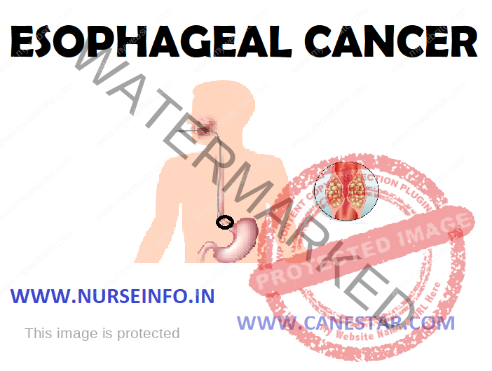
DEFINITION
Cancer is formed in tissues lining the esophagus. Two types of esophageal cancer are squamous cell carcinoma (cancer that begins in flat cells lining the esophagus) and adenocarcinoma (cancer that begins in cells that make and release mucus and other fluids).
CAUSES AND RISK FACTORS
- Smoking
- Heavy drinking
- Damage from acid reflux
ACID REFLUX RAISES RISK
This sphincter also prevents stomach contents from refluxing back into the esophagus. If stomach juices with acid and bile come into the esophagus, it causes indigestion or heartburn. E.g. reflux and gastroesophageal reflux disease (GERD)
- A medical history of other head and neck cancers increases the chance of developing a second cancer in the head and neck area, including esophageal cancer
- Plummer-Vinson syndrome (anemia and esophageal webbing)
- Radiation therapy
- Celiac disease predisposes towards squamous cell carcinoma
- Obesity
- Thermal injury as a result of drinking hot beverages
ESOPHAGEAL CANCER TYPES
- Adenocarcinoma is the most common type especially in white males. It starts in gland cells in the tissue, most often in the lower part of the esophagus near the stomach. The major risk factors include reflux and Barrett’s esophagus
- Squamous cell carcinoma or cancer, also called epidermoid carcinoma, begins in the tissue that lines the esophagus, particularly in the middle and upper parts. Risk factors include smoking and drinking alcohol.
STAGES OF CANCER
Stage 0: (carcinoma in situ): in stage 0, abnormal cells are found in the innermost layer of tissue lining the esophagus. These abnormal cells may become cancer and spread into nearby normal tissue. Stage 0 is also called carcinoma in situ
Stage I: in stage I, cancer has formed and spread beyond the innermost layer of tissue to the next layer of tissue in the wall of the esophagus.
Stage II: stage II esophageal cancer is divided into stage IIA and stage IIB, depending on where the cancer has spread
Stage IIA: cancer has spread to the layer of esophageal muscle or to the outer wall of the esophagus
Stage IIB: cancer may have spread to any of the first three layers of the esophagus and to nearby lymph nodes
Stage III: in stage III, cancer has spread to the outer wall of the esophagus and may have spread to tissues or lymph nodes near the esophagus
Stage IV: stage IV esophageal cancer is divided into stage IVA and stage IVB, depending on where the cancer has spread
Stage IVA: cancer has spread to nearby or distant lymph nodes
Stage IVB: cancer has spread to distant lymph nodes in other parts of the body
SIGNS AND SYMPTOMS
- Dysphagia
- Odynophagia (painful swallowing)
- Pain behind the sternum or in the epigastrium, often of a burning, heartburn
- Hoarse-sounding cough, a result of the tumor affecting the recurrent laryngeal nerve
- Nausea and vomiting, regurgitation of food, coughing
- Cough, fever
- If the disease has spread elsewhere, this may lead to symptoms related to this: liver metastasis could cause jaundice and ascites, lung metastasis could cause shortness of breath, pleural effusions, etc
DIAGNOSTIC EVALUATION
- Esophagoscopy: a procedure to look inside the esophagus to check for abnormal areas. An esophagoscope is inserted through the mouth or nose and down the throat into the esophagus. An esophagoscope is a thin, tube-like instrument with a light and a lens for viewing
- Endoscopic ultrasound (EUS): A procedure in which an endoscope is inserted into the body, usually through the mouth or rectum. A probe at the end of the endoscope is used to bounce high-energy sound waves off internal tissues or organs and make echoes. The echoes form a picture of body tissues called a sonogram. This procedure is also called endosonography
- CT with contrast
- EGI, endoscopy: this involves the passing of a flexible tube down the esophagus and examining the wall
- Biopsies taken of suspicious lesions are then examined histologically for signs of malignancy
- PET scan (positron emission tomography scan): a procedure to find malignant tumor cells in the body. A small amount of radionuclide glucose (sugar) is injected into a vein. The PET scanner rotates around the body and makes a picture of where glucose is being used in the body. Malignant tumor cells show up brighter in the picture because they are more active and take-up more glucose than normal cells does.
- Barium swallow: a series of X-rays of the esophagus and stomach. The patient drinks a liquid that contain barium (a silver-white metallic compound). The liquid coats the esophagus and stomach, and X-rays are taken. This procedure is also called an upper GI series
MANAGEMENT
Esophageal Stent
If the patient cannot swallow at all, an esophageal stent may be inserted to keep the esophagus patent; stents may also assist in occluding fistulas
- Laser therapy is the use of high-intensity light to destroy tumor cells; it affects only the treated area. This is typically done if the cancer cannot be removed by surgery. The relief of a blockage can help to reduce dysphagia and pain
- Photodynamic therapy: a type of laser therapy, involves the use of drugs that are absorbed by cancer cells. When exposed to a special light, the drugs become active and destroy the cancer cells
- Chemotherapy
- Radiotherapy is given before, during or after chemotherapy or surgery
Surgical Management
Esophagectomy
Surgery to remove some or most of the esophagus is called an esophagectomy. Often a small part of the stomach is removed as well. The upper part of the esophagus is then connected to the remaining part of the stomach. Part of the stomach is pulled up into the chest or neck to become the new esophagus. It may be done by two approaches:
- Open esophagectomy: many different approaches can be used in operating on esophageal cancer. For atransthoracic esophagectomy, the esophagus is removed with the main incisions in the abdomen and the chest. If the main incisions are in the abdomen and neck, it is called a transhiatal esophagectomy. Some approaches use incisions in the neck, chest and abdomen
- Minimally invasive esophagectomy: for some early cancers, the esophagus can be removed through several small incisions instead of 1 or 2 large incisions. The surgeon puts a scope (like a tiny telescope) through one of the incisions to see everything during the operation
NURSING MANAGEMENT
- Altered nutrition less than body requirement difficulty in swallowing secondary to disease condition
- Pain related to disease condition or surgery
- Anxiety related to disease condition, its treatment and prognosis
- Knowledge deficit related to treatment and prognosis
- Altered nutrition less than body requirement difficulty in swallowing secondary to disease condition
Intervention
- Assess the level of daily nutrition
- Assess the likes and dislikes of the patients
- Provide small and frequent diet to the patient
- If needed provide the food through nesogastric tube
- Maintain intake output chart
- Pain related to disease condition or surgery
Intervention
- Assess the level of pain
- Provide comfortable position to the patient
- Provide diversional therapy to the patient
- Administer analgesic as per the doctor’s order
- Anxiety related to disease condition, its treatment and prognosis
Interventions
- Assess the level of anxiety
- Diversional therapy provided to the patient
- Comfortable environment provided to the patient
- Answer each question of the patient
- Administer antianxiety drug as order by the doctor
- Knowledge deficit related to treatment and prognosis
Interventions
- Asses the level of knowledge related to disease condition
- Answer each question of the patient
- Clarify all doubts of the patient
- Give information regarding the disease and its treatment