Home Blog Page 22

INJURIES DUE TO IONIZING RADIATIONS

INJURIES DUE TO IONIZING RADIATIONS – General Considerations, Acute Radiation Syndrome, Damage to Embryo by Irradiation in Fetal Life and Local Radiation Injury

GENERAL CONSIDERATIONS

Ionizing radiations are either electromagnetic or particulate in nature. They are derived from natural or artificial radioactive isotopes, nuclear reactors, diagnostic and therapeutic equipment (e.g. X-rays, CT scan) and the complex shower of particles from the outer space known as cosmic rays. The radiations include alpha, beta, and gamma particles and electromagnetic waves.

Radiation injury is seen in survivors of nuclear war, after radiotherapy, persons engaged in industries involving radioactive materials, radiologists and radiotherapists. In some parts of the world, radioactive sand accounts for low dose continuous radiation. Radiation is harmful at all doses, the danger increasing with the rate and dose of exposure. The age group 10-19 years is most susceptible. The maximal permissible radiation exposure for occupationally exposed workers is 0.1 rem per week for the whole body.

Critical targets are the cellular nuclei.

The damage caused to the biological systems is of two types:

  1. Direct absorption of radiation energy results in the formation of H+ ion, OH ion and hydrogen peroxide which all interfere with enzyme systems.
  2. Direct injury to chromosomes lead to chromosomal breaks and abnormal cross links in the DNA or between the DNA and cellular proteins. Dividing cells are more susceptible to radiation injury. The effects may be: (1) delay in mitosis, (2) reduction in the number of dividing cells, and (3) chromosomal changes.

The effects of radiation vary according to the degree and nature of exposure. Massive radiation exposure causes immediate effects, while small repeated exposures induce response that may not be perceived even for years. When the dose is large and exposure is extensive over the body, the acute radiation syndrome results. Excessive dosage delivered locally gives rise to localized damage, e.g., radiation dermatitis, radiation nephritis etc.

Pathological changes

Radiation leads to arrest of mitotic activity, which progresses to cell damage and death. Tissues, which have a high rate of cell division such as bone marrow, intestinal epithelium and germinal cells of gonads, suffer most. Secondary complications include infection, hemorrhage and fibrosis. Loss of epithelium, neutropenia and depression of immune response favor bacterial invasion and septicemia. Infective lesions are most evident in the oropharynx and intestines. The brain and spinal cord are more sensitive to radiation than peripheral nerves.

ACUTE RADIATION SYNDROME

Acute exposure of the whole body to 300 cGy is fatal. Lethal doses produce maximal effects on the bone marrow and the intestinal epithelium. With high doses, intestinal lesions predominate over marrow toxicity. With still higher doses neurological features such as disorientation, convulsions and shock predominate. Except in the fulminant neurological type of injury, the classical form occurs in four phases:

Phase 1

This phase occurs within minutes of exposure and is characterized by nausea, vomiting and diarrhea. Reduction in lymphocytes may occur in 24-30 hours due to direct destruction of these cells.

Phase 2

There is a latent period of about a week after which the third stage develops.

Phase 3

During this critical phase there is marrow failure leading to neutropenia and thrombocytopenia, which result in secondary infection and hemorrhage. Fever, vomiting, diarrhea, oropharyngeal ulceration, and purpura may be seen.

Phase 4

The survivors enter the fourth (recovery) phase in which the symptoms subside.

Temporary sterility may develop and persist for up to one year. Risk of developing malignancies such as chronic myeloid leukemia, cancers of the breast, thyroid, lungs and digestive organs is considerably increased in such persons.

Treatment

It is supportive and aims at:

1. Correcting toxemia and infections,

2. Correcting fluid and electrolyte imbalances and

3. Treating hemorrhagic manifestations, profound anemia and neutropenia.

These patients are preferably managed in an aseptic environment. Adequate antibiotic therapy and transfusion of neutrophils and platelets are life saving. Hemopoetic factors such as filgrastim and sargamostim help in the recovery of blood cells. Blood stem cell transplantation and bone marrow transplantation may be may be needed later.

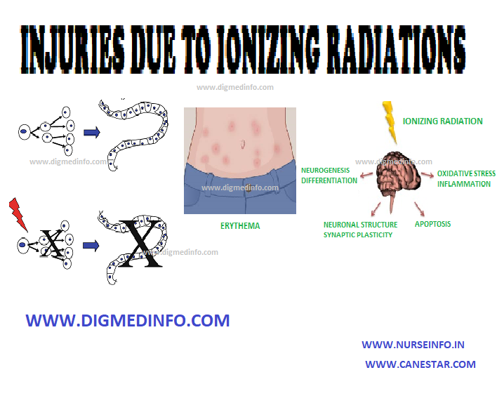
DAMAGE TO EMBRYO BY IRRADIATION IN FETAL LIFE

Irradiation during stage of implantation results in fetal death. During the stage of organogenesis, fetal malformation or abortion may result. After three months, by which time, the main organs would have formed, gross deformities are not produced, but stunting of growth, reduction of life span, sterility and tendency to develop leukemias and cancers have been noted. Radiation dose as low as 5 cGY can injure the growing fetus. The irradiation given out by modern X-ray machines for skiagram of the chest is only 1/100 cGY.

LOCAL RADIATION INJURY

Erythema develops over the area of exposure within days or weeks after acute exposure and this may progress to epidermolysis. Late complications are atrophy of the skin, subcutaneous fibrosis, telangiectasia, and later hyperkeratosis. These hyperkeratotic lesions may develop malignancy later.

INJURIES DUE TO IONIZING RADIATIONS – General Considerations, Acute Radiation Syndrome, Damage to Embryo by Irradiation in Fetal Life and Local Radiation Injury

HOOK WORM INFECTION

HOOK WORM INFECTION (Ancyclostomiasis) – General Characteristics, Life Cycle, Clinical Manifestations, Diagnosis, Treatment and Prevention

General Characteristics

Hookworm infection is prevalent in hot damp areas throughout the tropics and subtropics. Two species of hookworms – Ancylostoma duodenale and Necator americanus are seen to parasitize the small intestine of man. Ancylostoma is more harmful than Necator because it is more persistent in the environment and causes heavier blood loss. Ancylostoma is seen in the tropical and temperate zones whereas Necator is more widespread in the tropics.

Man is the natural host. The adults remain in the small intestine. Eggs are passed in feces. Adult worm of Ancylostoma duodenale is 8-13 mm long and is thread-like in thickness. The buccal cavity has four pointed hook-like teeth. Necator americanus is smaller than A. duodenale. The buccal cavity contains two chitinous cutting plates.

The worms attach to the intestinal villi of the jejunum and duodenum by their mouthparts. They produce bleeding points from which the blood passes through the alimentary canal of the worm continuously. This blood is required for the nutritive and respiratory functions of the worm.

The fertilized female can lay up to 20,000 eggs a day. The eggs measure 40-60 μ and are embryonated when passed. Necator americanus lays fewer eggs than A.duodenale, but the eggs are larger (60-70 μ) rarely two other species of hookworms- A. cyclonicum and A. braziliense may infect man. Ancylostoma cyclonicum is a hookworm of cat found in the Far East, which occasionally reaches maturity in humans. Ancylostoma braziliense is a hookworm of dogs and cats, which may also infect man but does not reach maturity.

Life Cycle

Eggs are passed in feces and within 5 days they hatch in warm moist soil. The rhabditiform larvae come out and they moult twice on the third and fifth days and develop into filariform larvae, which develop a sheath. These are infective to man. The larvae can persist in the soil for two months, feeding on bacteria and other organic matter. On coming into contact with the skin of man, the sheath is shed and the larvae penetrate into the subcutaneous tissues and enter the lymphatics and venules.

Within three days of entry into the skin, the larvae pass through the right side of the heart and the pulmonary capillaries to enter alveolar spaces. Foci of inflammation develop in the lungs. Then they enter the bronchi and pass up the trachea, larynx and back of the pharynx to be swallowed. In the oesophagus a third moulting occurs and a terminal buccal capsule is formed. In the small intestine they attach to the villi and grow into adults in 3-5 weeks. Average life-span of the worm is 2 years, but sometimes it may exceed 10 years.

Clinical Manifestations

Skin: The site of entry of the larvae becomes itchy and sodden. It ulcerates and becomes secondarily infected (ground itch). Common site for this lesion is the web of the toes.

Lungs: Larval migration through the lungs may lead to allergic symptoms, malaise, eosinophilia, dyspnea, cough and hemoptysis – Loeffler’s syndrome

Intestinal tract: Mild infections are usually asymptomatic. When the worm load is heavy, considerable blood loss occurs from the intestine. This leads to iron deficiency anemia, which is aggravated by coexistent malnutrition. Vague abdominal symptoms like pain resembling peptic ulcer or chronic intestinal amoebiasis may develop. Heavy infection, apart from severe anemia, leads to stunted growth, apathy, reduction of learning abilities in children, frequent absence from work and school, diminished productivity, impairment of immune responses, frequent respiratory infections and increased susceptibility to tuberculosis.

Diagnosis

The diagnosis is established by demonstrating the ova in feces. Ova of A. duodenale and N. americanus are indistinguishable. The feces gives positive reaction for occult blood. The worm load can be quantitated by egg counting using Kato-Katz cellophane thick smear technique or by Stoll’s method when there is heavy infection. Each worm produces 30 eggs per gram of feces per day. Identification of species is carried out byexamining the adult worms passed after a vermifuge.

Treatment

Specific treatment can be administered directly if the hemoglobin is above 7 g/dL. In severely anemic patients proper diet and iron supplements are given to raise the hemoglobin to 5 g/dL or more before administering the anthelmintic. Though this is a golden rule to be followed in outpatient practice, specific anthelmintic treatment can

be started much earlier using albendazole or mebendazole, which are considerably less toxic. Elimination of the worms leads to quicker recovery of the nutritional status as well.

Prophylaxis

Personal measures include the use of proper footwear and sanitary disposal of excreta.

HOOK WORM INFECTION (Ancyclostomiasis) – General Characteristics, Life Cycle, Clinical Manifestations, Diagnosis, Treatment and Prevention

HIGH ALTITUDE PULMONARY EDEMA

HIGH ALTITUDE PULMONARY EDEMA – Pathophysiology, Clinical Features, Diagnosis, Treatment and Prevention

This is a more serious complication compared to AMS. Young subjects in apparently good health are more affected. It may manifest in newcomers and also in subjects acclimatized to high altitudes, if they ascend the heights rapidly after long periods of stay at lower altitude. Altitudes above 2500 m can be harmful, but most of the cases occur at heights of 3050 to 4550 m. Heavy meal, physical exertion and too rapid an ascent precipitate the condition. Symptoms start within 6 to 48 hours of reaching the station.

Pathophysiology

Though pulmonary hypertension is invariably found as a consequence of hypoxia, only a small proportion of subjects develop acute pulmonary edema. High-altitude pulmonary edema is a noncardiogenic pulmonary edema associated with pulmonary hypertension and elevated capillary pressure. The pulmonary hypertension is secondary to the exaggerated pulmonary vasoconstriction induced by hypoxia. Sympathetic over activity, endothelial dysfunction, and hypoxemia are the proposed mechanisms. An immunogenetic basis also has been suggested because of higher incidence of HLA-DR6 and HLA-DQ4 antigens in susceptible individuals.

CLINICAL FEATURES

Many cases follow acute mountain sickness, but in some, pulmonary edema develops abruptly. Early symptoms are cough, tachypnea, dyspnea, and chest pain. These are soon followed by hemoptysis, cyanosis, frothy expectoration, and intense chest discomfort. Oliguria may develop.

The course is variable. In some, the pulmonary edema worsens while in others it may become sub acute and persist for a few days. In severe cases right-sided heart failure may follow and this may precede death.

Radiographic abnormalities include prominence of the pulmonary arteries, irregular patchy infiltration of one lung or bilateral nodular densities. Electrocardiogram reveals acute right ventricular strain due to pulmonary hypertension.

Diagnosis

High altitude pulmonary edema should be anticipated in healthy subjects who develop vague cerebral and respiratory symptoms on reaching high altitudes. Early recognition and treatment are necessary to avoid rapid deterioration or cardiac failure. Malingering has to be differentiated from this condition.

Treatment

Immediate treatment under field conditions is rest and administration of 100% oxygen at a rate of 4-6L/min for 15-30 min. Recompression using a portable hyperbaric bag will reduce symptoms if immediate descent is not possible. If oxygen is not available the patient should be evacuated to a lower camp, hospitalized and administered oxygen. Frusemide 40 mg should be given intravenously. Persistence of pulmonary edema is an indication for repeating frusemide. Nifedipine 10 mg initially followed by 20 mg repeated every 12 hours gives symptomatic relief. Physiological venesection by applying tourniquets proximally to the limbs helps to reduce pulmonary edema. Precipitating factors such as respiratory infection have to be looked for and treated appropriately.

Cases, which do not respond to the resuscitatory measures, should be evacuated to lower altitude urgently to prevent death.

Prevention

Proper training and conditioning for 1-2 weeks should be undertaken before reaching high altitudes. Ascent should be gradual to permit acclimatization. On reaching high altitudes, all unacclimatized persons should avoid physical exertion for 48-72 hours. Salmeterol inhalation 125 mg every 12 hours beginning 24 hours before the ascent is recommended to reduce incidence of high altitude pulmonary edema. Nifedipine 20 mg administered as a slow release preparation 8 h is also effective in preventing high attitude pulmonary edema. Those planning to ascent 3000 m should carry supply of oxygen and equipment sufficient for several days.

HIGH ALTITUDE PULMONARY EDEMA – Pathophysiology, Clinical Features, Diagnosis, Treatment and Prevention

HEATSTROKE (HEAT HYPERPYREXIA)

Definition, Etiology, Pathogenesis, Clinical features, Complications, Diagnosis, Treatment, Prognosis, Treatment and Prevention.

DISORDERS CAUSED BY HEAT

DEFINITION

Heatstroke follows exposure to heat and is characterized by hyperpyrexia owing to deranged heat regulatory mechanism. Heatstroke is defined clinically as a core body temperature that rises above 40°C and that is accompanied by hot, dry skin and central nervous system abnormalities such as delirium, convulsions, or coma.

ETIOLOGY

Heatstroke results from exposure to a high environmental temperature (classic,or nonexertional heatstroke) or from strenuous exercise (exertional heatstroke). Neuropsychiatric disturbances are prominent. Heatstroke generally occurs at temperatures ranging from 40 to 45o C, but may also occur at wet bulb temperatures as low as 30-35o C, especially if the humidity is high. The external temperature at which heatstroke occurs varies between individuals and therefore is not a reliable parameter for diagnosis. Precipitating factors include lack of acclimatization, unaccustomed and sustained heavy work at high temperatures, alcoholism, infections, dehydration, use of drugs like atropine, antihistamines, phenothiazines, antidepressants and diuretics, and obesity.

PATHOGENESIS

The systemic and cellular responses to heat stress include  thermoregulation, an acute-phase response whereby various cytokines and acute phase proteins are liberated, and the production of heat-shock proteins. Increased levels of heat-shock proteins in a cell induce a transient state of tolerance to heat stress, allowing the cell to survive. Thermoregulatory failure, exaggeration of the acute phase response, and alteration in the expression of heat-shock proteins may contribute to development of heatstroke.

Clinical features:

The onset is sudden or even abrupt with a rapid rise of body temperature to 41oC or more with acute neurological features such as confusion, restlessness, delirium, agitation, convulsions and coma. In a few patients there may be prodromata, including headache, giddiness, thirst, irrational behaviour and restlessness, and the onset is gradual.

The cardinal diagnostic features are hyperpyrexia, neuropsychiatric disturbances, and a hot and dry skin (hot dry man). The temperature (oral or rectal) varies from 41.5 to 44oC. It is preferable to take the rectal temperature in these cases. The patient has tachycardia and tachypnea. 25% of patients have hypotension. Electrocardiogram shows T wave changes suggestive of myocardial involvement.

Complications

The most serious complication of heatstroke is multi-organ dysfunction. This includes encephalopathy, rhabdomyolysis, acute renal failure, acute respiratory distress syndrome, myocardial injury, hepatocellular injury leading to jaundice, intestinal ischemia or infarction, pancreatic injury, and hemorrhagic complications, especially due to disseminated intravascular coagulation. Untreated, the condition is fatal owing to circulatory, hepatic or renal failure.

Diagnosis:

Diagnosis should be suspected in the presence of the three cardinal features, viz. hyperpyrexia, neurologic disturbances, and a hot and dry skin in a subject exposed to high ambient temperature. It is important to rule out falciparum malaria. Malaria may coexist with heatstroke but in cerebral malaria the peripheral blood will show signs of malaria hyperinfection. Other conditions like septicemia, typhus, pyogenic meningitis, encephalitis, pontine hemorrhage and closed head injuries have to be excluded.

Laboratory features

Laboratory features of heatstroke include neutrophil leucocytosis, thrombocytopenia, elevation of blood urea bilirubin, uric acid, and reduction of plasma bicarbonate, potassium, sodium, calcium and phosphorus. None of these are diagnostically specific. Therefore, diagnosis has to be clinical.

Treatment:

Heatstroke is a medical emergency. Treatment aims at the reduction of the rectal temperature to 39Oc within one hour, restoration of circulating blood volume and support of various organ system functions.

The patient is covered with wet sheets or towels and sprayed with cold water while air is blown or fanned over him to promote evaporative cooling. The patient should preferably be in the lateral recumbent position or hands and knees position to expose as much of skin as possible to the air. As an alternative the patient may be immersed in cold water. Immersion in ice water is not preferred because of the possibility of shivering and hypotension.

The limbs are gently massaged to promote circulation. When the rectal temperature falls to 39oC the patient is transferred to a cool room and watched for further rise of temperature. Recurrence is quite common and temperature regulation may remain unstable for weeks.

Rapid cooling to rectal temperatures below 38-39oC may precipitate convulsions, vomiting and hypotension. A mechanical body cooling unit is available in which cold water at 20oC is sprayed over the patient, while a blast of hot air at 50oC is blown from above. This prevents vasoconstriction and also augments evaporation. Prompt and effective cooling restores neurological functions in the majority of patients. Recovery of central nervous system function is a favorable prognostic sign.

Chlorpromazine (25-50 mg) administered intravenously is a useful adjunct during cooling. Antipyretics like acetaminophen and aspirin have no role and are contraindicated also.

If there is suspicion of malaria, antimalarials are given parenterally. Shock and cardiac failure have to be treated appropriately. The patient should also be observed and treated for renal failure due to rhabdomyolysis, disseminated intravascular coagulation and hepatic failure. Maintenance of hydration and electrolyte balance are important to prevent renal failure. Sequelae include headache, insomnia, giddiness, cerebellar dysfunction and even coma. Most of these clear up in the course of weeks or months.

Prognosis:

If left untreated, 50 -70% of subjects die of heatstroke. Early treatment reduces the mortality to 15-20%. Shock also carries a bad prognosis. Presence of other systemic diseases and extremes of age worsen the prognosis.

Prevention:

Heatstroke is a preventable disease. To prevent heatstroke, people should acclimatize themselves to heat, schedule outdoor activities during cooler times of the day, reduce level of physical activity, drink

additional water and consume extra amounts of salt – (12 to 15 g/day).

Definition, Etiology, Pathogenesis, Clinical features, Complications, Diagnosis, Treatment, Prognosis, Treatment and Prevention.

HANTA VIRUS

HANTA VIRUS – HFRS and HCPS and Treatment

General Characteristics

Hanta virus is a natural inhabitant of rodents and the virus is shed in their urine, feces and saliva. This virus affects the vascular endothelium and causes two major syndromes:

1. Hemorrhagic fever with renal syndrome (HFRS) and

2. Hantavirus cardiopulmonary syndrome (HCPS)

Infection is by aerosol inhalation of infective material. Incubation period is about 3 weeks.

The virus affecting the endothelium leads to immune response against viral antigens expressed on the endothelial cells of the heart, lungs, kidney and lymphoid organs. The immune reactions are mediated by T lymphocytes and macrophages. TNF-α and IL-1 beta increase capillary permeability. Nitric oxide released by TNF leads to vasodilation and shock.

HFRS: Nonspecific constitutional symptoms occur followed by hemorrhagic manifestations along with renal lesions.

Shock, oliguria and DIC follow. Survivors enter a diuretic phase by day 10-14. Thrombocytopenia, leukocytosis with shift to the left, increase in the number of circulating immunoblasts (up to 10% of circulating lymphocytes) and increase in serum levels or LDH should suggest the diagnosis. Specific tests include the demonstration of IgM and IgG antibodies by ELISA.

HCPS:

Constitutional symptoms lasting for 3-7 days are followed by hypotension and shock. Pulmonary edema, cardiac arrhythmias and cardiac arrest may occur. Renal lesions may develop leading to oliguria lasting for 3-8 days, and later followed by polyuria.

TREATMENT

Treatment is supportive with I V fluids, ionotropes, mechanical ventilation and blood products. Ribavarin has been used in usual doses- with variable results.

HANTA VIRUS – HFRS and HCPS and Treatment

GIARDIASIS

GIARDIASIS – General Characteristics, Transmission, Pathogenesis, Clinical Features, Diagnosis and Treatment

General Characteristics

Giardiasis is infection of the small intestine by the flagellate protozoon—Giardia intestinalis (G. lamblia). In the tropics and subtropics, this parasite is very widely prevalent, particularly in children.

Giardia lamblia is a flat pear-shaped organism 12-18μ in diameter with eight flagella, two nuclei and a large ventral sucker placed anteriorly. These flagellates are found in large numbers in the duodenum and jejunum in between the villi, and attached to them by their suckers.

Occasionally biopsy specimens may show giardia between villi. With the aid of the flagella, giardia moves actively and this helps to identify the organisms in freshstools or duodenal aspirates. Cysts are formed which are passed in feces.

Transmission

Transmission occurs by ingesting the cysts along with food and water. Infection rates are high in closed communities. New immigrants and travellers may get the infection from endemic areas. Hypochlorhydria and hypogammaglobulinemia predispose to heavy infection.

Pathogenesis

Though the exact mechanism is not known, it is possible that a large number of flagellates adhering to the mucosal surface may reduce the area for absorption. In addition, irritation of the brush border of the villi, alteration in bacterial flora in the small intestine; and invasion of the submucosa have been suggested as contributing factors. Many cases show increased intestinal motility.

Clinical Features

Presenting symptoms range from mild abdominal discomfort to explosive diarrhea. Some infections may be asymptomatic but the majority is subacute or chronic. Nonspecific symptoms include failure to thrive, abdominal pain, epigastric distress, intermittent diarrhea and general ill-health. Features of malabsorption may occur, especially in children and this should suggest the possibility of giardiasis.

In some outbreaks explosive diarrhea with watery foul smelling stool may be the presenting symptom. Giardiasis is a frequent cause for traveller’s diarrhea in endemic regions. The acute attack lasts for 3-4 days but in children the course may be prolonged for weeks or months and it results in malabsorption state. Many cases go into spontaneous remission and carrier state develops in them.

Diagnosis

Giardiasis is a common cause of diarrhea in children and vague abdominal discomfort in adults. Fresh feces should be examined for identifying the motile flagellate. Presence of the active flagellate in fresh stool or in the duodenal aspirate confirms the diagnosis. In many cases mixed infection with Giardia and other intestinal pathogens like

E. histolytica or helminths is common. Clinical severity does not always correlate with the number of parasites seen in feces. In mild infections, repeated examination and concentration methods (formol-ether concentration method or Zinc sulphate floatation) may be required to

establish the diagnosis. In some cases, when feces do not show the parasites, duodenal aspiration has to be done for recovering them. Presence of cysts indicates the carrier state. These can be identified by examining iodine-stained preparations of feces.

Treatment

The drug of choice is metronidazole in a dose of 15 mg/kg/day given for seven days. In 80-90% of cases the infection is eliminated. In resistant cases this course can be repeated. Single administration of metronidazole in a dose of 30 mg/kg repeated after a week is also effective and it may be more suited for mass treatment on account of better patient compliance. Chloroquine 250 mg thrice a day for 5 days and tinidazole are also effective. Giardiasis can be prevented by improving food hygiene and environmental sanitation. Heavy chlorination of water supply is required for killing giardia cysts.

GIARDIASIS - General Characteristics, Transmission, Pathogenesis, Clinical Features, Diagnosis and Treatment
GIARDIASIS – General Characteristics, Transmission, Pathogenesis, Clinical Features, Diagnosis and Treatment

GENITAL HERPES

GENITAL HERPES (HERPES SIMPLEX VIRUS) – General Characteristics, Cytology, Treatment and Prophylaxis

General Characteristics

Several viruses may be transmitted by sexual contact. Important among them are herpes simplex, human papilloma virus, molluscum contagiosum, hepatitis B virus, cytomegalovirus, Marburg virus, and AIDS.

HERPES GENITALIS

Herpes genitalis is caused by herpes simplex virus (HSV) type 2 and less frequently by HSV type 1. It is moderately infectious. The virus remains dormant in the presacral sensory ganglion cells. Periodically it moves into the skin through the axons of sensory nerves to produce recurrent attacks of herpes simplex locally. Factors which produce the reappearance of herpes are not clear. In many western countries herpes genitalis is emerging as the commonest among the STD. The disease is transmitted by sexual intercourse. The virus is shed from the lesions for up to 2 weeks after apparent healing. Infection of the baby during birth leads to neonatal herpes.

Clinical Features

The incubation period is 2-4 days or rarely longer. The onset of local lesions may be preceded by burning pain in the S1 to S5 dermatomes.

The lesions occur in the external genitalia of men and in the anal canal of homosexuals. Over the penis, it starts with erythema followed by the appearance of papules or vesicles which soon rupture to form shallow painful ulcers. Ultimately, crusts form which dry up and fall off leaving healed scars which fade. Tender inguinal adenopathy develops in the primary attack but not with recurrences. The lesions are infective throughout. Recurrences are generally much milder.

In women the primary lesion and recurrent attacks differ from each other considerably. Primary attack is severe, whereas, subsequent attacks are milder. Lesions occur usually in the vulva and adjacent skin areas. Morphology of the lesion is the same as in men. During the primary attack retention of urine may develop due to extreme pain on micturition. Herpes virus infection is directly or indirectly related to the development of carcinoma of the cervix, vagina, or vulva in later life as the virus is oncogenic. HSV 2 can also act as a cofactor with human papilloma virus for invasive cervical cancer.

Dissemination of the infection is rare in adults, but if it occurs it can be fatal. The skin, the central nervous system, and other internal organs may be affected. HSV2 can also act as a cofactor with human papilloma virus for invasive cervical cancer.

The newborn may acquire herpes during delivery if the mother has active lesions in the genitalia. Lesions develop in the baby usually within two weeks after birth. These occur more commonly with primary maternal herpes than with recurrent attacks. Both HSV-1 and HSV-2 may be responsible but the latter is more common. The lesions in the newborn include meningoencephalitis or generalized herpetic lesions involving several organs. If not treated, mortality and morbidity are very high.

DIAGNOSIS

Laboratory diagnosis is established by isolation of the virus from the lesions. Serological tests using blood include complement fixation test which becomes positive about a week after infections. In primary infection, the first sample is seronegative, the second sample shows a marked rise in titer. In recurrent herpes both samples give the same low titer. Herpes virus can be demonstrated by electron microscopy of vesicle fluid or tissue specimens.In herpes simplex encephalitis the CSF shows antibodies.

Cytology

Papanicolaou’s stained smears from vesicles show multinucleated giant cells with eosinophilic inclusions. In meningoencephalitis CSF shows changes. CT scan, MRI imaging and brain biopsy are employed to diagnose herpetic encephalitis.

Treatment and Prophylaxis

Treatment options for genital herpes:

First clinical episode: Treatment duration-7 days

Acyclovir 200 mg orally 5 times daily or 400 mg t.i.d or

Valacyclovir 1 g orally b.i.d or Famciclovir 250 mg t.i.d.

Recurrent infection: Treatment duration-5 days.

Acyclovir (oral) 200 mg 5 times daily or 400 mg t.i.d or

800 mg b.d or Valaciclovir 500 mg b.d or 1g once daily or

Famciclovir 125 mg twice daily

Suppressive therapy: The condition tends to recur often on cessation of treatment. If the recurrence is too frequent, >6 times a year, suppressive therapy is indicated for prolonged periods.

Acyclovir 400 mg bid or

Valaciclovir 500 mg or 1 g or

Famciclovir 250 mg bid continuously.

Recommended regimen for severe disease is acyclovir 5-10 mg/kg IV every 8 hours for 5-7 days or until clinical resolution is attained.

Severe herpes with coinfection with HIV: Acyclovir 400 mg orally 3-5 times daily until clinical resolution is attained.

Neonatal infection: Acyclovir 10 mg/kg IV 3 times daily for 10-21 days.

If the mother develops primary attack of herpes genitalis after the 36th week of pregnancy, caesarean section is indicated before the rupture of the membranes to avoid infection of the baby and the baby has to be watched for 2-4 weeks for the development of generalized herpes.

GENITAL HERPES (HERPES SIMPLEX VIRUS) – General Characteristics, Cytology, Treatment and Prophylaxis

GAS GANGRENE

GAS GANGRENE (Clostridial myonecrosis – General Characteristics, Pathogenesis, Clinical Features, Diagnosis and Treatment

General Characteristics

Gas gangrene is caused by Clostridium perfringens (C.welchii), C. septicum, C. novyi (C. oedematiens) C.histolyticum and C. sordelli. These clostridia are commensals in human gut and soil. They produce local necrosis and distant lethal effects.

Pathogenesis

Necrosis of tissue brought about by anoxia, ischemia or injury is complicated by clostridial infection which may be exogenous (introduced along with the wounds) or endogenous (derived from the intestinal tract). The organisms multiply locally and produce toxins which diffuse out and lead to generalized toxemia. The affected tissues are avascular and appear as if cooked.

Clinical Features

After an incubation period of 1-4 days the wound becomes swollen and tender and exudes brownish foul-smelling fluid. Due to the presence of gas, crepitus may be felt. Blebs filled with purplish foul-smelling fluid develop. These blebs rupture and ulcerate. General symptoms such as restlessness and fever develop and the patient may rapidly go into shock and anuria.

Diagnosis

The condition has to be diagnosed clinically. The organism can be identified by culture. X-ray of the affected part shows gas in the tissues. Gas in the tissues may occur in other conditions such as surgical emphysema and infections by streptococci, staphylococci and E. coli.

Treatment

Early institution of therapy is most important for success. Shock is treated on the lines of septic shock. Penicillin which is the drug of choice is given intravenously in a dose of 2 mega units 2 hourly. Other antibiotics may be tried depending on the sensitivity tests. Metronidazole and gentamicin are effective in most cases. Antitoxin is available for intravenous use (anti-gas gangrene serum—AGGS). It should be given in a dose of 40,000 units initially to be followed by 20,000 to 40,000 units repeatedly till the condition improves. Keeping the patient under hyperbaric oxygen (3 atmosphere pressure) remarkably improves the result of therapy and this should be instituted wherever it is available. Surgical excision of dead tissue and removal of foreign bodies should be undertaken after administration of AGGS. Gas gangrene can be prevented by taking proper care of the wound and early recognition of the condition.

GAS GANGRENE (Clostridial myonecrosis – General Characteristics, Pathogenesis, Clinical Features, Diagnosis and Treatment

GALACTOSEMIA

GALACTOSEMIA – Definition, Diagnosis  and Treatment

DEFINITION

Normally lactose present in milk is converted into glucose and galactose by intestinal lactase. Galactose is absorbed and converted to glucose in the liver. Conversion into glucose is defective in galactosemia and hence the concentration of galactose-1-phosphate and galactose increase in blood and tissues such as liver, brain, kidneys, intestines, and lens.

Galactosemia is transmitted as an autosomal recessive trait. In the fully developed form, classical galactosemia presents with cataract, mental retardation, hepatic cirrhosis, and death in early life. The enzyme galactose-1-phosphate uridyl transferase is deficient. The severe form manifests within a few days of birth with intolerance to milk, vomiting, refusal to feed, and failure to thrive. Jaundice, hepatomegaly, and hepatic dysfunction develop early. Cataracts develop within weeks or months. As the child grows up, mental deficiency becomes evident. Recurrent bacterial infections (E. coli) are common and may be fatal.

In the atypical form of galactosemia, the enzyme galactokinase is deficient. Galactokinase is required to convert galactose into galactose-1-phosphate and in its absence galactose accumulates in blood and tissues. The only complication in this disorder is cataract formation.

Diagnosis

Urine shows galactose. Diagnosis is established by demonstrating the deficiency of galactose-1-phosphate uridyl transferase in erythrocytes.

TREATMENT

Treatment consists of dietary measures to avoid milk and milk products, and these results in dramatic improvement. Even in advanced cases dietary management leads to regression of symptoms except the cataract.

GALACTOSEMIA – Definition, Diagnosis  and Treatment
GALACTOSEMIA – Definition, Diagnosis  and Treatment

FOLIC ACID (Pterolglutamic Acid)

FOLIC ACID (Pterolglutamic Acid) – Causes of Folate Deficiency and Treatment

Folic acid molecule consists of a pteridine ring, paraamino benzoic acid and l-glutamic acid. The free form does not occur commonly in nature. Foods and tissues contain several folate compounds   (pteroylpolyglutamates).

The main form present in human plasma and CSF is 5-methyltetrahydropteroyl monoglutamate. Rich dietary sources are yeast, liver, nuts, green vegetables, and chocolate. Folic acid is lost by boiling the articles of food in a large volume of water. Average requirement is 200-300 μg/day. The vitamin is absorbed in the proximal small intestine after hydrolysis of polyglutamates. Human body can store about 6-10 mg, especially in the liver.

Megaloblastic anemia has developed in volunteers after 10-20 weeks of dietary deprivation. Dietary deficiency is widely prevalent in India. It is seen most in pregnant women and children. The main reason was deficiency of folates, vit B12 and other deficiencies such as Vit B6 together or singly

Causes of Folate Deficiency

1. Dietary inadequacy of animal foods and green vegetables.

2. Malabsorption states.

3. Increased demands due to pregnancy, lactation, growth and intercurrent illnesses in children.

4. Alcohol intake increases folate requirements.

5. Accelerated erythropoiesis as in hemolytic anemia.

6. Other disorders like tuberculosis, rheumatoid disease, and malignancy increase folate requirements.

7. Several drugs interfere with the metabolism of folates, e.g., amethopterine, pyrimethamine, trimethoprim, hydantoins, oral contraceptives.

8. Folate losses may occur in several disorders of skin and the intestinal tract.

9. Inborn errors of metabolism may interfere with the utilization of folate.

Folate deficiency leads to megaloblastic anemia and similar changes in several rapidly proliferating tissues. In the body the metabolic pathways of folates and vitamin B12 are closely interrelated. In many cases of nutritional anemias, combined deficiencies of iron, folate and to a smaller extent B12 operate. Normal serum folate levels are 4-10 ng/mL and red cell folate is above 100 ng/mL.

Folic acid deficiency occurring in the early period of gestation when the neural tube of the embryo develops, may result in the higher incidence of neural tube defects such as spina bifida and anencephaly. Supplementation of 400 mcg of folate starting from 28 days before and continuing for 28 days after conception reduce the risks of neural tube defects substantially.

Treatment

Dietary deficiency can be corrected by giving 5-10 mg of folic acid orally, in addition to the dietary improvement. Pregnant and lactating women, children and alcoholics require prophylactic supplementation. In poor communities routine folic acid supplementation in pregnancy (1 mg/day) reduces the incidence of premature births. Premature infants should be given folate supplementation.

Folate and vitamin B12 deficiencies are known to elevate serum homocysteine levels and this may lead to abnormal thrombotic tendency.

FOLIC ACID (Pterolglutamic Acid) - Causes of Folate Deficiency and Treatment
FOLIC ACID (Pterolglutamic Acid) – Causes of Folate Deficiency and Treatment
Nurse Info