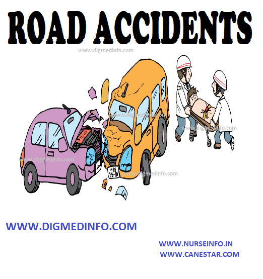
ROAD ACCIDENTS – General Considerations, Injuries Caused by Road Accident and Management At the Site of Accident
General Considerations
In modern times mortality and morbidity caused by road accidents is on the rise. This is common in all countries of the world where fast traffic on the road is increasing.
The accidents may involve the vehicles or the pedestrians using the road. The major types of accidents are collision between vehicles, loss of control of the vehicle and collision with hard objects or overturning. The other type of accidents involves knocking down pedestrians or occupants of lighter vehicles such as two wheelers and light motor vehicles. In general the major damage and passenger injury occurs to the lighter vehicle.
The major factors contributing to road accidents can be classified as given below:
A. Causes attributable to the vehicles
1. Speed of the vehicle- The rate and severity of the accident increases with increasing speed of either vehicle. With the introduction of modern highways speed-related injuries have become much more common
2. Mechanical defects in the vehicle, which lead to failure of the controls.
3. Exceeding the safety limits prescribed for the vehicle in terms of speed, loading and maintenance.
B. Causes attributable to the driver
1. Fatigue and sleep- long driving exceeding 6 hours of continuous driving leads to driver fatigue, delay in the reflexes and tendency to sleep.
2. Drunkenness: Consumption of alcohol and less commonly other narcotic drugs impairs the efficiency of the driver
3. Inattention, non-compliance with signals and rash driving
4. Inexperience of the driver
Most of the major collision accidents occur on highways, especially in the early hours of the morning when driver fatigue and somnolence are maximal. Rarely accidents may occur due to organic disease in the driver such as epilepsy, cardiovascular disease, strokes and others.
Injuries to Pedestrians
Walking on highways and fast traffic roads is associated with injury to the pedestrians. Elderly people, obese individuals, persons with movement disorders, alcoholics and children are involved more.
INJURIES CAUSED BY ROAD ACCIDENTS
Vehicular Accidents
Trauma to the driver and passengers due to the impact and damage and deformation to the vehicle are usually serious. This leads to injuries which may be instantaneously fatal. In motor car accidents, the luggage kept unsecured in the passenger compartment fly out and cause further missile- like injuries to the passengers. Trauma to vital parts such as head, chest cage, spine, bones, abdomen and major blood vessels, intracranial injuries, intracranial bleeding, tension pneumothorax, rupture of major blood vessels like aorta, injuries to the heart, rupture of solid organs and hollow viscera in the abdomen and pelvis and exsanguinating bleeding ( both external and internal ) are the causes of death at the site of accident or within a few hours. Pedestrian injuries cause death due to damage to vital structures, especially intracranial bleeding, chest injuries, intra abdominal injuries and exsanguination.
MANAGEMENT AT THE SITE OF ACCIDENT
Effective first aid helps to save life and reduce morbidity and delayed mortality. Mainly this consists of maintenance of the airway and attention to ventilation, tourniquets to arrest external hemorrhage, immobilization of parts of the body such as the neck and spine to avoid further damage to the vital neural structures, covering open chest injuries which interfere with ventilation and rapid transport to the nearest hospital with adequate facilities. It is the duty of all medical men to give first aid in such a situation. It is to be remembered that an obvious injury may distract the attention of the doctor from other invisible injuries such as internal bleeding or rupture of viscera which can cause death.
When major accidents involving buses occur, a large member of persons will be simultaneously affected and this may cause a heavy strain on the medical facilities. In this situation, a senior medical officer has to inspect the victims and using the principles of ‘triage’, arrange for appropriate management. In such an emergency all medical personnel have to participate in the emergency management.
All road traffic accidents invite legal procedures which have to be complied with. General guidelines to reduce road traffic accidents and their consequences
1. Education of the public on the safe use of roads and motor vehicles
2. Strict compliance with road traffic rules
3. Avoidance of alcohol and drugs before driving
4. Avoid driver fatigue and somnolence
5. Avoid the presence of unsecured luggage in the passenger compartment
6. Use the seat belts as prescribed.
7. Enforcement of traffic regulations by the appropriate authorities