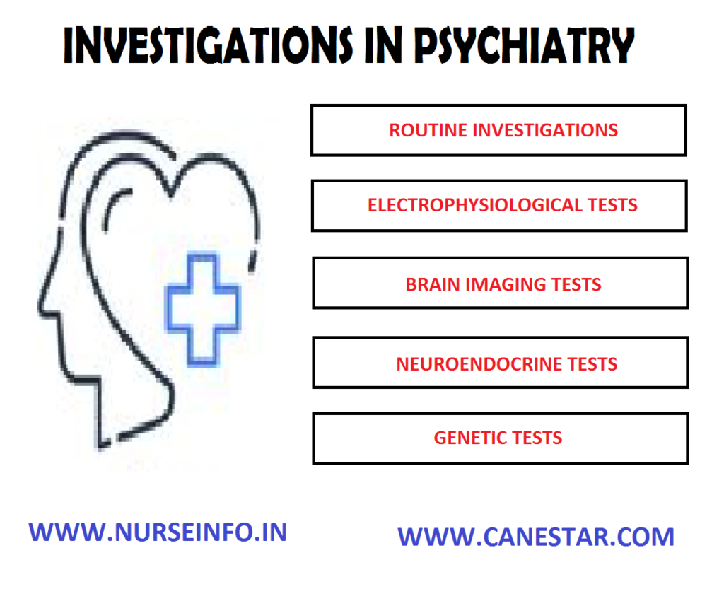
PSYCHIATRIC INVESTIGATION – Routine Tests and Diagnostic Procedures used to Detect Altered Brain Function (Mental Health Nursing)
The growing awareness of various physical conditions which can produce psychiatric symptoms and the increased use of biological therapies have made it mandatory that appropriate physical investigations should be carried out before starting any treatment and during it. They serve diagnostic, basal screening and monitoring purposes
ROUTINE TESTS
- A complete hemogram (total and differential blood count, haemoglobin, ESR) and urinalysis are the basic minimum of routine test. Leucopenia and agranulocytosis are associated with certain medications. Treatment with lithium and neuroleptic malignant syndrome are often associated with leukocytosis
- Fasting and post-prandial blood sugar, chest X-ray and an EEG are often considered routine tests
- An EEG is necessary for monitoring cardiac effects of certain drugs
- Serum electrolytes (sodium, potassium chlorides, bicarbonates, calcium, etc.) are sometimes needed as basal routine investigations. An electrolyte imbalance causes various neuropsychiatric symptoms like delirium
- Liver function tests: serum glutamic oxaloacetic transaminase (SGOT), serum glutamic-pyruvic transaminase (SGPT), serum alkaline phosphatase, prothrombin time, serum bilirubin levels and serum proteins (total and differential) are some common liver function tests. Liver function tests are done for all alcoholic patients
- Renal function tests: blood urea, serum creatinine, and creatinine clearance, thyroid function tests (T3, T4 and TSH) and ECG are routinely done on patients prior to starting lithium therapy
- An ECG and chest X-ray are usually done before a patient is posted for ECT
DIAGNOSTIC PROCEDURES used to DETECT ALTERED BRAIN FUNCTION
Several diagnostic procedures are used to detect alternation in biologic function that may contribute to psychiatric disorders
Electroencephalography (EEG)
Technique: electrodes are placed on the scalp in a standardized position. Amplitude and frequency of beta, alpha, theta and delta brain waves are graphically recorded on paper by ink markers for multiple areas of the brain surface
Purpose: it measures brain electrical activity; identifies dysrhythmia, asymmetries, or suppression of brain rhythms; used in the diagnosis of epilepsy, neoplasm stroke, metabolic or degenerative disease
Computed Tomography (CT)
Technique: series of radiographs that are computer constructed into slices of the brain that can be stacked by the computer giving a three-dimensional image
Purpose: measures accuracy of brain structure to detect possible lesion, abscesses, areas of infarction or aneurysm. CT has also identified various anatomic differences in clients, with schizophrenia, organic mental disorder and bipolar disorder
Magnetic Resonance Imaging (MRI)
Technique: a magnetic field surrounding the head induces brain tissue to emit radio waves that are computerized to provide clear and detailed construction of sectional images of the brain. No radiation or contrast medium is used
Purpose: measures anatomic and biochemical status of various segments of the brain; detects brain edema, ischemia, infection, neoplasm, trauma and other changes such as demyelination. Morphological differences between the brains of clients with schizophrenia and those of control subjects have been noted
Brain Electrical Activity Mapping (BEAM)
Technique: uses computed tomographic techniques to display data derived from EEG recordings of brain electrical activity that can be sensory evoked by specific stimuli, such as flash of light or a sudden sound, or cognitive evoked by specific mental tasks
Purpose: measures brain electrical activity; used largely in research to represent statistical relationship between individuals and groups or between two populations of subject (e.g. client with schizophrenia vs. control subjects)
Positron Emission Tomography (PET)
Technique: an injected radioactive substance travels to the brain and shows up as a bright spot on the scan; different substances are taken up by the brain in different amounts, depending on the type of tissues and the level of activity
Purpose: measures specific brain functioning, such as glucose metabolism, oxygen utilization, blood flow, and of particular interest in psychiatry neurotransmitter/receptor interaction
Single Photon Emission Computed Tomography (SPECT)
Technique: the technique is similar to PET but a longer acting radioactive substance must be used to allow time for a gamma camera to rotate about the head and gather the data, which are then assembled on the computer into a brain image
Purpose: measures various aspects of brain functioning as with PET; has also been used to image activity or cerebrospinal fluid circulation


