EMERGENCY CONDITIONS – Shock (Etiology, Pathophysiology, Signs and Symptoms, Diagnostic Evaluation and Management)
SHOCK
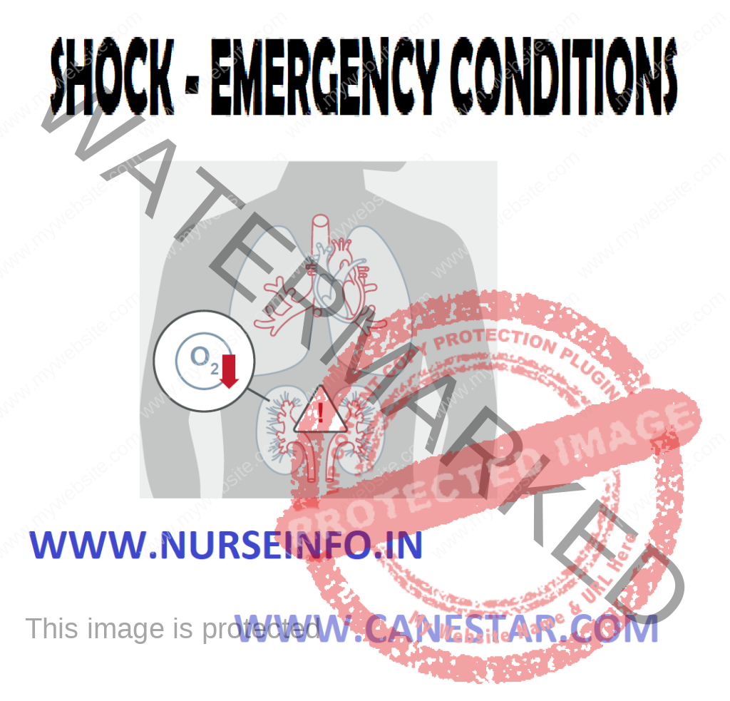
Clinical syndrome characterized by decreased tissue perfusion and impaired cellular metabolism resulting in an imbalance between the supply and demand for oxygen and nutrients
ETIOLOGY AND PATHOPHYSIOLOGY
- Cardiogenic shock occurs when either systolic or diastolic dysfunction of the pumping action of the heart results in compromised cardiac output (CO).
Precipitating causes of cardiogenic shock include myocardial infarction (MI), cardiomyopathy, blunt cardiac injury, severe systemic or pulmonary hypertension, cardiac tamponade, and myocardial depression from metabolic problems.
Hemodynamic profile will demonstrate an increase in the pulmonary artery wedge pressure (PAWP) and pulmonary vascular resistance
SIGNS AND SYMPTOMS
Tachycardia, hypotension, a narrowed pulse pressure, tachypnea, pulmonary congestion, cyanosis, pallor, cool and clammy skin, decreased capillary refill time, anxiety, confusion, and agitation.
- Hypovolemic shock occurs when there is a loss of intravascular fluid volume
Absolute hypovolemia results when fluid is lost through hemorrhage, gastrointestinal (GI) loss (e.g. vomiting, diarrhea), fistula drainage, diabetes insipidus, hyperglycemia, or diuresis.
Relative hypovolemia results when fluid volume moves out of the vascular space into extravascular space (e.g., interstitial or intracavitary space) and this is called third spacing
The physiologic consequences of hypovolemia include a decrease in venous return, preload, stroke volume and CO resulting in decreased tissue perfusion and impaired cellular metabolism.
Clinical manifestations depend on the extent of injury or insult, age and general state of health and may include anxiety, an increase in heart rate, CO, and respiratory rate and depth, and a decrease in stroke volume, PAWP, and urine output.
- Neurogenic shock is a hemodynamic phenomenon that can occur within 30 minutes of a spinal cord injury at the fifth thoracic (T5) vertebra or above an last up to 6 weeks, or in response to spinal anesthesia.
Immediate reaction causes massive vasodilation, release of vasoactive mediators, and an increase in capillary permeability resulting in fluid leaks from the vascular space into the interstitial space
Clinical manifestations can include anxiety, confusion, dizziness, chest pain, incontinence, swelling of the lips and tongue, wheezing, stridor, flushing, pruritus, urticaria and angioedema.
- Septic shock is the presence of sepsis with hypotension despite fluid resuscitation along with the presence of tissue perfusion abnormalities
In severe sepsis and septic shock, the initiated body response to an antigen is exaggerated resulting in an increase in inflammation and coagulation, and a decrease in fibrinolysis
Endotoxins from the microorganisms cell wall stimulate the release of cytokines and other proinflammatory mediators that act through secondary mediators such as platelet-activating factor.
Clinical presentation for sepsis is complex. Patients will usually experience a hyperdynamic state characterized by increased CO. Persistence of a high CO beyond 24 hours is ominous and often associated with hypotension and multiple organ dysfunction syndrome (MODS). Initially patients will hyperventilate as a compensatory mechanism, resulting in respiratory alkalosis followed by respiratory acidosis and respiratory failure. Other clinical signs include alteration in neurologic status, decreased urine output, and GI dysfunction.
STAGES OF SHOCK
- Compensatory Stage
Decrease in circulating blood volume
Sympathetic nervous system stimulated, release catecholamines (epinephrine and norepinephrine), bronchodilation and increased cardiac output occurs. To maintain blood pressure; increase heart rate and contractility increases in peripheral vasoconstriction due to stimulation of beta adrenergic fibers (cause vasoconstriction of blood vessels of skin and abdominal viscera) and increase in heart rate and contractility.
Renin-angiotensin release of aldosterone-reabsorb H2O and sodium. Get fluid shift from interstitial to capillaries due to decrease in hydrostatic pressure in capillaries
Shunting blood from the lungs-ventilation-perfusion mismatch
Circulation maintained, but only sustained short time without harm to tissues
- Progressive Stage
Altered capillary permeability (3rd spacing)
In the lungs: alveolar or pulmonary edema, ARDS, increased pulmonary artery pressures
Cardiac output decreases and coronary perfusion is decreased. Decreased myocardial perfusion-arrhythmias and myocardial ischemia
Kidneys: elevated BUN and creatinine
Metabolic acidosis, anaerobic metabolism and kidneys cannot excrete acids and reabsorb bicarbonate
GI-ischemia causes ulcers and GI bleed
Liver: cannot eliminate waste products, elevated ammonia and lactate, bilirubin (jaundice) bacteria released in bloodstream
Hematologic: disseminated intravascular coagulopathy (DIC)
- Refractory Stage
Anaerobic metabolism starts. Lactic acid build-up
Increased capillary blood leak, worsens hypotension and tachycardia, also get cerebral ischemia
Get profound hypotension and hypoxemia
Cellular death leads, tissue, death, vital organs fail and death occurs (lungs, liver and kidneys result in accumulation of waste products. One organ failure leads to another.
Recovery unlikely
DIAGNOSTIC EVALUATION
- Blood: RBC, hemoglobin and hematocrit
- Arterial Blood Gases: respiratory alkalosis and metabolic acidosis
- Electrolyte (Na level increased early, decreased later if hypotonic fluid given) K decrease later increase K with cellular breakdown and renal failure
- BUN and creatinine increased, specific gravity increased then fixed at 1.010
- Blood cultures: identify causative organism in septic shock
- Cardiac enzymes: diagnosis of cardiogenic shock
- Glucose: increased early then decreased
- DIC screen: fibrinogen level, platelet count, PTT and PT, thrombin time
- Lactic acid: increased
- Liver enzymes: ALT, AST and GGT increased
MANAGEMENT
- General management strategies for a patient in shock begin with ensuring that the patient has a patient airway and oxygen delivery is optimized. The cornerstone of therapy for septic, hypovolemic and anaphylactic shock is volume expansion with the administration of the appropriate fluid
- It is generally accepted that isotonic crystalloids, such as normal saline, are used in the initial resuscitation of shock. If the patient does not respond to 2 to 3 L of crystalloids, blood administration and central venous monitoring maybe instituted
- The primary goal of drug therapy for shock is the correction of decreased tissue perfusion
Sympathomimetic drugs cause peripheral vasoconstriction and are referred to as vasopressor drugs (e.g. epinephrine and norepinephrine)
The goals of vasopressor therapy are to achieve and maintain a mean arterial pressure (MAP) of 60 to 65 mm Hg and the use of these drugs is reserved for patients unresponsive to other therapies
The goal of vasodilator therapy, as in vasopressor therapy, is to maintain Mean arterial pressure at 60 mm Hg or greater
Vasodilator agents most often used are nitroglycerin (in cardiogenic shock) and nitroprusside)
COLLABORATIVE CARE
Cardiogenic Shock
- Overall goal is to restore blood flow to the myocardium by restoring the balance between oxygen supply and demand
- Definitive measures include thrombolytic therapy, angioplasty with stenting, emergency revascularization and valve replacement
- Care involves hemodynamic monitoring, drug therapy (e.g. diuretics to reduce preload), and use of circulatory assist devices (e.g. intra-aortic balloon pump, ventricular assist device)
Hypovolemic Shock
- The underlying principles of managing patients with hypovolemic shock focus on stopping the loss of fluid and restoring the circulating volume
- Fluid replacement is calculated using a 3:1 rule (3 ml of isotonic crystalloid for every 1 ml of estimated blood loss)
Septic Shock
- Patients in septic shock require large amounts of fluid replacement, sometimes as much as 6 to 10 L of isotonic crystalloids and 2 to 4 L of colloids, to restore perfusion
- Vasopressor drug therapy maybe added and vasopressin maybe given to patient’s refractory to vasopressor therapy
- Intravenous corticosteroids are recommended for patients who require vasopressor therapy, despite fluid resuscitation, to maintain adequate BP
- Antibiotics are early component of therapy and are started after obtaining cultures
- Drotrecogin alpha, a recombinant form of activated protein C, has demonstrated promise in treating patients with severe sepsis.
- Glucose levels should be maintained at less than 150 mg/dl
- Stress ulcer prophylaxis with histamine (H2)-receptor blockers and deep vein thrombosis prophylaxis with low dose unfractionated heparin or low molecular weight heparin are recommended
Neurogenic Shock
- Treatment of neuogenic shock is dependent on the tissue
In spinal cord injury, general measures to promote spinal stability are initially used
Definitive treatment of the hypotension and bradycardia involves the use of vasopressor and atropine respectively
Fluids are administered cautiously as the cause of the hypotension is generally not related to fluid loss
The patient is monitored for hypothermia
Anaphylactic Shock
- Epinephrine is the drug of choice to treat anaphylactic shock
- Diphenhydramine is administered to block the massive release of histamine
- Maintaining a patent airway is critical and the use of nebulization with bronchodilators is highly effective
- Endotracheal intubation or cricothyroidotomy maybe necessary
- Aggressive fluid replacement, predominantly with colloids, is necessary
- Intravenous corticosteroids maybe helpful in anaphylactic shock if significant hypotension persists after 1 to 2 hours of aggressive therapy
NURSING MANAGEMENT
- Acute Intervention
The role of the nurse in shock involves
Monitoring the patient’s ongoing physical and emotional status to detect subtle changes in the patient’s condition
Planning and implementing nursing interventions and therapy
Evaluating the patient’s response to therapy
Providing emotional support to the patient and family and
Collaborating with other members of the health team when warranted by the patient’s condition
NURSING CARE
- Neurologic status, including orientation and level of consciousness, should be assessed every hour or more often.
- Heart rate, rhythm, BP, central venous pressure and PA pressures including continuous cardiac output should be assessed at least every 15 minutes.
- The patient’s ECG should be continuously monitored to detect dysrhythmias that may result from the cardiovascular and metabolic derangements associated with shock. Heart sounds should be assessed for the presence of an S3 and S4 sound or new murmurs. The presence of an S3 sound in an adult usually indicated heart failure.
- The respiratory status of the patient in shock must be frequently assessed to ensure adequate oxygenation, detect complications early and provide data regarding the patient’s acid base status.
- Pulse oximetry is used to continuously monitor oxygen saturation.
- Arterial blood gases (ABGs) provide definitive information on ventilation and oxygenation status, and acid base balance.
- Most patients in shock will be intubated and on mechanical ventilation.
- Hourly urine output measurements assess the adequacy of renal perfusion and a urine output of less than 0.5 ml/kg/hour may indicate inadequate kidney perfusion.
- BUN and serum creatinine values are also used to assess renal function.
- Tympanic or pulmonary arterial temperatures should be obtained hourly if temperature is elevated or subnormal, otherwise every 4 hours.
- Capillary refill should be assessed and skin monitored for temperature, pallor, flushing, cyanosis and diaphoresis
- Bowel sounds should be auscultated at least every 4 hours and abdominal distention should be assessed
- If a nasogastric tube is inserted, drainage should be checked for occult blood as should stools
- Oral care for the patient in shock is essential and passive range of motion should be performed three or four times per day
- Anxiety, fear and pain may aggravate respiratory distress and increase the release of catecholamines
- The nurse should talk to the patient, even if the patient is intubated, sedated and paralyzed or appears comatose. If the intubated patient is capable of writing, a pencil and paper should be provided.