CEREBROVASCULAR ACCIDENT (STROKE) – Etiology, Risk Factors, Signs and Symptoms, Diagnostic Evaluation and Management
- A cerebrovascular accident is also called a CVA, brain attack, or stroke. It occurs when blood flow to a part of the brain is suddenly stopped and oxygen cannot get to that part. This lack of oxygen may damage or kill the brain cells. Death of a part of the brain may lead to loss of certain body functions controlled by that affected part and it last longer than 24 hours
- A transient ischemic attack (TIA) – also called a mini stroke, is a brief episode of symptoms similar to those have in a stroke. A transient ischemic attack is caused by a temporary decrease in blood supply to part of brain. It last less than five minutes.
ETIOLOGY AND TYPES
- Ischemic Stoke
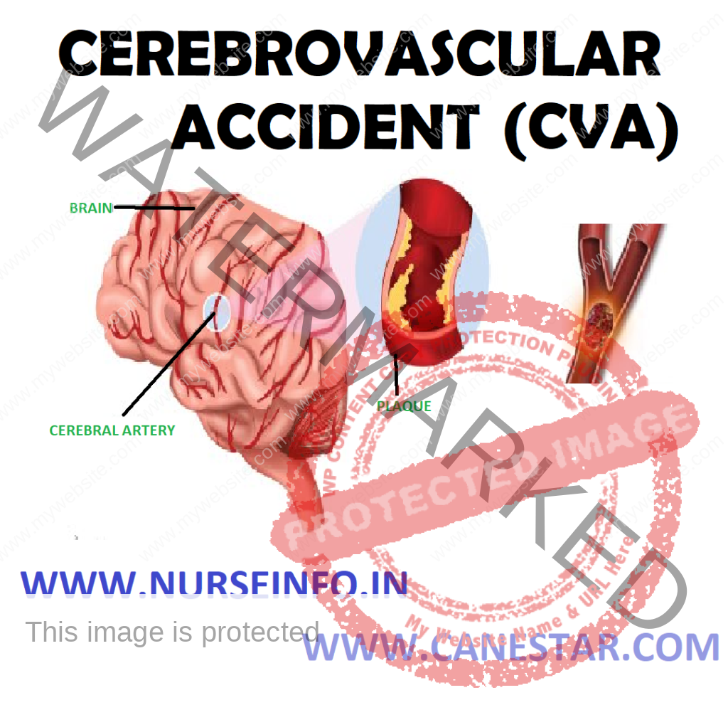
Any ischemic stroke occurs when a blood clot blocks a blood vessel, preventing blood and oxygen from getting to a part of the brain. When a clot forms somewhere else in the body and gets lodged in a brain blood vessel, it is called an embolic stroke. When the clot forms in the brain blood vessel, it is called a thrombotic stroke.
- Hemorrhagic Stroke
- A hemorrhagic stroke occurs when a blood vessel ruptures, or hemorrhages, which then prevents blood from getting to part of the brain. The hemorrhage may occur in a blood vessel in the brain, or in the membrane that surrounds the brain. It maybe of the following types:
- Intracerebral hemorrhage: in an intracerebral hemorrhage, a blood vessel in the brain bursts and spills into the surrounding brain tissue, damaging brain cells. Brain cells beyond the leak are deprived of blood and damaged. High blood pressure, trauma, vascular malformations, use of blood-thinning medications and other conditions may cause intracerebral hemorrhage
- Subarachnoid hemorrhage: in a subarachnoid hemorrhage, an artery on or near the surface of brain bursts and spills into the space between the surface of brain and skull. This bleeding is often signaled by a sudden, severe headache. A subarachnoid hemorrhage is commonly caused by the rupture of an aneurysm, a small sack-shaped or berry-shaped outpouching on an artery in the brain.
RISK FACTORS
- High blood pressure
- Cigarette smoking or exposure to second hand smoke
- High cholesterol level
- Diabetes
- Overweight or obese
- Physical inactivity
- Obstructive sleep apnea
- Cardiovascular disease, including heart failure, heart defects, heart infection or abnormal heart rhythm
- Use of some birth control pills or hormone therapies that include estrogen
- Heavy drinking
- Use of drugs such as cocaine and methamphetamines
- Having regular checkups after being diagnosed with preeclampsia
- Personal or family history of stroke, heart attack or TIA
- Being age 55 or older
- Race-Black has higher risk of stroke than people of other races
- Gender-stroke is more common in women than men, and more deaths from stroke occur in women
PATHOPHYSIOLOGY
- Due to thrombosis or embolism, some neurons die because of lack of oxygen and nutrients —- infarction of the cerebral vessels known as stroke —- tissue injury triggers an inflammatory response which increases intracranial pressure —- the injury disrupts metabolism leading to changes in ionic transport, localized acidosis, and free radical formation —- calcium, sodium and water accumulate in the injured cells and excitatory neurotransmitters are released —- continued cell cellular injury and swelling both occurs resulting to further cell damage —– Brain Death
- Impaired cerebral tissue perfusion (hemorrhagic) —- infarction of the cerebral vessels known as stroke —- space-occupying blood clots put more pressure on the brain tissues —- the regulatory mechanisms of the brain attempt to maintain equilibrium by increasing BP and ICP —- the ruptured cerebral vessels may constrict to limit blood loss however; this vasospam will result to further ischemia and necrosis of brain tissues —- Brain Death
SIGNS AND SYMPTOMS
- Difficulty walking
- Dizziness
- Loss of balance and coordination
- Difficulty speaking or understanding others who are speaking
- Numbness or paralysis in the face, leg, or arm, most likely on just one side of the body
- Blurred or darkened vision
- A sudden headache, especially when accompanied by nausea, vomiting, or dizziness
DIAGNOSTIC EVALUATION
- Physical examination
- Personal and family history of heart disease, TIA or stroke
- Blood tests: to evaluate the clotting time, bleeding time, etc
- Computerized tomography scan: brain imaging plays a key role in determining a stroke and what type of stroke maybe experiencing. A CT scan uses a series of X-rays to create a detailed image of brain. A CT scan can show a brain hemorrhage, tumors, strokes and other conditions. A dye is injected into blood vessels to view blood vessels to view blood vessels in neck and brain in greater detail
- Magnetic resonance imaging: an MRI uses powerful radio waves and magnets to create a detailed view of brain. An MRI can detect brain tissue damaged by an ischemic stroke and brain hemorrhages
- Carotid ultrasound: in this test, sound waves create detailed images of the inside of the carotid arteries in neck. This test shows buildup of fatty deposits (plaques) and blood flow in carotid arteries
- Cerebral angiogram: in this test, a thin, flexible tube (catheter) is inserted through a small incision, usually in groin, and guides it through major arteries and into carotid or vertebral artery. A dye is injected into blood vessels to make them visible under X-ray imaging. This procedure gives a detailed view of arteries in brain and neck
- Echocardiogram: this imaging technique uses sound waves to create a picture of heart. It can help to find the source of blood clots
MANAGEMENT
Prevention
There are many risk factors for having a stroke. Correspondingly, there are many measures that can be taken to help prevent them. These preventive measures are similar to the actions that you would take to help prevent heart disease, and include the following:
- Maintain normal blood pressure
- Limit saturated fat and cholesterol intake
- Refrain from smoking and drink alcohol in moderation
- Control diabetes
- Maintain a healthy weight
- Get regular exercise
- Eat a diet rich in vegetables and fruits
Medical Management
- Aspirin, an antithrombotic drug, is an immediate treatment after an ischemic stroke to reduce the likelihood of having another stroke. Aspirin prevents blood clots from forming.
- Other blood-thinning drugs, such as heparin, warfarin, or aspirin in combination with extended release dipyridamole may also be used, but these are not usually used in the emergency room setting.
- Intravenous injection of tissue plasminogen activator (TPA): some people who are having an ischemic stroke can benefit from an injection of a recombinant tissue plasminogen activator (TPA), also called alteplase, usually given through a vein in the arm. This potent clot-busting drug needs to be given within 4.5 hours after stroke symptoms begin if it is given into the vein. This drug restores blood flow by dissolving the blood clot causing stroke
- Carotid endarterectomy: in the carotid endarterectomy, a surgeon removes fatty deposits (plaques) from carotid arteries. In this procedure, a small incision along the front of neck, opens carotid artery, and removes fatty deposits that block the carotid artery.
- Angioplasty and stents: in an angioplasty, a surgeon inserts a catheter with a mesh tube and balloon on the tip into an artery in groin and guides it to the blocked carotid artery in neck. Surgeon inflates the balloon in the narrowed artery and inserts a mesh tube into the opening to keep artery from becoming narrowed after the procedure.
- Surgical clipping: a surgeon places a tiny clamp at the base of the aneurysm, to stop blood flow to it. This can keep the aneurysm from bursting
- Coiling (endovascular embolization): in this procedure, a surgeon inserts a catheter into an artery in groin and guides it to brain using X-ray imaging. Then guides tiny detachable coils into the aneurysm (aneurysm coiling). The coils fill the aneurysm, which blocks blood flow into the aneurysm and causes the blood to clot
NURSING MANAGEMENT
Nursing Diagnosis
- Ineffective cerebral tissue perfusion related to interruption of blood flow
Interventions
- Determine factors related to individual situation, cause for coma, decreased cerebral perfusion and potential for increased ICP
- Monitor and document neurological status frequently and compare with baseline
- Monitor vital signs, i.e. hypertension/hypotension, compare BP readings in both arms, heart rate and rhythm, auscultate for murmurs, respirations, noting patterns and rhythm, e.g. periods of apnea after hypervenitilation, Cheyne-Stokes respiration.
- Evaluate pupils, noting size, shape, equality, light reactivity
- Document changes in vision, e.g., reports of blurred vision, alternations in visual field and perception
- Assess higher functions, including speech, if patient is alert
- Position with head slightly elevated and in neutral position
- Maintain bedrest, provide quiet environment, and restrict visitors as indicated. Provide rest periods between care activities, limit duration of procedures
- Prevent straining at stool, holding breath
- Assess for nuchal rigidity, twitching, increased restlessness, irritability, onset of seizure activity
- Administer supplemental oxygen as indicated
- Administer medications as indicated: alteplase, anticoagulants, e.g., warfarin sodium, low-molecular weight heparin, antiplatelet agents, aspirin, dipyridamole, ticlopidine. Antihypertensives, peripheral vasodilators, e.g., cyclandelate, papaverine, isoxsuprine, steroids, e.g., dexamethasone
- Prepare for surgery, as appropriate, e.g., endarterectomy, microvascular bypass, cerebral angioplasty
- Monitor laboratory studies as indicated, e.g., prothrombin time (PT), activated partial thromboplastin time (aptt) time, dilantin level
- Impaired physical mobility related to neuromuscular abnormality
- Assess functional ability of impairment initially and on a regular basis
- Change positions at least every 2 hour (supine, sidelying) and possibly more often if placed on affected side.
- Position in prone position once or twice a day if patient can tolerate
- Prop extremities in functional position, use footboard during the period of flaccid paralysis. Maintain neutral position of head.
- Use arm sling when patient is in upright position, as indicated.
- Evaluate use and need for positional aids and splints during spastic paralysis, place pillow under axillae to abduct arm, elevate arm and hand
- Observe affected side for color, edema, or other signs of compromised circulation
- Inspect skin regularly, particularly over bony prominences. Gently massage any reddened areas and provide aids such as sheepskin pads as necessary
- Begin active/passive range of motion exercise to all extremities
- Assist to develop sitting balance (e.g. raise head of bed, assist to sit on edge of bed, having patient sue the strong arm to support body weight and strong leg to move affected leg, increase sitting time) and standing balance (e.g. put flat walking shoes on patient, support patient’s lower back with hands while positioning own knees outside patient’s knees, assist in using parallel bars/walkers).
- Get patient up in chair as soon as vital signs are stable, except following cerebral hemorrhage
- Pad chair seat with foam or water-filled cushion, and assist patient to shift weight at frequent intervals
- Provide egg-crate mattress, water-bed, flotation device, or specialized beds (e.g. kinetic), as indicated
- Disturbed sensory perceptions related to disturbed sensory reception and neuromuscular dysfunction
Interventions
- Observe behavioral responses e.g., hostility, crying, inappropriate affect, agitation, hallucination
- Eliminate extraneous noise and stimuli as necessary
- Speak in calm, quiet voice, using short sentences. Maintain eye contact
- Reorient patient frequently to environment, staff, and procedures
- Evaluate for visual deficits. Note loss of visual field, changes in depth perception (horizontal/vertical planes), and presence of diplopia
- Approach patient from visually intact side. Leave light on, position objects to take advantage of intact visual fields. Patch affected eye if indicated
- Assess sensory awareness, e.g. differentiation of hot/cold, dull/sharp, position of body parts/muscle, joint sense
- Stimulate sense of touch; e.g. give patient objects to touch, grasp
- Protect from temperature extremes, assess environment for hazards. Recommend testing warm water with unaffected hand
- Ineffective coping related to situational crisis and cognitive perceptual changes
Interventions
- Assess extent of altered perception and related degree of disability. Determine functional independence measure score
- Identify meaning of the loss, dysfunction and change to patient. Note ability to understand events, provide realistic appraisal of situation
- Determine outside stressors, e.g. family, work, social, future nursing/healthcare needs
- Encourage patient to express feelings, including hostility or anger, denial, depression sense of disconnectedness
- Note whether patient refers to affected side as ‘it’ or denies affected side and says it is ‘dead’
- Identify previous methods of dealing with life problems. Determine presence and quality of support systems
- Emphasize small gains either in recovery of function or independence
- Support behaviors and efforts such as increased interest, participation in rehabilitation activities
- Monitor for sleep disturbance, increased difficulty concentrating and statements of inability to cope, lethargy, and withdrawal
- Refer for neuropsychological evaluation and/or counseling if indicated
- Self-care deficit related to neuromuscular impairment and decreases strength and endurance
Interventions
- Assess abilities and level of deficit (0-4 scale) for performing ADLs
- Avoid doing things for patient that patient can do for self, but provide assistance as necessary
- Be aware of impulsive behavior and actions suggestive of impaired judgment
- Maintain a supportive, firm attitude. Allow patient sufficient time to accomplish task.
- Provide positive feedback for efforts and accomplishments
- Create plan for visual deficits that are present, e.g. place food and utensils on the tray related to patient’s unaffected side, situate the bed so that patient’s unaffected side is facing the room with the affected side to the wall, position furniture against wall and out of travel path
- Provide self-help devices, e.g. button/zipper hook, knife-fork combinations, long-handled brushes, extensions for picking things up from floor, toilet riser, leg bag for catheter, shower chair
- Assist and encourage good grooming and makeup habits
- Encourage family member to allow patient to do as much as possible for self
- Assess patient’s ability to communicate the need to void and ability to use urinal, bedpan. Take patient to the bathroom at frequent and periodic intervals for voiding if appropriate
- Identify previous bowel habits and re-establish normal regimen. Increase bulk in diet, encourage fluid intake, increased activity
- Risk for impaired swallowing related to neuromuscular dysfunction
Intervention
- Review individual pathology and ability to swallow, noting extend of paralysis, clarity of speech, facial, tongue involvement, ability to protect airway and episodes of coughing or choking, presence of adventitious breath sounds, amount and character of oral secretions
- Have suction equipment available at bedside, especially during early feeding efforts
- Promote effective swallowing, e.g. schedule activities, medications to provide a minimum of 30 min rest before eating, provide pleasant environment free of distractions, assist patient with head control and support, and position based on specific dysfunction
- Place patient in upright position during and after feeding as appropriate
- Provide oral care based on individual need prior to meal
- Season food with herbs, spices, lemon juice, etc. according to patient’s preference, within dietary restrictions
- Place food of appropriate consistency in unaffected side of mouth
- Touch parts of the cheek with tongue blade and apply ice to weak tongue
- Feed slowly, allowing 30-45 min for meals
- Offer solid foods and liquids at different times
- Maintain upright position for 45-60 min after eating
- Maintain accurate intake output, record calorie count
- Encourage participation in exercise
- Administer IV fluids and or tube feedings
- Coordinate multidisciplinary approach to develop treatment plan that meets individual needs
- Knowledge deficit related to lack of exposure and cognitive limitation
Interventions
- Evaluate type and degree of sensory-perceptual involvement
- Include family in discussions and teaching
- Discuss specific pathology and individual potentials
- Identify signs and symptoms requiring further follow-up, e.g. changes or decline in visual, motor, sensory function, alternation in mentation or behavioral responses, severe headache
- Review current restrictions or limitations and discuss planned resumption of activities (including sexual relations)
- Provide written instructions and schedules for activity, medication, important facts
- Encourage patient to refer to lists communication or notes instead of depending on memory
- Discuss plans for meeting self-care needs
- Refer to discharge planner, home care supervisor, visiting nurse
- Suggest patient reduce or limit environmental stimuli, especially during cognitive activities
- Recommend patient seek assistance in problem-solving process and validate decisions, as indicated
- Review importance of balanced diet, low in cholesterol and sodium if indicated. Discuss role of vitamins and other supplements
- Refer to reinforce importance of follow-up care by rehabilitation team, e.g. physical, occupational, speech, vocational therapists.