
CARDIOMYOPATHY – Etiology, Signs and Symptoms, Diagnostic Evaluation, Treatment and Management
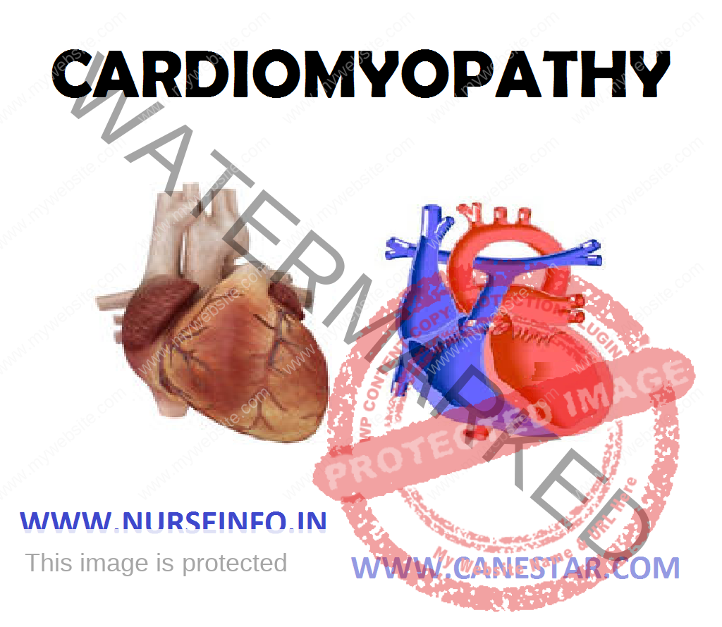
Cardiomyopathy is a weakening of the heart muscle or associated with other problems with the heart muscle. It may be associated with heart failure, endocarditis or other heart problems which after the normal architecture of heart. Most patients with cardiomyopathy have heart failure
ETIOLOGY
In the broadest sense, “cardiomyopathy” (CM) refers to heart disease resulting from a primary abnormality of the myocardium (heart muscle). There are three types:
- Dilated cardiomyopathy (also called ‘congestive’ cardiomyopathy)
- Restrictive cardiomyopathy
- Hypertrophic cardiomyopathy
Dilated cardiomyopathy is a condition in which the heart becomes weak and the chambers get large. As a result, the heart cannot pump enough blood out to the body. The heart with dilated cardiomyopathy is striking in appearance. Dilation (enlargement) of all chambers (both atria and both ventricles). The total size of the heart is typically huge (cardiomegaly). The myocardium becomes ‘flabby’ and loses its ability to contract. Naturally, the heart chambers will lose their pumping function and the heart will ultimately undergo failure. Since blood flow within the chambers is sluggish, intracardiac mural thrombi are prone to form on the inner walls of the atria and ventricles. Pieces of these thrombi may break off and embolize to the lungs (pulmonary emboli), or any other organ and tissue (systemic emboli). This may lead to infarction of these organs.
Hypertrophic cardiomyopathy (HCM) is a condition in which the heart muscle becomes thick. The thickening makes it harder for blood to leave the heart. This type of cardiomyopathy is usually passed down through families. This is a disease of younger people (mean age 26). It is a genetically inherited disease. The classic anatomic feature is the profound hypertrophy of the myocardium of the left ventricle. The part of the LV wall that forms the interventricular septum (IVS) is more hypertrophic than the lateral part of the LV wall. This extra-thickened interventricular septum is referred to as asymmetric septal hypertrophy (ASH). The IVS can become so hypertrophied that it bulges into the lumen of the LV, thereby decreasing the volume of the LV chamber.
Restrictive cardiomyopathy is a group of disorders. Restrictive cardiomyopathy can either be idiopathic or can be caused by diseases that deposit abnormal substances within the myocardium. The classic example is amyloidosis, whereby the abnormal amyloid protein accumulates within the myocardium, resulting in stiffness.
Peripartum cardiomyopathy occurs during pregnancy or in the first 5 months afterwards.
SIGNS AND SYMPTOMS
- Breathlessness with exertion or even at rest
- Swelling of the legs, ankles and feet
- Bloating of the abdomen due to fluid buildup
- Fatigue
- Irregular heartbeats that feel rapid, pounding or fluttering
- Dizziness, lightheadedness and fainting
- Palpitations (fluttering in the chest due to abnormal heart rhythms)
- Fainting (usually caused by irregular heart rhythms or abnormal responses of the blood vessels during exercise
- Chest pain or pressure (occurs usually with exercise or physical activity but can also occur will rest or after meals)
DIAGNOSTIC EVALUATION
- The health care provider may hear abnormal sounds, called murmurs, when listening to your heart with a stethoscope
- A physical exam may also reveal:
Enlarged spleen and enlarged heart size with atrophy
- The following tests may be performed:
Blood culture and sensitivity (to detect bacteria)
Chest X-ray
Complete blood count (may show mild anemia)
CT scan of the chest
Echocardiogram (ultrasound of the heart)
ECG
TREATMENT
- When possible, the cause of cardiomyopathy is treated. Medicines and lifestyle changes are often needed to treat the symptoms of heart failure, angina, and abnormal heart rhythms
Different procedures or surgeries may also be used:
- A defibrillator sends an electrical pulse to stop life-threatening abnormal heart rhythms
- A pacemaker treats a slow heart rate or helps both sides of your heart beat at the same time
- Coronary artery bypass (CABG) surgery or angioplasty can improve blood flow to the damaged or weakened heart muscle
- Heart transplant is used when all other treatments have failed
MANAGEMENT
- The overall goals of treatment for cardiomyopathy are to manage your signs and symptoms, prevent your condition from worsening, and reduce your risk of complications.
- Angiotensin-converting enzyme (ACE) inhibitors to improve your heart’s pumping capability, such as enalapril (Vasotec), lisinopril (Zestril, Prinivil), ramipril (Altace) and captopril (Capoten).
- Angiotensin receptor blockers (ARBs) for those who cannot take ACE inhibitors, such as losartan (Cozaar) and valsartan (Diovan)
- Beta blockers to improve heart function, such as carvedilol (Coreg) and metoprolol (Lopressor, Toprol-XL)
- Digoxin (Lanoxin) this drug, also referred to as digitalis, increases the strength of your heart muscle contractions. It also tends to show the heartbeat. Digoxin reduces heart failure symptoms and improves your ability to live with cardiomyopathy
- Diuretics: often called water pills, diuretics make you urinate more frequently and keep fluid from collecting in your body. Commonly prescribed diuretics for heart failure include bumetanide (Bumex) and furosemide (Lasix). The drugs also decrease fluid in your lungs, so you can breathe more easily. One diuretic, spironolactone (Aldactone), may also be helpful in treating scarring of your heart tissue.
- Another option for some people with dilated cardiomyopathy is a special pacemaker that coordinates the contractions between the left and right ventricles (biventricular pacing). In people who may be at risk of serious arrhythmias, drug therapy or an implantable cardioverter-defibrillator (ICD) may be an option. An ICD is a small device – about the size of a box of matches – implanted in your chest to continuously monitor your heart rhythm and deliver electrical shocks when needed to control abnormal, rapid heartbeats. The device can also work as a pacemaker.
Surgical Management
- Septal myectomy: this is an open-heart operation in which the surgeon removes part of the thickened, overgrown heart muscle wall (septum) that separates the two bottom heart chambers (ventricles). Removing the part of this overgrown muscle improves blood flow and reduces mitral regurgitation. Myectomy is used if medications do not relieve symptoms. Most people who have symptoms and undergo myectomy have no further symptoms. This type of surgery is available only in medical centers that specialize in the treatment of hypertrophic cardiomyopathy.
- Septal ablation: also called septal alcohol ablation, this is a treatment in which a small portion of the thickened heart muscle is destroyed by injecting alcohol through a catheter into the artery supplying blood to it. There are possible complications with this procedure, including heart block – a disruption of the heart’s electrical system – which requires implantation of a pacemaker. The long term success of this procedure is not yet known, but it is becoming more commonly used.
- Pacemaker implantation: a pacemaker is a small electronic device inserted under your skin that sends electrical signals to your heart to monitor and regulate your heartbeat. Surgery to implant the pacemaker is usually performed during local anesthesia and typically takes less than three hours. Pacemaker implantation is generally not as effective as surgical options, but it is sometimes used in older people who want to avoid more invasive procedures
- Implantable cardioverter-defibrillator (ICD): this is a pager-sized device implanted in your chest like a pacemaker. An ICD continuously monitors your heartbeat. If a life-threatening arrhythmia occurs, the ICD delivers precisely calibrated electrical shocks to restore a normal heart rhythm. A small number of people with hypertrophic cardiomyopathy are at risk of sudden cardiac death because of abnormal heart rhythms. In these high-risk individuals, many doctors recommend the implantation of an ICD.
- Heart transplant and ventricular assist devices (VADs): If you have severe cardiomyopathy and medications cannot control your symptoms, a heart transplant may be an option. Because of the shortage of donor hearts, even people who are critically ill may have a long wait before having a heart transplant. In some cases, a mechanical heart assist device can help critically ill people as they wait for an appropriately matched donor. These devices, known as ventricular assist devices (VADs), can help blood circulate through your heart for months or even years.
Nursing Management
- Acute pain related to an impaired ability of blood vessels to supply oxygen to the tissues
- Activity intolerance related to compromised oxygen transport system secondary to heart muscle dysfunction
- Risk for ineffective breathing pattern related to decreased respiratory depth secondary to pain
Interventions
- Bed rest is important because it reduces myocardial oxygen demand and usually continues until the following criteria are met:
Temperature remain normal without the use of salicylates
Resting pulse rate remains less than 100 beats/min
ECG tracings show no manifestations of myocardial damage
Pericardial friction rub is not present
- Obtain a clear description of the pain or discomfort. Identify the source of greatest discomfort as a focus for intervention
- Administer analgesics as needed and use salicylates around the clock. Balance rest and activity according to the degree of pain and activity tolerance.
- Provide psychosocial support while patient is confined to hospital or home with restrictive intravenous therapy
- If patient received surgical treatment, provide postsurgical care and instruction
- After surgery, monitor patient’s temperature; a fever may be present for weeks
- A high-protein, high-carbohydrate diet helps maintain adequate nutrition in the presence of fever and infection
- Oral hygiene every 4 hours; small, attractive meal servings and foods that are not overly rich, sweet or greasy stimulate the appetite
- Instruct the client about how to reduce exposure to infection as follows:
Take good care of the teeth and gums; obtain prompt dental care for cavities and gingivitis
Prophylactic medication may be needed before invasive dental procedures, and individualized evaluation for prophylaxis medication is also needed
Avoid people who have an upper respiratory tract infection
Assess for signs and symptoms of organ damage, such as stroke (CVA, brain attack), meningitis, heart failure, myocardial infarction, glomerulonephritis, and splenomegaly
Instruct patient and family about activity restrictions, medications and signs and symptoms of infection
Refer to home care nurse to supervise and monitor intravenous antibiotic therapy at home
CARDIOMYOPATHY – Etiology, Signs and Symptoms, Diagnostic Evaluation, Treatment and Management
