
BRONCHITIS – Etiology, Risk Factors, Pathophysiology, Clinical Manifestations, Complications, Diagnostic Evaluations and Management
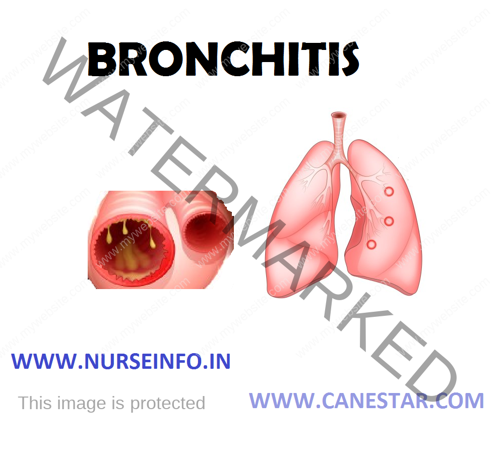
Bronchitis is a term that describes inflammation of the bronchial tubes (bronchi and the smaller branches termed as bronchioles) that results in excessive secretions of mucus into the tubes, leading to tissue swelling that can narrow or close off bronchial tubes.
ETIOLOGY
- Acute bronchitis (also known as a chest cold) is typically a fairly minor illness that causes symptoms for a few days to a couple of weeks. It usually resolves on its own with rest without leaving any obvious long-term consequences. Viruses, such as influenza, respiratory syncytial virus (RSV), and rhinoviruses cause the majority of cases of acute bronchitis, while the remainder are caused by bacteria (for example, mycoplasma, pneumococcus) or short-term exposure to chemical irritants (for example, tobacco smoke, gastric reflux contents, inhaled solvents).
- Chronic bronchitis is a cough with mucus for at least three months in each of the two consecutive years. It can indicate other health problems as well as lead to permanent damage to lungs. It may occur due to deficiency of alpha-1 antitripsinogen enzyme.
RISK FACTORS
- Cigarette smoke: people who smoke or who live with a smoker are at higher risk of both acute bronchitis and chronic bronchitis
- Low resistance: this may result from another acute illness, such as cold, or from a chronic condition that compromises the immune system. Older adults, infants, and young children have greater vulnerability to infection.
- Exposure to irritants: risk of developing bronchitis is greater if exposed to certain lung irritants, such as grains or textiles, or are exposed to chemical fumes.
PATHOPHYSIOLOGY
Cigarette smoking RTI (Respiratory Tract Infection) Environmental pollutants —- inflammation —- bradykinin, histamine, prostaglandin —- ↑ capillary permeability —- fluid/cellular exudation —- edema of mucous membrane —- hypersecretion of mucus —- persistent cough
CLINICAL MANIFESTATIONS
For either acute bronchitis or chronic bronchitis, signs and symptoms may include:
- Cough
- Production of mucus (sputum), which can be clear, white, yellowish-gray or green in color
- Fatigue
- Slight fever and chills
- Chest discomfort
COMPLICATIONS
Although a single episode of bronchitis usually is not the cause for concern, it can lead to pneumonia in some people. Repeated bouts of bronchitis may signal:
- Chronic bronchitis
- Asthma
- Bronchiectasis
- Cystic fibrosis
- Tuberculosis
- Sinusitis
- Dyspnea, sometimes severe
- Respiratory failure
- Pneumonia
- Cor pulmonale
- Pneumothorax
- Polycythemia
- COPD
- Emphysema
- Chronic advancement of the disease and
- High mortality rate
DIAGNOSTIC EVALUATION
During the first few days of illness, it can be difficult to distinguish the signs and symptoms of bronchitis from those of a common cold
- Chest X-ray: a chest X-ray can help determine pneumonia or another condition that may explain cough
- Sputum culture: this test checks for the presence of bacteria in sputum produced when coughed. It is helpful in determining whether whooping cough or other illnesses would be helped by antibiotics
- Pulmonary function test: during a pulmonary function test, you blow into a device called a spirometer, which measures how much air your lungs can hold and how quickly you can get air out of your lungs. This test checks for signs of asthma or emphysema.
MANAGEMENT
The goal of treatment for bronchitis is to relieve symptoms and ease breathing. In most cases, acute bronchitis requires only self-care treatments, such as:
- Getting more rest
- Taking over-the-counter pain medications
- Drinking fluids
- Breathing in warm, moist air
Medications
- Antibiotics: such as fluoroquinolones, macrolides, sulfonamides, tetracyclines
- Cough medicine: such as expectorants, mucolytics etc
- Bronchodilators: such as theophyllin, etc. to dilate the bronchus and ease the sputum to come out
- Steroids: such as prednisone, methylprednisone to reduce the inflammatory reaction and thus decrease the bronchial swelling and secretions that, in turn, allows better airflow because of reduced airway obstruction.
- PDE4 inhibitors are a class of anti-inflammatory agents for exacerbations of COPD. It is primarily for exacerbations that involve excessive bronchitis and mucus production. There is currently only one agent called roflumilast a pill taken once per day.
Nursing Management
Nursing Diagnosis
- Impaired Gas Exchange related to altered oxygen supply
Interventions
- Assess respirations: quality, rate, pattern, depth, and breathing effort
- Assess for life-threatening problems (i.e. respiratory arrest, flail chest, sucking chest wound)
- Auscultate lung sounds: also assess for the presence of jugular vein distension (IVD) or tracheal deviation
- Assess for signs of hypoxemia
- Monitor vital signs
- Assess for changes in orientation and behavior
- Monitor ABGs
- Place the patient on continuous pulse oximetry
- Assess skin color for developmental of cyanosis, especially circumoral cyanosis
- Provide supplemental oxygen, via 100% O2 non-rebreather mask
- Prepare the patient for intubation
- Ineffective Airway Clearance related to tracheobronchial obstruction
Interventions
- Assess airway for patency by asking the patient to state his name
- Inspect the mouth, neck and position of trachea for potential obstruction
- Auscultate lungs for presence of normal or adventitious lung sounds
- Assess respiratory quality, rate, depth, effort and pattern
- Assess for mental status changes
- Assess changes in vital signs
- Monitor arterial blood gases (ABGs)
- Administer supplemental oxygen
- Position patient’s head with bed at 45 degrees (if tolerated)
- Assist patient with coughing and deep breathing techniques (positioning, incentive spirometry, frequent position changes)
- Prepare for placement of endotracheal or surgical airway (i.e. cricothyroidectomy, tracheostomy).
- Confirm placement of the artificial airway
- Impaired Gas Exchange related to altered oxygen supply
Interventions
- Assess respirations: quality, rate, pattern, depth and breathing effort
- Assess for life-threatening problems (i.e. respiratory arrest, flail chest, sucking chest wound)
- Auscultate lung sounds. Also assess for the presence of jugular vein distention (JVD) or tracheal deviation
- Assess for signs of hypoxemia
- Monitor vital signs
- Assess for changes in orientation and behavior
- Monitor ABGs
- Place the patient on continuous pulse oximetry
- Assess skin color for development of cyanosis, especially circumoral cyanosis
- Provide supplemental oxygen, via 100% O2 non-rebreather mask
- Prepare the patient for intubation
BRONCHITIS – Etiology, Risk Factors, Pathophysiology, Clinical Manifestations, Complications, Diagnostic Evaluations and Management
