Home Blog Page 59

HYPERPARATHYROIDISM

HYPERPARATHYROIDISM – Etiology, Risk Factors, Signs and Symptoms, Complications and Management

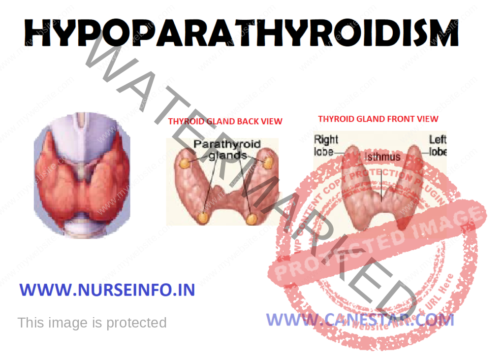
Hyperparathyroidism is an excess of parathyroid hormone in the bloodstream due to over activity of parathyroid glands. These oval, grain-of-rice-sized glands are located in your neck. The parathyroid glands produce parathyroid hormone, which helps maintain an appropriate balance of calcium in the bloodstream and in tissues that depend on calcium for proper functioning.

ETIOLOGY

  • Primary hyperparathyroidism: it occurs because of some problem with one or more of the four parathyroid glands: a noncancerous growth (adenoma) on a gland is the most common cause, enlargement (hyperplasia) of two or more parathyroid glands accounts for most other cases. A cancerous (malignant) tumor is a rare cause of primary hyperparathyroidism
  • Secondary hyperparathyroidism: it is the result of another condition that lowers calcium levels
  • Severe calcium deficiency
  • Severe vitamin D deficiency
  • Chronic kidney failure: kidneys convert vitamin D into a form that body can use. If kidneys function poorly, useable vitamin D may decline and calcium levels drop. Chronic kidney failure is the most common cause of secondary hyperparathyroidism

RISK FACTORS

  • Woman who has gone through menopause
  • Severe calcium or vitamin D deficiency
  • Inherited disorder, such as multiple endocrine neoplasia, type I, which usually affects multiple glands
  • Radiation treatment for cancer that has exposed neck to radiation a drug most often used to treat bipolar disorder

SIGNS AND SYMPTOMS

  • Fragile bones that easily fracture (osteoporosis)
  • Kidney stones
  • Excessive urination
  • Abdominal pain
  • Tiring easily or weakness
  • Depression or forgetfulness
  • Bone and joint pain
  • Frequent complaints of illness with no apparent cause
  • Nausea, vomiting or loss of appetite

DIAGNOSTIC EVALUATION

  • Blood tests: if the result of a blood test indicates elevated calcium in your blood
  • Bone mineral density test (bone densitometry): the most common test to measure bone mineral density is dual energy X-ray absorptiometry, or a DXA scan. This test uses special X-ray devices to measure how many grams of calcium and other bone minerals are packed into a segment of bone
  • Urine tests: a 24-hour collection of urine can provide information on how well kidneys function and how much calcium is excreted in urine. This test may help in judging the severity of hyperparathyroidism or diagnosing a kidney disorder causing hyperparathyroidism.
  • Ultrasound: ultrasound uses sound waves to create images of your parathyroid glands and surrounding tissue. A small device held against your skin (transducer) emits high-pitched sound waves and records the sound wave echoes as they reflect off internal structures. A computer converts the echoes into images on a monitor.
  • Sestamibi scan: sestamibi is a specifically designed radioactive compound that is absorbed by overactive parathyroid glands and can be detected on computerized tomography (CT) scans. A small dose of the compound is injected into your bloodstream before the imaging test is done

COMPLICATIONS

  • Osteoporosis: the loss of calcium often results in osteoporosis, or weak, brittle bones that fracture easily.
  • Kidney stones: the excess of calcium in blood may cause small, hard deposits of calcium and other substances to form in kidneys. A kidney stone usually causes significant pain as it passes through the urinary tract
  • Cardiovascular disease: although the exact cause-and-effect link is unclear, high calcium levels are associated with cardiovascular conditions, such as high blood pressure (hypertension) and certain types of heart disease
  • Neonatal hyperparathyroidism: severe, untreated hyperparathyroidism in pregnant women may cause dangerously low levels of calcium in newborns

MANAGEMENT

Your doctor may recommend no treatment and regular monitoring if:

  • Your calcium levels are only slightly elevated
  • Your kidneys are functioning normally
  • Your bone density is normal or only slightly below normal
  • You have no other symptoms that may improve with treatment
  • If you choose this watch-and-wait approach, you will likely need a test to check your blood-calcium levels at least twice a year and have other monitoring tests done at least once a year

Surgical Management

Surgery is the most common treatment for primary hyperparathyroidism and provides a cure in at least 90 percent of all cases. A surgeon will remove only those glands that are enlarged or have a tumor (adenoma). If all four glands are affected, a surgeon will likely remove only three glands and perhaps a portion of the fourth – leaving some functioning parathyroid tissue

Complications from surgery are not common. Risks include:

  • Damage to nerves controlling the vocal cords
  • Long-term low calcium levels requiring the use of calcium and vitamin D supplements

Drugs

  • Calcimimetics: a calcimimetic is a drug that mimics calcium circulating in the blood. Therefore, the drug may trick the parathyroid glands into releasing less parathyroid hormone. This drug is solid as cinacalcet (sensipar). The Food and Drug Administration approved cinacalcet to treat hyperparathyroidism caused by chronic kidney disease or parathyroid cancer
  • Hormone replacement therapy: for women who have gone through menopause and have signs of osteoporosis, hormone replacement therapy may help bones retain calcium. This treatment, usually a combination estrogen and progestin, does not address the underlying problems with the parathyroid glands. Prolonged use of hormone replacement therapy can increase the risk of cardiovascular disease and some cancers. Work with your doctor to evaluate the risks and benefits to help you decide what is best for you.
  • Bisphosphonates: bisphosphonates also prevent the loss of calcium from bones and may lessen osteoporosis caused by hyperparathyroidism

LIFESTYLE AND HOME REMEDIES

  • Monitor how much calcium and vitamin D you get in your diet. The Institute of Medicine recommends 1,000 milligrams (mg) of calcium a day for adult’s ages 19 to 50 and men ages 51-70. That calcium recommendation increases to 1,200 mg a day if you are a woman age 51 or older or a man age 71 or older. The Institute of Medicine also recommends 600 international units (IUs) of vitamin D a day for adults ages 19 to 70 and 800 IUs a day for adults age 71 and older.
  • Drink plenty of water. Drink six to eight glasses of water daily to lessen the risk of kidney stones
  • Exercise regularly. Regular exercise, including strength training, helps maintain strong bones.
  • Do not smoke. Smoking may increase bone loss as well as increase your risk of a number of serious health problems
  • Avoid calcium-raising drugs. Certain medications, including some diuretics and lithium, can raise calcium levels

NURSING MANAGEMENT

  • Protect the patient from injury
  • Monitor for possible complications
  • Provide patient education independent
  • Adjust activities and reduce intensity
  • Provide positive atmosphere, while acknowledging the difficulty of situation for the client
  • Assist patients with activities/monitor clients use of assistive device such as walker
  • Helps minimize frustration, rechanneling of energy
  • To protect client from injury
  • To remove cause of hypersecretion of parathormones
  • Loop diuretic used with normal saline to cause diuresis and to reduce calcium levels
  • Obtain baseline serum potassium, calcium, phosphate, and magnesium levels before treatment
  • Provide at least 3 liters of fluid per day, including cranberry or prune juice, to increase urine acidity and help prevent calculus formation
  • Take safety precaution to minimize the risk for injury from fall
  • Schedule care to allow the patient with muscle weakness as much rest as possible
  • Provide comfort measures to alleviate bone pain
  • Administer antacids, as appropriate to prevent pelvic ulcers
  • Auscultate the lungs regularly. Check for signs of pulmonary edema in the patient receiving large amounts of normal saline solution, especially if he has pulmonary or cardiac disease
  • Check for elevated serum calcium levels if the patient is receiving cardiac glycosides
  • Assess the patient for parathyroid poisoning, musculoskeletal changes, and renal impairment
  • Observe the patient for signs of pain and monitor him for effectiveness of analgesics and comfort measures
HYPERPARATHYROIDISM – Etiology, Risk Factors, Signs and Symptoms, Complications and Management

For More Medical Surgical Nursing, CLICK HERE

For More Psychiatric Nursing, CLICK HERE

NURSING MODEL

NURSING MODEL

Nursing focuses on the individual’s response to potential or actual health problems. Under the nursing model, human behavior is viewed from a holistic perspective.

Nursing View of Behavioral Deviations

  • Behavior is viewed on a continuum from healthy adaptive responses to maladaptive responses that indicate illness.
  • Each individual is predisposed to respond to life events in unique ways. These predispositions are biological, psychological, socio cultural, and the sum of the person’s heritage and past experiences.
  • Behavior is the result of combining the predisposing factors with precipitating stressors. Stressors are life events that the individual perceives as challenging, threatening or demanding. The nature of the behavioral response depends on the person’s primary appraisal of the stressor and his secondary appraisal of the coping resources available to him.
  • A stressor that has primary impact on physiological functioning also affects the person’s psychological and socio-cultural behavior. For instance, a man who has a myocardial infarction may also become severely depressed, because he fears he will lose his ability to work. On the other hand, the patient who enters the psychiatric inpatient unit with major depression may be suffering from malnutrition and dehydration because of his refusal to eat or drink. The holistic nature of nursing encompasses all of these facets of behavior and incorporated them into patient care planning.

Nursing Process

Nursing intervention may take place at any point on the continuum. Nursing diagnosis may focus on behavior associated with a medical diagnosis or other health behavior that the patient wishes to change.

A nurse may practice primary prevention by intervening in a potential health problem, secondary prevention by intervening in an actual acute health problem or tertiary prevention by intervening to limit the disability caused by actual chronic health problem. The nursing assessment of the patient includes presenting complaints, past history, family history, personal history, occupational history, sexual history, physical examination and mental status examination. Additional data may be collected for significant others and by reviewing the systems. A nursing diagnosis is then formulated and based on this diagnosis; planning and interventions are carried out. Finally, evaluation will be done to find out the effectiveness of nursing interventions.

Providing nursing care is a collaborative effort, with both the nurse and the patient contributing ideas and energy to the therapeutic process.

Summary of Selected Nursing Theories

Peplau’s Theory

Peplau proposed an interpersonal theory applicable to nursing practice in general and to psychiatric-mental health nursing in particular. It focuses primarily on the nurse-patient relationship. Peplau’s theory describes, explains, predicts and to some extent, permits control of the sequence of events occurring in the nurse-patient relationship.

Peplau describes the interpersonal aspects of nursing as a process consisting of four phases. These are orientation, identification, exploitation and resolution phases.

While working with the patient through these phases, the nurse assumes six roles: resource person, technical expert, teacher, leader, surrogate parent and a counselor.

Peplau’s theory continues to apply to today’s nursing scene, especially with respect to long-term psychiatric care in outpatient and home health settings.

Orem’s Theory

Dorothea E Orem’s theory is based on the premise that people need a composite of self-care actions to survive. Self-care actions consist of all behaviors performed by people to maintain life and health. The capacity of the patient and the patient’s family to perform self-care is called self-care agency. Orem states that a need for nursing care exists if the patient’s self –care demand exceeds the patient’s self-care agency. Thus, the goal of nursing is to meet the patient’s self-care demands until the patient and his family are able to do so.

Orem’s theory describes three types of self-care:

  • Universal self-care behaviors, required to meet physiological and psychosocial needs.
  • Developmental self-care behaviors, required to undergo normal human development.
  • Health deviation self-care behaviors, required to meet patient’s needs during health deviations.

The classification of self-care behaviors in this manner helps to ensure complete assessment of the patient’s self-care agency.

Assessment focuses on the patient’s self-care demand, self-care agency and self-care deficits. A plan is formulated from the information obtained in the assessment that indicates the nursing approach needed to meet the patient’s needs. It can be categorized as follows:

  • Wholly compensatory, in which the patient does not participate behaviorally in self- care.
  • Partially compensatory, in which the patient and nurse participate behaviorally in meeting the patient’s self-care needs.
  • Educative-developmental, in which the patient meets self-care needs with minimal nursing assistance.

To implement the required nursing approach, the nurse uses one of the five behaviors: acting or doing for the patient, guiding, supporting, providing and teaching.

Roger’s Theory

Roger’s model focuses on the individual as a unified whole in constant interaction with the environment. The unitary person is viewed as an energy field that is more than as well as different from the sum of the biological, physical, social and psychological parts. In Roger’s model, nursing is concerned with the unitary person as a synergistic phenomenon.

Nursing science is devoted to the study of nature and direction of unitary human development. Nursing practice helps individuals achieve maximum well-being within their potential.

Roy’s Theory

According to Callista Roy’s theory, the goal of nursing is to promote the patient’s adaptation in health and illness. This goal is achieved through the nurse’s efforts to change, manipulate or block stress-producing stimuli that may impinge on the patient. The theory assumes that this kind of nursing intervention assists the patient to cope more effectively through reducing stress.

Roy’s theory assumes that all human beings have adaptive systems and change in response to stimuli. If the change does not promote the person’s integrity then the change can be considered maladaptive.

The nursing process used in Roy’s theory involves two levels of assessment. The first level includes observation of behavior related to the four adaptive modes: Physiologic, self-concept, role function and interdependence. These four modes represent methods used by the patient to adapt. The second level of assessment consists of identifying focal, contextual and residual stimuli. The focal stimulus represents the immediate dominant stimulus affecting the patient, such as injury, stress or illness. Contextual stimuli include the environment, the patient’s family and all other background factors related to the focal stimulus. Residual stimuli consist of the patient’s previous background, beliefs, attitudes and traits.

According to Roy’s theory, a person’s adaptation level is a function of focal, contextual and residual stimuli. When a person encounters stresses form these stimuli that surpassinnate and acquired mechanisms to cope effectively, the person behaves ineffectively as demonstrated by one or more of the adaptive modes. At this point, nursing intervention is required. This emphasizes on the patient’s behavior, stimuli determining the patient’s behavior, stimuli determining the patient’s behavior, and the nurse intervening in some way to interfere with the stimuli.

NURSING MODEL
NURSING MODEL

INVESTIGATIONS IN PSYCHIATRY

INVESTIGATIONS IN PSYCHIATRY

Investigations are useful to detect alteration in biologic function and to screen for medical disorders causing psychiatric symptoms.

Routine Investigations

  • A complete hemogram (total and differential blood count, hemoglobin, ESR) and urinalysis are the basic routine tests. Leucopenia and agranulocytosis are associated with certain medications (Clozapine). Treatment with lithium and neuroleptic malignant syndrome are often associated with leukocytosis.
  • Renal function tests: Treatment with lithium
  • Liver function tests: For all alcoholic patients treatment with carbamazepine, valproate and benzodiazepines
  • Serum electrolytes: Dehydration, treatment with carbamazepine,  antipsychotics, lithium
  • Blood glucose: Routine screen above 35 years age
  • Thyroid function test: Depression, treatment with lithium and carbamazepine
  • Electrocardiogram (ECG): Above 35 years of age, treatment with lithium, antidepressants, ECT, antipsychotics
  • HIV testing: IV drug users suggestive sexual history, AIDS, dementia
  • VDRL: Suggestive sexual history
  • Serum CPK: Neuroleptic malignant syndrome (markedly increased levels)
  • Chest X-ray: Before treatment with ECT
  • Drug level estimation: Drug levels are indicated to test for therapeutic blood levels, for toxic blood levels and for testing drug compliance. Examples are lithium (0.6 – 1.6 mEq/L), carbamazepine (6-12 mg/mL), valproate (50-100 mg/mL), haloperidol (8-18 ng/mL), tricyclic antidepressants (Imipramine 200-250 ng/mL, nortriptyline 50-150 ng/mL), benzodiazepines, barbiturates.

Electrophysiological Tests

Electroencephalogram (EEG): Measures brain electrical activity, identifies dysrhythmias and asymmetries, used in the diagnosis of seizures, dementia, neoplasm, stroke, metabolic or degenerative disease.

        Polysomnography/sleep studies: Used in the diagnosis of sleep disorders and seizures.

Brain Imaging Tests (Cranial)

  • Computed tomography (CT) scan: Measures accuracy of brain structure to detect possible lesions, abscesses, areas of infarction or aneurysm. CT scan also identifies various anatomic differences in patients with schizophrenia, organic mental disorder and bipolar disorder.
  • Magnetic resonance imaging (MRI) scan: Measures the anatomic and biochemical status of various segments of the brain; detects brain edema, ischemia, infection, neoplasm, trauma and other changes such as demyelination used in the diagnosis of dementia, to detect morphological changes in schizophrenia patients.
  • Other tests are positron emission tomography (PET).

Neuroendocrine Tests

Commonly used neuroendocrine tests are dexamethasone suppression test, TRH stimulation test, serum prolactin levels, serum 17-hydroxycorticosteriod, serum melatonin levels.

Genetic tests

Cytogenetic work-up is advised in some cases of mental retardation.

INVESTIGATIONS IN PSYCHIATRY

HOLISTIC MODEL (PSYCHIATRIC NURSING)

HOLISTIC MODEL

The holistic view of the patient, with the body and soul seen as inseparable, and the patient viewed as a member of a family and community was central to Nightingale’s view of nursing. The primary goal of nursing is to help patients develop strategies to achieve harmony within themselves and with others, nature and the world. Integrative functioning of the patient is physical, emotional, intellectual, social and spiritual dimensions are emphasized. Each person is considered as a whole, with many factors contributing to health and illness.

Major Concepts

Five major concepts are generally accepted as premises of holistic health care philosophy:

  • First, each person is multidimensional; one’s physical, emotional, intellectual, social and spiritual dimensions are in constant interaction with each other:
  • The physical dimension involves everything associated with one’s body, both internal and external.
  • The emotional dimension consists of affective states and feelings, including motor behavior associated with emotion, the experienced aspect of emotion, and the physiological mechanisms that underlie emotion
  • The intellectual dimension includes receptive functions, memory and learning, cognition and expressive functions.
  • The social dimension is based on social intersection and relationships, more so the global concept of culture.
  • The spiritual dimension is that aspect of a person from which meaning in life is determined; through which transcendence over the ordinary is possible.
  • The second premise of holistic care philosophy is that the environment makes significant contributions to the nature of one’s existence. Each person’s environment consists of many factors that are influential in that person’s quality of life. Consequently, people cannot be fully understood without consideration of environmental factors such as family relationships, culture, and physical surroundings.  Individuals interact with their unique environments through all dimensions, based on subjective experience as well as external stimuli.
  • The third premise is that each person experiences development across his life cycle; in each stage of life, the individual experiences and confronts different issues or similar issues in different ways. One’s experience of each stage of life, forms the basis for further development as one moves through the life cycle.
  • Fourth, the holistic healthcare model maintains that stress is a primary factor in health and illness. Any event or circumstance can act as a stressor. Regardless of the source, stress has an impact on the whole person. Examples of stressors directly affecting the physical dimension include stressors associated with genetic factors, physiological processes, and body image. Emotional stress may result from any experience or situation. Examples include poor physical conditions, perceived social inequities, a significant loss, intellectual incompetence, and a sense of meaninglessness. Stressors affecting the intellectual dimension may include factors that interfere with receptive functions, memory and learning, cognitive functions, and expressive functions. Social stressors may arise from interactions and relationships with other people, as well as from more general societal and cultural factors. Stressors affecting the spiritual dimension include all such factors that interfere with one’s ability to meet spiritual needs.
  • Fifth, people are ultimately responsible for the directions their lives take and the lifestyles they choose. Within a holistic framework, people are viewed as active participants in and contributors to their health status; they are willing to learn from illness and strive towards healthier choices.

Recognizing all human dimensions encourages a balanced and whole view of a person. Each facet of an individual is important and contributes to the quality of life experience. All dimensions are intricately interwoven, and the person as a whole functioning organism is more than the simple combination of dimensions. The holistic model emphasizes that all the dimensions of the individual should be considered when planning and instituting care.

HOLISTIC MODEL (PSYCHIATRIC NURSING)

HYPERTHYROIDISM

HYPERTHYROIDISM – Etiology, Pathophysiology, Symptoms, Complications, Clinical Features, Diagnostic Evaluation and Management

Hyperthyroidism is a condition in which thyroid gland produces too much of the hormone thyroxine, characteristic by elevated T3  and T4 level that result in accelerate body’s metabolism, causing sudden weight loss, a rapid or irregular heartbeat, sweating, and nervousness or irritability.

ETIOLOGY

  • Grave’s disease: Grave’s disease, an autoimmune disorder in which antibodies produced by immune system stimulate thyroid to produce too much T4, is the most common cause of hyperthyroidism. It may lead to graves ophthalmopathy and graves dermopathy.
  • Hyper functioning thyroid nodules (toxic adenoma, toxic multinodular goiter, Plummer’s disease): hyperthyroidism occurs when one or more adenomas of thyroid produce too much T4.
  • Thyroiditis: thyroid gland can become inflamed for unknown reasons. The inflammation can cause excess thyroid hormone stored in the gland to leak into bloodstream. It may be subacute thyroiditis or postpartum thyroiditis.

Pathophysiology

Any disturbance in the normal homeostatic mechanism at hypothalamus and pituitary gland leads to hypersecretion of thyroid hormone. It may occur due to certain tumor or a defect of thyroid gland that may lead to hypersecretion of T3 and T4.  Hypersecretion of T3 and T4 lead to hypermetabolism state.

Hypersecretion of T3 and T4 —- increase degradation of protein, carbohydrate and fat —- increase fuel consumption —- increase BMR —- increasing rate of catabolism —- weight loss and excess heat production —- heat intolerance —- palpitation and excessive sweating

SYMPTOMS

Hyperthyroidism can mimic other health problems, which may make it difficult to diagnose. It can also cause a wide variety of signs and symptoms.

  • Sudden weight loss
  • Rapid heartbeat (tachycardia)
  • Increased appetite
  • Nervousness anxiety and irritability
  • Tremor
  • Sweating
  • Changes in menstrual patterns
  • Increased senstitivity to heat
  • Changes in bowel patterns
  • An enlarged thyroid gland (goiter)
  • Fatigue, muscle weakness
  • Difficulty sleeping
  • Skin thinning
  • Fine, brittle hair
  • Exophthalamus
  • Fast tendon reflex
  • Reduce eye movement

COMPLICATIONS

  • Heart problems: these include a rapid heart rate, a heart rhythm disorder called atrial fibrillation and congestive heart failure
  • Brittle bones: untreated hyperthyroidism can also lead to weak, brittle bones (osteoporosis) because of over secretion of calcitonine

Eye problems: Grave’s ophthamlopathy characterized by bulging, red or swollen eyes, sensitivity to light, and blurring or double vision.

CLINICAL FEATURES

The characteristic symptoms of Graves’ eye disease feature the inflammation of the eye tissues. The eyes are painful, red and watery – particularly in sunshine or wind. The covering of the eye is inflamed and swollen. The lids and tissues around the eyes are swollen with fluid. The eyeballs bulge out of their sockets. Because of eye muscle swelling, the eyes are unable to move normally and there may be blurred or double vision. Some patients have decreased color vision as well.

  • Red, swollen skin: people with Graves’ disease develop Graves’ dermopathy, which affects the skin, causing redness and swelling, often on the shins and feet
  • Thyrotoxic crisis: this leads to a fever, a rapid pulse and even delirium

DIAGNOSTIC EVALUATION

  • Radioactive iodine (RAI) uptake test: High in Graves’ disease and toxic nodular goiter, low in thyroiditis
  • Serum T4  and T3: increased in hyperthyroidism. Normal T4 with elevated T3 indicates thyrotoxicosis
  • Thyroid-stimulating hormone (TSH): Suppressed
  • Thyroglobulin: increased
  • TRH stimulation: hyperthyroidism is indicated if TSH fails to rise after administration of TRH
  • Thyroid T3 uptake: normal to high
  • Protein-bound iodine: increased
  • Serum glucose: elevated (related to adrenal involvement)
  • Plasma cortisol: low levels (less adrenal reserve)
  • Alkaline phosphatase and serum calcium: increased
  • Liver function tests: abnormal
  • Electrolytes: hyponatremia may reflect adrenal response or dilutional effect in fluid replacement therapy. Hypokalemia occurs because of GI losses and diuresis
  • Serum catecholamines: decreased
  • Urine creatinine: increased
  • ECG: atrial fibrillations, shorter systole time, cardiomegaly, heart enlarged with fibrosis and necrosis
  • Needle or open biopsy: may be done to determine cause hyperthyroidism, differentiate cysts or tumors, diagnose enlargement of thyroid gland
  • Thyroid scan: differentiates between Graves’ disease and Plummer’s disease, both of which result in hyperthyroidism

Physiologic state – hyperthyroidism, untreated

Serum TSH               low

Serum free T4           high

Serum T3                   high

24-h radioiodine     high

Uptake

Physiologic state – hyperthyroidism, T3 toxicosis

Serum TSH               low

Serum free T4           normal  

Serum T3                   high

24-h radioiodine     Normal or high

Uptake

Physiologic state – Primary hypothyroidism, untreated 

Serum TSH               low or normal

Serum free T4           low

Serum T3                   low or normal

24-h radioiodine     low or normal 

Uptake

Physiologic state – Hypothyroidism, secondary to pituitary disease 

Serum TSH               low or normal

Serum free T4           low

Serum T3                   low or normal

24-h radioiodine     low or normal 

Uptake

Physiologic state – Euthyroid, on exogenous thyroid hormone 

Serum TSH               normal

Serum free T4           normal on T4, low on T3

Serum T3                   high on T3, normal on T4

24-h radioiodine     low

Uptake

MANAGEMENT

Beta blockers

Beta blockers offer prompt relief of the adrenergic symptoms of hyperthyroidism such as tremor, palpitations, heat intolerance, and nervousness. Propranolol has been used most widely, but other beta blockers can be used. Nonselective beta blockers such as propranolol are preferred because they have a more direct effect on hypermetabolism. Therapy with propranolol should be initiated at 10 to 20 mg every six hours. The dose should be increased progressively until symptoms are controlled. Calcium channel blockers such as diltiazem can be used to reduce heart rate in patiens who cannot tolerate beta blockers

Iodides

Iodides block the peripheral conversion of thyroxine (T4) to triiodothyronine (T3) and inhibit hormone release. Iodides also are used as adjunctive therapy before emergency nonthyroid surgery, if beta blockers are unable to control the hyperthyroidism, and to reduce gland vascularity before surgery for Graves’ disease. Iodides are not used in the routine treatment of hyperthyroidism because of paradoxical increases in hormone release that can occur with prolonged use. Organic iodide radiographic contrast agents (e.g. iopanoic acid or ipodate sodium) are used more commonly than the inorganic iodides (e.g. potassium iodide). The dosage of either agent is 1 g per day for up to 12 weeks.

Antithyroid Drugs

Antithyroid drugs act principally by interfering with the organification of iodine, thereby suppressing thyroid hormone levels. Methimazole and propylthiouracil are the two agents available which are commonly used.

Methimazole

Methimazole usually is the drug of choice in nonpregnant patients because of its lower cost, longer half-life, and lower incidence of hematologic side effects. The starting dosage is 15 to 30 mg per day, and it can give in conjunction with a beta blocker. The beta blockade can be tapered after four to eight weeks and the methimazole adjusted. Maintenance dosage of 5 to 10 mg per day

Propyithiouracil

PTU is preferred for pregnant women because methimazole has been associated with rare congenital abnormalities. The starting dosage of PTU is 100 mg three times per day with a maintenance dosage of 100 to 200 mg daily

Radioactive Iodine

Radioactive iodine is the treatment of choice for most patients with Graves’ disease and toxic nodular goiter. It is inexpensive, highly effective, easy to administer, and safe. There has been reluctance to use radioactive iodine in women of childbearing years because of the theoretical risk of cancer of the thyroid, leukemia, or genetic damage in future offspring

Potential side effects from treatments for hypothyroidism

Treatment and Side Effects

Radioactive iodine – underactive thyroid (hypothyroidism) requiring lifelong thyroid hormone replacement

Most people who are treated with radioactive iodine will eventually develop hypothyroidism

Transiently sore thyroid gland

Anti-thyroid pills – rash, usually requiring stopping the drug (one in twenty risk)

Agranulocytosis: low white blood cells leading to infection risk (one in five hundred risks)

Surgery – underactive thyroid (hypothyroidism) requiring lifelong thyroid hormone replacement

Damage to nerves controlling vocal chords

Damage to parathyroid glands (glands responsible for maintaining calcium in the blood)

Surgical risks (Typically less than 1% with experienced surgeons)

Thyroidectomy: removal of thyroid gland, it may be subtotal thyroidectomy, hemithyroidectomy or total thyroidectomy

NURSING MANAGEMENT

  1. Decrease cardiac output related to uncontrolled hyperthyroidism and hypermetabolic state

Interventions

  • Monitor BP lying, sitting, and standing, if able. Note widened pulse pressure
  • Monitor central venous pressure (CVP), if available
  • Investigate reports of chest pain or angina
  • Assess pulse and heart rate while patient is sleeping
  • Monitor ECG, noting rate and rhythm. Document dysrhythmias
  • Auscultate heart sounds, note extra heart sounds, development of gallops and systolic murmurs
  • Monitor temperature, provide cool environment, limit bed linens or clothes, administer tepid sponge baths
  • Observe signs and symptoms of severe thirst, dry mucous membranes, weak or thready pulse, poor capillary refill, decreased urinary output, and hypotension
  • Weigh daily. Encourage chair-rest or bedrest. Limit unnecessary activities
  • Fatigueness related to hypermetabolic state or altered body chemistry

Intervention

  • Monitor vital signs, noting pulse rate at rest and when active
  • Note development of tachypnea, dyspnea, pallor, and cyanosis
  • Encourage patient to restrict activity and rest in bed as much as possible
  • Provide for quiet environment, cool room, decreased sensory stimuli, soothing colors, quiet music
  • Encourage patient to restrict activity and rest in bed as much as possible
  • Provide comfort measures: touch therapy or massage, cool showers. Patient with dyspnea will be most comfortable sitting in high Fowler’s position
  • Provide for diversional activities that are calming, e.g. reading, radio, television
  • Avoid topics that irritate or upset patient. Discuss ways to respond to these feelings
  • Disturbed thought processes related to altered sleep pattern

Intervention

  • Assess thinking process. Determine attention span, orientation to place, person, or time.
  • Note changes in behavior
  • Assess level of anxiety
  • Reorient to person, place, or time as indicated
  • Provide quiet environment; decreased stimuli, cool room, dim lights
  • Reorient to person, place, or time as indicated
  • Provide, calendar, room with outside window, alter level of lighting to simulate day or night
  • Present reality concisely and briefly without challenging illogical thinking
  • Administer medication as indicated: sedatives, antianxiety agents, antipsychotic drugs
  • Provide safety measures. Pad side rails, close supervision, applying soft restraints as last resorts as necessary
  • Risk for imbalanced nutrition: less than body requirements related to nausea, vomiting, diarrhea

Intervention

  • Monitor daily food intake. Weigh daily and report losses
  • Encourage patient to eat and increase number of meals and snacks. Give or suggest high-calorie foods that are easily digested
  • Provide a balance diet, with six meals per day
  • Avoid foods that increase peristalsis and fluids that cause diarrhea
  • Administer medications as indicated: glucose, vitamin B complex, insulin
  • Consult with dietitian to provide diet high in calories, protein, carbohydrate and vitamins
  • Anxiety related to CNS stimulation (hypermetabolic state)

Intervention

  • Observe behavior indicative of level of anxiety
  • Monitor physical responses, noting palpitations, repetitive movements, hyperventilation, and insomnia
  • Stay with patient, maintaining calm manner. Acknowledge fear and allow patient’s behavior to belong to patient
  • Describe and explain procedures, surrounding environment, or sounds that may be heard by patient
  • Speak in brief statements. Use simple words
  • Discuss with patient reasons for emotional liability and psychotic reaction
  • Reduce external stimuli: place in quiet room, provide soft, soothing music; reduce bright lights, reduce number of persons contacting patient
  • Reinforce expectation that emotional control should return as drug therapy progresses
  • Administer antianxiety agents or sedatives and monitor effects
  • Impaired tissue integrity related to alternation in protective mechanism of eye

Intervention

  • Encourage use of dark glasses when awake and taping the eyelids shut during sleep as needed. Suggest use of sunglasses or eye patch. Moisten conjunctiva often with isotonic eye drops
  • Elevate the head of the bed and restrict salt intake if indicated
  • Instruct patient in extraocular muscle exercises if appropriate
  • Provide opportunity for patient to discuss feelings about altered appearance and measures to enhance self-image
  • Administer medications as indicated: adrenocorticotropic hormone (ACTH), prednisone, methylcellulose drops
  • Knowledge deficit regarding condition, prognosis, treatment, self-care and discharge needs

Intervention

  • Provide information appropriate to individual situation
  • Provide information about signs and symptoms of hypothyroidism and the need for continuing follow-up care
  • Identify stressors and discuss precipitators to thyroid crises: personal or social and job concerns, infection, pregnancy
  • Monitor CBC periodically
  • Identify signs and symptoms requiring medical evaluation: fever, sore throat, and skin eruptions
  • Discuss drug therapy, including need for adhering to regimen, and expected therapeutic and side effects
  • Explain need to check with physician before taking other prescribed or OTC drugs
  • Review need for nutritious diet and periodic review of nutrient needs. Tell patient to avoid caffeine, artificial preservatives
  • Emphasize importance of planned rest periods

Other Possible Nursing Diagnosis

  • Imbalanced nutrition less than body requirements related intake less than metabolic needs secondary to excessive metabolic rate
  • Risk for injury related to tremors
  • Risk for hyperthermia related to lack of metabolic compensatory mechanisms secondary to hyperthyroidism
  • Activity intolerance related to fatigue, exhaustion secondary to excessive metabolic rate
  • Diarrhea related to increased peristalsis secondary to excessive metabolic rate
  • Impaired comfort related to heat intolerance and profuse diaphoresis
HYPERTHYROIDISM – Etiology, Pathophysiology, Symptoms, Complications, Clinical Features, Diagnostic Evaluation and Management

HYPOPARATHYROIDISM

HYPOPARATHYROIDISM – Etiology, Risk Factors, Diagnostic Evaluation, Complication and Management 

Hypoparathyroidism is decreased function of the parathyroid glands with under production of parathyroid hormone. This can lead to low levels of calcium in the blood, often causing cramping and twitching of muscles or tetany (involuntary muscle contraction), and several other symptoms.

Hypoparathyroidism is a condition in which body secretes abnormally low levels of parathyroid hormone (parathormone). This hormone plays a key role in regulating and maintaining a balance of body’s levels of two minerals, calcium and phosphorous. This leads to abnormally low ionized calcium levels in blood and bones and to an increased amount of phosphorus.

ETIOLOGY

  • Acquired hypoparathyroidism: this is most common cause of hypoparathyroidism develops after accidental damage to or removal of the parathyroid glands during surgery. Surgery maybe a treatment for diseases of the nearby thyroid gland, or for throat cancer or neck cancer.
  • Hereditary hypoparathyroidism (DeGeorge syndrome): in this form, either the parathyroid glands are not present at birth, or they do not work properly.
  • Autoimmune disease: in this condition in which immune system creates antibodies against the parathyroid tissues, and treats them as antigen. In the process, the parathyroid glands stop manufacturing their hormone.
  • Extensive cancer radiation treatment of face or neck, which can result in destruction of parathyroid glands
  • Low levels of magnesium in blood, which can affect the function of parathyroid glands. Normal magnesium levels are required for optimum secretion of parathyroid hormone.

RISK FACTORS

  • Recent neck surgery, particularly if the thyroid was involved
  • A family history of hypoparathyroidism
  • Having certain autoimmune or endocrine conditions, such as Addison’s disease – a condition characterized by a deficit in hormone production by the adrenal glands

DIAGNOSTIC EVALUATION

  • History: It begins with the diagnostic process by taking a medical history and asking about symptoms, such as muscle cramps or tingling of the extremities, recent surgeries, particularly operations involving the thyroid or neck
  • Physical examination, looking for signs that suggest hypoparathyroidism, such as facial muscle twitching
  • Blood tests will reveal low blood – calcium level, low parathyroid hormone level, high blood – phosphorous level, and low blood – magnesium level
  • Electrocardiogram (ECG): this test involves taking electrical readings of heart’s activity. It can detect arrhythmias associated with hypocalcemia, and in turn, hypoparathyroidism
  • Urine test: evaluation  of a sample of urine can show whether body is excreting too much calcium
  • X-rays and bone density tests these can determine whether abnormal calcium levels have affected the strength of bones

COMPLICATION

  • Tetany: these cramp-like spasms of hands and fingers may be prolonged and painful. Tetany also may include muscle discomfort and twitches or spasms of the muscles of face, throat or arms. When these spasms occur in your throat, they can interfere with breathing, creating a possible emergency.
  • Paresthesias: these are characterized by sensory symptoms of odd, tingling sensations or pins and needles feelings in your lips, tongue, fingers and feet
  • Loss of consciousness with convulsions (grand mal seizures)
  • Malformation of the teeth
  • Impaired kidney function
  • Heart arrhythmias and fainting
  • Stunted growth (short stature)
  • Slow mental development (or mental retardation) in children
  • Calcium deposits in the brain
  • Cataracts

MANAGEMENT

Treatments and Drugs

Oral calcium carbonate tablets

Vitamin D can help your body absorb calcium and eliminate phosphorous. The forms of vitamin D called ergocalciferol or calcitriol are recommended most often because they have a longer duration of action or are more potent than are other forms of this vitamin. Often, the required doses of vitamin D are much higher than are those used for a typical daily vitamin supplement.

Diet

  • Rich in calcium. This includes dairy products, green leafy vegetables, broccoli, kale, and fortified orange juice and breakfast cereals.
  • Low in phosphorous-rich items. This means avoiding carbonated soft drinks, which contain phosphorous I the form of phosphoric acid. Eggs and meats also tend to be high in phosphorous.
  • Intravenous infusion: in some cases, when immediate relief of symptoms are required, administer of calcium by intravenous (IV) infusion
  • Regular monitoring, regularly assessment of blood to monitor levels of calcium and phosphorous. Initially, these tests will be weekly to monthly
  • Nutrition and supplements
  • Following these nutritional tips may help reduce symptoms of hypoparathyroidism
  • Eliminate all potential food allergens, including dairy, wheat (gluten), soy, corn, preservatives and food additives
  • Eat calcium rich foods, including beans, almonds, and dark green leafy vegetables (such as spinach)
  • Avoid refined foods, such as white breads, pastas, and sugar
  • Eat fewer red meats and more lean meats, cold water fish, tofu (soy), or beans for protein
  • Use healthy cooking oils, such as olive oil or vegetable oil
  • Reduce or eliminate trans-fatty acids, found in commercially baked goods such as cookies, crackers, cakes and doughnuts. Also avoid French fries, onion rings, processed foods
  • Limit carbonated beverages. They are high in phosphates, which can leach calcium from your bones
  • Avoid coffee and other stimulants, alcohol and tobacco
  • Drink 6-8 glasses of filtered water daily
  • Drink soy milk for bone health unless allergic to soy
  • Exercise moderately at least 30 minutes daily, 5 days a week
  • A multivitamin daily, containing the antioxidant vitamins A, C, E, the B-complex vitamins, and trace minerals such as magnesium, calcium, zinc and selenium
  • Ipriflavone (soy isoflavones) standardized extract, 200 mg 3 times a day, for bone loss. Ipriflavone can interfere with many different medications and may not be suitable for certain people with immune deficiencies
  • Omega-3 fatty acids, such as fish oils, 1-2 capsules or 1-2 tablespoonful of oil daily, to help decrease inflammation and support healthy metabolism. Omega-3 fatty acids can have a blood-thinning effect and may increase the effect of blood-thinning drugs, such as warfarin (Coumadin) and aspirin.

NURSING MANAGEMENT

  • Activity intolerance related to fatigue and decreased cognitive process
  • Constipation related to decreased gastrointestinal function
  • Knowledge deficit related to exposure to information about treatment program for lifelong thyroid replacement therapy
  • Ineffective breathing pattern related to depression of ventilation
  • Disturbed thought process related to metabolic disorder and cardiovascular and respiratory status changes
HYPOPARATHYROIDISM – Etiology, Risk Factors, Diagnostic Evaluation, Complication and Management
HYPOPARATHYROIDISM – Etiology, Risk Factors, Diagnostic Evaluation, Complication and Management 

HEMOTHORAX

HEMOTHORAX – Etiology, Pathophysiology, Signs and Symptoms, Diagnostic Evaluation and Management

A hemothorax is a condition that results from blood accumulating in the pleural cavity.

ETIOLOGY

  • Penetrating trauma
  • Blunt trauma
  • Broken ribs
  • Shearing forces
  • Violet compression of pleural cavity

PATHOPHYSIOLOGY

Due to trauma (blunt or penetrating injury) to the thorax (resulting in) —- rupture of the serious membrane or covering the lungs (this rupture allows) —- blood to spill into the pleural space (leading to) —- hemothorax

SIGNS AND SYMPTOMS

  • Tachypnea
  • Dyspnea
  • Cyanosis
  • Decreased or absent breath sounds on affected side
  • Tracheal deviation to unaffected side
  • Dull resonance on percussion
  • Unequal chest rise
  • Tachycardia
  • Hypotension
  • Pale, cool, clammy skin
  • Possibly subcutaneous emphysema
  • Narrowing pulse pressure

DIAGNOSTIC EVALUATIONS

  • Chest X-ray
  • CT scan
  • Pleural fluid analysis
  • Thoracentesis

MANAGEMENT

  • Thoracostomy: a hemothorax is managed by removing the source of bleeding and by draining the blood already in the thoracic cavity. Blood in the cavity can be removed by inserting a drain (chest tube) in a procedure called tube thoracostomy
  • Thrombolytic agents have been used to break up clot in tubes or when the clot becomes organized in the pleural space. However, this is risky as it can lead to increased bleeding and the need for reoperation

COMPLICATIONS

  • Collapsed lung, leading to respiratory failure
  • Death
  • Fibrosis or scarring of the pleural membranes
  • Infection of the pleural fluid (empyema)
  • Pneumothorax
  • Shock
HEMOTHORAX - Etiology, Pathophysiology, Signs and Symptoms, Diagnostic Evaluation and Management
HEMOTHORAX – Etiology, Pathophysiology, Signs and Symptoms, Diagnostic Evaluation and Management

GASTRIC CANCER

GASTRIC CANCER – Causes and Risk Factors, Pathophysiology, Signs and Symptoms, Stages, Diagnosis and Management

INTRODUCTION

Gastric cancer was once the second most common cancer in the world. In most developed countries, however, rates of stomach cancer have declined dramatically over the past half century. Men have higher incidence of gastric cancer than women. Most of these deaths occur in people older than 40 years of age.

Gastric cancer: tumors in the stomach can be benign or malignant. Gastric cancer is a disease in which tumors are found in the stomach. Stomach cancer is common throughout the world and affects all races, it is more common in men than women, and has its peak age range between 40 and 60 years old. If it is not diagnosed quickly, it may spread to other parts of your stomach as well as to other organs. There are twice as many males with this disease than females.

Stomach cancer usually begins in cells in the inner layer of the stomach. Overtime, the cancer may invade more deeply into the stomach wall. A stomach tumor can grow through the stomach’s outer layer into nearby organs, such as the liver, pancreas, esophagus, or intestine.

Causes/Risk Factors of gastric Cancer

  • Helicobacter pylori infection: H.pylori is a bacterium that commonly infects the inner lining (the mucosa) of the stomach. Infection with H.pylori can cause stomach inflammation and peptic ulcers.
  • Long-term inflammation of the stomach: people who have conditions associated with long-term stomach inflammation (such as the blood disease pernicious anemia) are at increased risk of stomach cancer
  • Smoking: smokers are more likely than nonsmokers to develop stomach cancer. Heavy smokers are most at risk
  • Family history: close relatives (parents, brothers, sisters, or children) of a person with a history of stomach cancer are somewhat more likely to develop the disease themselves. If many close relatives have a history of stomach cancer, the risk is even greater
  • Poor diet, lack of physical activity
  • Obesity people who eat a diet high in foods that are smoked, salted, or pickled have an increased risk for stomach cancer
  • A lack of physical activity may increase the risk of stomach cancer

PATHOPHYSIOLOGY

Most gastric cancer is adenocarcinomas and occurs in portion of stomach —- the tumors infiltrate the surrounding mucosa —- penetrating the wall of the stomach and adjacent organs and structures —- the liver, pancreas esophagus and duodenum are often affected —- metastasis through lymph to the peritoneal cavity occurs later in disease

SIGNS AND SYMPTOMS

Stomach cancer is often asymptomatic or it may cause only nonspecific symptoms. By the time symptoms occur, the cancer has often reached an advanced stage and may have also metastasized. Stomach cancer can cause the following signs and symptoms.

STAGE 1 (Early)

  • Indigestion or a burning sensation (heartburn)
  • Loss of appetite, especially for meat
  • Abdominal discomfort or irritation

Stage 2 (Middle)

  • Weakness and fatigue
  • Bloating of the stomach, usually after meals

Stage 3 (Late)

  • Abdominal pain in the upper abdomen
  • Nausea and occasional vomiting
  • Diarrhea and constipation
  • Weight loss
  • Bleeding (vomiting blood or having blood in the stool) which will appear as black. This can lead to anemia.
  • Dysphagia this feature suggests a tumor in the cardiac or extension of the gastric tumor into the esophagus

DIAGNOSIS

  • Physical examination: abdomen for fluid, swelling, or other changes. Also will check for swollen lymph nodes
  • Endoscopy: uses a thin, lighted tube (endoscope) to look into your stomach
  • Biopsy: an endoscope has a tool for removing tissue
  • Computed tomography or CT scanning of the abdomen may reveal gastric cancer, but is more useful to determine invasion into adjacent tissues, or the presence of spread to local lymph nodes
  • Gastroscopic examination: this involves insertion of a fiber optic camera into the stomach to visualize it

MANAGEMENT

Treatment for stomach cancer may include surgery, chemotherapy, and radiation therapy

Surgery: surgery is the most common treatment. The surgeon removes part or all of the stomach, as well as surrounding lymph nodes, with the basic goal of removing all cancer and a margin of normal tissue.

  • Endoscopic mucosal resection (EMR) is a treatment for early gastric cancer (tumor only involves the mucosa. In this procedure, the tumor, together with the inner lining of stomach (mucosa), is removed from the wall of the stomach using an electrical wire loop through the endoscope. The advantage is that it is a much smaller operation than removing the stomach
  • Endoscopic submucosal dissection (ESD) is a similar technique used to resect a large area of mucosa in one piece. If the pathologic examination of the resected specimen shows incomplete resection or deep invasion by tumor, the patient would need a formal stomach resection
  • Surgery to remove stomach cancer the types of operation have to remove stomach cancer will depend on which part of the stomach the cancer is in. if cancer is near the area where stomach joins food pipe (esophagus) may need part of food pipe removed as well.
  • Gastric bypass procedures (GBP) – in this stomach is divided into a small upper  pouch and a much larger lower ‘remnant’ pouch and then re-arranges the small intestine to connect to both
  • Radiation therapy is the use of high-energy rays to damage cancer cells and stop them from growing. When used, it is generally in combination with surgery and chemotherapy, or used only with chemotherapy in cases where the individual is unable to undergo surgery

COMPLICATIONS OF ABDOMINAL SURGERY

  • Infection: infection of the incisions or of the inside of the abdomen (peritonitis, abscess) may occur due to release of bacteria from the bowel during the operation
  • Venous thromboembolism: any injury, such as a surgical operation, causes the body to increase the coagulation of the blood
  • Hemorrhage: many blood vessels must be cut in order to divide the stomach and to move the bowel. Any of these may later begin bleeding, either into the abdomen (intra-abdominal hemorrhage), or into the bowel itself (gastrointestinal hemorrhage)
  • Hernia: a hernia is an abnormal opening, either within the abdomen or through the abdominal wall muscles. An internal hernia may result from surgery and re-arrangement abdominal of the bowel, and is a cause of bowel obstruction
  • Bowel obstruction: abdominal surgery always results in some scarring of the bowel, called adhesions. A hernia, either internal or through the abdominal wall, may also result

Preoperative Management

  • Preoperative assessment: the patient’s preoperative physiological status is a major factor in determining outcome after major surgery. Although scoring systems including a variety of parameters have been evaluated, the previous medical history and concurrent morbidity remain the strongest predictors
  • Past medical history: a detailed medical history and physical examination is a prerequisite to the assessment of any anesthetic and operative risk. Cardiorespiratory disease has been identified as the commonest coexisting disease in patient presenting for esophagectomy. Pre-existing ischemic heart disease, poorly controlled hypertension, and pulmonary dysfunction are all associated with increased operative morbidity, particularly in the elderly and following upper abdominal and thoracic surgery. The efficacy of any medication prescribed for cardiorespiratory conditions should be evaluated at an early stage
  • Social habits: smoking is a significant etiological factor in preoperative morbidity. All patients must be encouraged to stop smoking preoperatively
  • Preoperative investigations: the minimum preoperative investigations for all patients undergoing gastric or esophageal surgery should include baseline hematological and biochemical profiles, arterial blood gases on air, pulmonary functions tests, a resting electrocardiogram, and a chest X-ray
  • Nutritional status: obesity is associated with increased operative risk

Psychological Preparation

All patients should be counseled about treatment options, paying particular attention to the results and limitations of surgery. A clear description of the preoperative period should be given. An assessment of pretreatment symptoms on quality of life of the patient should be carefully undertaken as there is accumulating evidence of quality of life scores having an independent effect on outcome

Thromboembolic Prophylaxis

Appropriate measures should be taken against the risk of thromboembolic complications. Antithromboembolic stockings, low molecular weight heparin, and preoperative calf compression should be employed

  • Antibiotic prophylaxis: broad spectrum antibiotic prophylaxis  should be administered preoperatively
  • Blood cross match: four units of blood should be cross matched prior to surgery. Transfusion however should be avoided if at all possible as the immunological suppressive effect can adversely affect survival

Postoperative Management

  • Meticulous attention to the maintenance of fluid balance and respiratory care are essential in the immediate postoperative period
  • Pain control
  • Pulmonary physiotherapy
  • Early mobilization is important in the prevention of venous thrombosis and pulmonary embolism
  • Promote pulmonary ventilation
  • Provide adequate analgesic during few days
  • Encourage ambulation
  • Promote nutrition and family education
  • Add food in small amount at frequent interval until well tolerated
  • Monitor weight regularly

STOMACH CANCER (GASTRIC) PREVENTION

Gastric cancer can sometimes be associated with known risk factors for the disease. Many risk factors are modifiable though not all can be avoided

  • Diet and lifestyle: excessive salt intake has been identified as a possible risk factor for gastric cancer. Having a high intake of fresh fruits and vegetables may be associated with a decreased risk of gastric cancer. Studies have suggested that eating foods that contain beta-carotene and vitamin C may decrease the risk of gastric cancer, especially if intake of micronutrients is inadequate
  • Pre-existing conditions: infection with a certain bacteria, Helicobacter pylori, is associated with an increased risk of gastric cancer. Long standing reflux of gastric contents and the development of an abnormal cellular lining is also associated with an increased risk of cancer at the junction of the stomach and esophagus
  • Cancer-fighting foods
  • Citrus fruits: it is no secret that oranges, tangerines and clementines bring us vitamin C; they are among the richest sources of this critical vitamin

NURSING MANAGEMENT

Nursing assessment: careful selection of the varying therapeutic modalities is essential. Such selection should consider not only the nature of the symptoms to be relieved but also the general medical and psychological status of the patient. Decisions should be taken in the context of the predicted prognosis and the effect of any treatment intervention on quality of life

Nursing Diagnosis

Preoperative

  • Acute pain related to the growth of cancer cells
  • Anxiety related to plan surgery
  • Imbalanced nutrition less than body requirements related to nausea, vomiting and no appetite
  • Activity intolerance related to physical weakness

Postoperative

  • Ineffective breathing pattern related to the influence of anesthesia
  • Acute pain related to interruption of the body secondary to invasive procedures or surgical intervention
  • Imbalanced nutrition less than body requirements related to fasting status
  • Risk for infection related to an increased susceptibility secondary to the procedure

Intervention

  • Encourage the patient to eat small and frequent portions of nonirritating foods to decrease gastric irritation
  • Food supplements should be high in calories as well as vitamin A and C and iron to enhance tissue repair
  • The nurse administers analgesic as prescribed
  • A continuous infusion of an Opioid may be necessary for severe pain
  • The nurse help the patient express fears, concern grief and diagnosis
  • Encourage the patient to participate in treatment decisions
GASTRIC CANCER – Causes and Risk Factors, Pathophysiology, Signs and Symptoms, Stages, Diagnosis and Management

ESOPHAGEAL CANCER

ESOPHAGEAL CANCER – Definition, Etiology, Types, Stages, Signs and Symptoms, Diagnostic Evaluation and Management

Esophageal cancer is malignancy of the esophagus. Esophageal tumors usually lead to dysphagia (difficulty swallowing), pain and other symptoms. Small and localized tumors are treated surgically with curative intent. Large tumors tend not to be operable and hence are treated with palliative care; their growth can still be delayed with chemotherapy, radiotherapy or a combination of the two. In some cases chemo and radiotherapy can render these large tumors operable. Prognosis depends on the extent of the disease and other medical problems, but is generally fairly poor.

DEFINITION

Cancer is formed in tissues lining the esophagus. Two types of esophageal cancer are squamous cell carcinoma (cancer that begins in flat cells lining the esophagus) and adenocarcinoma (cancer that begins in cells that make and release mucus and other fluids).

CAUSES AND RISK FACTORS

  • Smoking
  • Heavy drinking
  • Damage from acid reflux

ACID REFLUX RAISES RISK

This sphincter also prevents stomach contents from refluxing back into the esophagus. If stomach juices with acid and bile come into the esophagus, it causes indigestion or heartburn. E.g. reflux and gastroesophageal reflux disease (GERD)

  • A medical history of other head and neck cancers increases the chance of developing a second cancer in the head and neck area, including esophageal cancer
  • Plummer-Vinson syndrome (anemia and esophageal webbing)
  • Radiation therapy
  • Celiac disease predisposes towards squamous cell carcinoma
  • Obesity
  • Thermal injury as a result of drinking hot beverages

ESOPHAGEAL CANCER TYPES

  • Adenocarcinoma is the most common type especially in white males. It starts in gland cells in the tissue, most often in the lower part of the esophagus near the stomach. The major risk factors include reflux and Barrett’s esophagus
  • Squamous cell carcinoma or cancer, also called epidermoid carcinoma, begins in the tissue that lines the esophagus, particularly in the middle and upper parts. Risk factors include smoking and drinking alcohol.

STAGES OF CANCER

Stage 0: (carcinoma in situ): in stage 0, abnormal cells are found in the innermost layer of tissue lining the esophagus. These abnormal cells may become cancer and spread into nearby normal tissue. Stage 0 is also called carcinoma in situ

Stage I: in stage I, cancer has formed and spread beyond the innermost layer of tissue to the next layer of tissue in the wall of the esophagus.

Stage II: stage II esophageal cancer is divided into stage IIA and stage IIB, depending on where the cancer has spread

Stage IIA: cancer has spread to the layer of esophageal muscle or to the outer wall of the esophagus

Stage IIB: cancer may have spread to any of the first three layers of the esophagus and to nearby lymph nodes

Stage III: in stage III, cancer has spread to the outer wall of the esophagus and may have spread to tissues or lymph nodes near the esophagus

Stage IV: stage IV esophageal cancer is divided into stage IVA and stage IVB, depending on where the cancer has spread

Stage IVA: cancer has spread to nearby or distant lymph nodes

Stage IVB: cancer has spread to distant lymph nodes in other parts of the body

SIGNS AND SYMPTOMS

  • Dysphagia
  • Odynophagia (painful swallowing)
  • Pain behind the sternum or in the epigastrium, often of a burning, heartburn
  • Hoarse-sounding cough, a result of the tumor affecting the recurrent laryngeal nerve
  • Nausea and vomiting, regurgitation of food, coughing
  • Cough, fever
  • If the disease has spread elsewhere, this may lead to symptoms related to this: liver metastasis could cause jaundice and ascites, lung metastasis could cause shortness of breath, pleural effusions, etc

DIAGNOSTIC EVALUATION

  • Esophagoscopy: a procedure to look inside the esophagus to check for abnormal areas. An esophagoscope is inserted through the mouth or nose and down the throat into the esophagus. An esophagoscope is a thin, tube-like instrument with a light and a lens for viewing
  • Endoscopic ultrasound (EUS): A procedure in which an endoscope is inserted into the body, usually through the mouth or rectum. A probe at the end of the endoscope is used to bounce high-energy sound waves off internal tissues or organs and make echoes. The echoes form a picture of body tissues called a sonogram. This procedure is also called endosonography
  • CT with contrast
  • EGI, endoscopy: this involves the passing of a flexible tube down the esophagus and examining the wall
  • Biopsies taken of suspicious lesions are then examined histologically for signs of malignancy
  • PET scan (positron emission tomography scan): a procedure to find malignant tumor cells in the body. A small amount of radionuclide glucose (sugar) is injected into a vein. The PET scanner rotates around the body and makes a picture of where glucose is being used in the body. Malignant tumor cells show up brighter in the picture because they are more active and take-up more glucose than normal cells does.
  • Barium swallow: a series of X-rays of the esophagus and stomach. The patient drinks a liquid that contain barium (a silver-white metallic compound). The liquid coats the esophagus and stomach, and X-rays are taken. This procedure is also called an upper GI series

MANAGEMENT

Esophageal Stent

If the patient cannot swallow at all, an esophageal stent may be inserted to keep the esophagus patent; stents may also assist in occluding fistulas

  • Laser therapy is the use of high-intensity light to destroy tumor cells; it affects only the treated area. This is typically done if the cancer cannot be removed by surgery. The relief of a blockage can help to reduce dysphagia and pain
  • Photodynamic therapy: a type of laser therapy, involves the use of drugs that are absorbed by cancer cells. When exposed to a special light, the drugs become active and destroy the cancer cells
  • Chemotherapy
  • Radiotherapy is given before, during or after chemotherapy or surgery

Surgical Management

Esophagectomy

Surgery to remove some or most of the esophagus is called an esophagectomy. Often a small part of the stomach is removed as well. The upper part of the esophagus is then connected to the remaining part of the stomach. Part of the stomach is pulled up into the chest or neck to become the new esophagus. It may be done by two approaches:

  • Open esophagectomy: many different approaches can be used in operating on esophageal cancer. For atransthoracic esophagectomy, the esophagus is removed with the main incisions in the abdomen and the chest. If the main incisions are in the abdomen and neck, it is called a transhiatal esophagectomy. Some approaches use incisions in the neck, chest and abdomen
  • Minimally invasive esophagectomy: for some early cancers, the esophagus can be removed through several small incisions instead of 1 or 2 large incisions. The surgeon puts a scope (like a tiny telescope) through one of the incisions to see everything during the operation

NURSING MANAGEMENT

  • Altered nutrition less than body requirement difficulty in swallowing secondary to disease condition
  • Pain related to disease condition or surgery
  • Anxiety related to disease condition, its treatment and prognosis
  • Knowledge deficit related to treatment and prognosis
  1. Altered nutrition less than body requirement difficulty in swallowing secondary to disease condition

Intervention

  • Assess the level of daily nutrition
  • Assess the likes and dislikes of the patients
  • Provide small and frequent diet to the patient
  • If needed provide the food through nesogastric tube
  • Maintain intake output chart
  • Pain related to disease condition or surgery

Intervention

  • Assess the level of pain
  • Provide comfortable position to the patient
  • Provide diversional therapy to the patient
  • Administer analgesic as per the doctor’s order
  • Anxiety related to disease condition, its treatment and prognosis

Interventions

  • Assess the level of anxiety
  • Diversional therapy provided to the patient
  • Comfortable environment provided to the patient
  • Answer each question of the patient
  • Administer antianxiety drug as order by the doctor
  • Knowledge deficit related to treatment and prognosis

Interventions

  • Asses the level of knowledge related to disease condition
  • Answer each question of the patient
  • Clarify all doubts of the patient
  • Give information regarding the disease and its treatment
ESOPHAGEAL CANCER - Definition, Etiology, Types, Stages, Signs and Symptoms, Diagnostic Evaluation and Management
ESOPHAGEAL CANCER – Definition, Etiology, Types, Stages, Signs and Symptoms, Diagnostic Evaluation and Management

PULMONARY STENOSIS

PULMONARY STENOSIS – Etiology, Risk Factors, Signs and Symptoms, Diagnostic Evaluations and Management

Pulmonary valve stenosis is a condition in which the flow of blood from heart to lungs is slowed by a deformity or near pulmonary valve. In pulmonary valve stenosis, one or more of the leaflets may be defective or too thick, or the leaflets may not separate from each other properly. If this happens, the valve does not open correctly, restricting blood flow.

ETIOLOGY

  • Pulmonary valve stenosis usually occurs when the pulmonary valve does not grow properly during fetal development
  • Other heart abnormalities also are often present at birth (congenital) in babies who have pulmonary valve stenosis. It is not known what causes the valve to develop abnormally.

Other Contributing Conditions

Sometimes other medical conditions or having an artificial valve can cause the condition in older people.

  • Carcinoid syndrome: this syndrome is a combination of signs and symptoms, including flushing of the skin and diarrhea. Carcinoid syndrome results from the release of a chemical, serotonin, from growths called carcinoid tumors located in the digestive system. People with carcinoid syndrome may develop problems with their heart valves from the serotonin.
  • Rheumatic fever: this is a complication of an infection caused by Streptococcus bacteria, such as strep throat or scarlet fever. Rheumatic fever may injure the heart valves.

RISK FACTORS

Because most causes of pulmonary valve stenosis develop before birth, there are not many known risk factors. However, certain conditions can increase your risk of developing pulmonary valve stenosis, including:

  • Carcinoid syndrome
  • Rheumatic fever
  • Noonan’s syndrome

SIGNS AND SYMPTOMS

  • Pulmonary valve stenosis symptoms vary, depending on the extent to which the valve is obstructed. People with mild pulmonary stenosis will usually not have any symptoms. Those with more significant stenosis often first notice symptoms while exercising.
  • Pulmonary valve stenosis signs and symptoms may include:

Heart murmur – an abnormal whooshing sound heard using a stethoscope, caused by turbulent blood flow

Shortness of breath, especially during exertion

Chest pain

Loss of consciousness

Fatigue

Palpitations

DIAGNOSTIC EVALUATIONS

A variety of tests to confirm the diagnosis:

  • Electrocardiogram: an electrocardiogram records the electrical activity of heart each time it contracts. During this procedure, patches with wires are placed on chest, wrists and ankles. The electrodes measure electrical activity, which is recorded on paper. This test helps determine if the muscular wall of right ventricle is thickened (ventricular hypertrophy).
  • Echocardiography: echocardiography use high-pitched sound waves to produce an image of the heart. Sound waves bounce off the heart and produce moving images that can be viewed on a video screen. This test is useful for checking the structure of the pulmonary valve, the location and severity of the narrowing and the functioning of the right ventricle of the heart.
  • Other imaging tests: magnetic resonance imaging and CT scans are sometimes used to confirm the diagnosis of pulmonary valve stenosis.
  • Cardiac catheterization: during this procedure, doctor inserts a thin, flexible tube into an artery or vein in groin and weaves it up to the heart or blood vessels. A dye is injected through the catheter to make blood vessels visible on X-ray pictures. Doctors also use cardiac catheterization to measure the blood pressure in the heart chambers and blood vessels.

MANAGEMENT

A physician may prescribe medications that make it easier for blood to flow through the heart’s chambers. Examples of medications prescribed may include:

  • Prostaglandins to  improve blood flow
  • Blood thinners to reduce clotting
  • Antiarrhythmic that prevents irregular heart rhythms

SURGICAL MANAGEMENT

  • Some cases of pulmonary stenosis are mild and do not require treatment except for routine checkups. However, if the case is more serious, it may need either balloon valvuloplasty or oper-heart surgery
  • The decision to perform a balloon valvuloplasty or open-heart surgery depends on the extent to which the pulmonary valve is obstructed. Pulmonary stenosis is classified as mild, moderate or severe, depending on a measurement of the blood pressure difference between the right ventricle and pulmonary artery.

Balloon Valvuloplasty

This technique, which tends to be the first choice for treatment, uses cardiac catheterization to treat pulmonary valve stenosis. During this procedure, doctor threads a small tube through a vein in leg and up to heart. An uninflated balloon is placed through the opening of the narrowed pulmonary valve. Then he inflates the balloon, opening up the narrowed pulmonary valve and increasing the area available for blood flow. The balloon is then removed.

Open-Heart Surgery

  • Balloon valvuloplasty cannot be used for cases of pulmonary stenosis that occur above the pulmonary valve (supravalvular) or below the valve (subvalvular). Open-heart surgery is required for these types of stenosis and occasionally for valvular stenosis.
  • During this surgery, doctor repairs the pulmonary artery or the valve to allow blood to pass through more easily. In certain cases, doctor may replace the pulmonary valve with an artificial valve.
  • Some people with pulmonary stenosis have other congenital heart defects, and these may be repaired at the time of surgery. As with balloon valvuloplasty, there is a slight risk of bleeding, infection or blood clots associated with the surgery.

NURSING MANAGEMENT

Nursing Diagnosis

  • Ineffective airway clearance related to bronchoconstriction, increased mucus production, ineffective cough, possible bronchopulmonary infection
  • Risk for infection related to compromised pulmonary function, retained secretions and compromised defense mechanism
  • Impaired gas exchange related to chronic pulmonary obstruction due to destruction of alveolar capillary membrane
  • Imbalanced nutritional status less than body requirements related to increased work of breathing, air swallowing, drug effects with resultant wasting of respiratory and skeletal muscles
  • Activity intolerance related to compromised pulmonary function, resulting in shortness of breath and fatigue
  • Ineffective coping related to the stress of living with chronic disease, loss of independence, depression, anxiety disorder

Nursing Interventions

  • Improving airway clearance:

Eliminate all pulmonary irritants, particularly cigarette smoking

Cessation of smoking usually results in less pulmonary irritation, sputum production, and cough

Keep patient’s room as dust-free as possible

Add moisture to indoor environment

Administer bronchodilators to control bronchospasm and assist with raising sputum

Assess for side effects – tremulousness, tachycardia, cardiac dysrhythmias, central nervous system stimulation, and hypertension

Auscultate the chest after administration of aerosol bronchodilator to assess for improvement of aeration and reduction of adventitious breath sounds.

Observe if patient has reduction in dyspnea

Monitor serum theophylline level as ordered, to ensure therapeutic level and prevent toxicity

Use postural drainage position to aid in clearance of secretions, because mucopurulent secretions are responsible for airway obstruction

Encourage the patient to take high-liquid fluids

Provide inhalation-nebulized water to humidify bronchial tree and liquefy sputum

  • Controlling infection:

Recognize early manifestations of respiratory infection – increased dyspnea, fatigue, and change in color, amount and character of sputum, nervousness, irritability, low-grade fever

Obtain sputum for smear and culture

Administer prescribed antimicrobial tree, thus clearing the airway

Advice to live in proper ventilated room

Advice to take medicine regularly as prescribed by doctor

Advice to live in hygienic place, i.e. free from dust, etc

  • Improving gas exchange

Watch for restlessness, aggressiveness, anxiety, or confusion; central cyanosis and shortness of breath at rest, which frequently is caused by acute respiratory insufficiency and may signal respiratory failure

Give low-flow oxygen therapy as ordered to correct hypoxemia in a controlled manner and minimize carbon dioxide retention

Be prepared to assist with intubation and mechanical ventilation if acute respiratory failure and rapid carbon dioxide retention occur

Advice to live in ventilated room and contact with fresh air daily

Advice to do breathing exercise as prescribed by doctors

  • Improving nutrition:

Take nutritional history, weight, and anthropometric measurements

Encourage frequent small meal if patient is dyspneic, even small increase in abdominal contents may press on diaphragm and impede breathing

Avoid food which causes abdominal discomfort

Give supplemental oxygen therapy while patient is eating to relieve dyspnea, as directed

Monitor body weight regularly

  • Increased activity tolerance:

Encourage the patient for daily exercise and discuss about walking, bicycling, swimming and its benefits

Encourage patient to carry out regular exercise program to increase physical endurances

Train patient in energy conservation techniques

Educate about the benefits of exercise to patient as well as to family

  • Enhancing coping:

Understand that the constant shortness of breath and fatigue make the patient irritable, anxious and depressed, with feeling of helplessness/hopelessness

Assess the patient for reactive behavior (anger, depression, and acceptance)

Strengthen the patient’s self-image

Allow the patient to express feelings and retain the mechanisms of dental and repression

Support spouse/family members

PULMONARY STENOSIS – Etiology, Risk Factors, Signs and Symptoms, Diagnostic Evaluations and Management

PULMONARY STENOSIS – Etiology, Risk Factors, Signs and Symptoms, Diagnostic Evaluations and Management
Nurse Info