FEVER OF UNKNOWN ORIGIN (FUO) – Definition, Etiology, Diagnosis and Investigations
Syn: Pyrexia
of Unknown Origin (PUO) FUO is the term used to denote fever that does not
resolve spontaneously in the period expected for self limited infections and
whose cause could not be ascertained even after reasonable investigations.
DEFINITION
FUO was
defined by Petersdorf and Beeson in 1961 as temperature > 38.3oC
(101oF), lasting for more than 3 weeks and failure to reach a
diagnosis, even after 1 week of inpatient investigations. A subsequent
definition in 2003 stated that failure to get a diagnosis after 3 days of inpatient
investigations or three outpatient visits should qualify for the term FUO.
ETIOLOGY
A. Infections: 30–50% is caused by infections.
These include the following:
1. Occult
tuberculosis: Extrapulmonary tuberculosis affecting the lymph nodes,
intestines, mesentery, vertebrae, joints, liver, pleura, pericardium, and genital
organs may remain latent without any local symptoms; but causing fever. The
local lesion may become evident only after several weeks or months.
2.
Intra-abdominal infections: Renal, retroperitoneal paraspinal,
subdiaphragmatic, and pelvic abscesses may remain silent and cause diagnostic problems.
Complicated urinary tract infection and infections of the biliary tree may also
present as FUO.
3. Other
infections: Infective endocarditis, brucellosis osteomyelitis, dental and sinus
infections, enteric fever with atypical presentation, malaria, amebiasis,
syphilis, leprosy, leshmaniasis and prostatis may present as FUO.
4. Viral
infections: Infectious mononucleosis and cytomegalovirus infection may present
as FUO.
5. Fungal
infections: Examples are histoplasmosis and cryptococcosis.
B. Malignancy: These form about 20% Leading malignancies
presenting as FUO are:
1.
Leukemias, lymphomas and multiple myeloma 2. Solid tumours like renal cell carcinoma,
liver, colon stomach and pancreatic cancers.
D. Miscellaneous (About 20%): Inflammatory bowel disease,
cirrhosis of the liver, granulomatous hepatitis, sarcoidosis, atrial myxoma, thyroiditis,
thyrotoxicosis, drug reactions, hemolytic anemias, hematomas, deep vein
thrombosis ( DVT) and pulmonary embolism.
Undiagnosed: About 15 % may remain undiagnosed.
Factitious fever: This is elevation of temperature,
self induced by patients with psychological problems – mostly young women
working in health profession. They may induce disease or may devise methods to
make the thermometer record higher temperature even when they are normal.
Nosocomial FUO: This term denotes fever above 38.3oC,
(101o F) lasting for more than 72 hours, developing in a patient who
is afebrile on admission to hospital. Most common causes are pneumonia, urinary
tract infection, catheter related infections, Clostridium difficile colitis, Cytomegalovirus
infection, sinusitis, septic thrombophlebitis and pulmonary embolism.
Neutropenic FUO: This is fever more than 38.3Oc
(101o F) lasting for more than 72 hours in a patient with absolute
neutrophil count less than 500/cmm or expected to reach that level in 1-2 days.
Most commonly this is produced by bacterial infections – bacteremias, pneumonia
and soft tissue infections. Infection of the perianal area is also common. If
neutropenia is prolonged, fungal and viral infections also supervene.
Human immunodeficiency virus (HIV) associated
FUO: Fever is a
common accompaniment of HIV infection when it leads to immunodeficiency. The
most common infective agents are Mycobacterium tuberculosis, Mycobacterium
avium complex, Pneumocystis carinii, disseminated cryptococcosis, toxoplasmosis
and nocardiosis. Uncomplicated HIV infection itself can be the cause of
prolonged fever.
DIAGNOSIS
Careful
attention to history and a complete physical examination, repeated when
necessary may reveal the etiology in many. Enquire about possible recent exposure
to sexually transmitted disease, injected illicit drug use, which may
predispose to hepatitis B and C, HIV, and infective endocarditis. Travel
history may give a clue. Occupational history may be significant and give
evidence of exposure to birds (psittacosis) or animals (toxoplasmosis, brucellosis).
Examination ocular fungi may reveal etiological clues – Roth spots in infective
endocarditis, choroid tubercles in miliary tuberculosis and CMV retinitis. It
is important to reassess the patient clinically at regular intervals. Look
carefully for skin rash, skin nodules, conjunctival petechiae, clubbing and
splinter hemorrhage. Digital rectal examination may suggest the cause
occasionally – perianal disease suggests inflammatory bowel disease, local
sepsis and abscess, rectal carcinoma, prostatitis, prostatic malignancy.
Even though
the first clinical examination may be negative, repetition at weekly intervals
or (shorter) will bring out diagnostic clues and therefore this is an important
diagnostic procedure.
Investigations: Investigations must be individualized
based on the most probable suspected diagnosis. Differential diagnosis will
depend upon the geographical area, age of the patient and co-morbid conditions.
In general non invasive investigations should be done first before resorting to
invasive investigations such as biopsy, endoscopy and others.
A complete blood count and
erythrocyte sedimentation rate (ESR) is to be done in all cases. ESR above 80 mm/hr is seen
in tuberculosis, malignancy, connective tissue diseases and temporal arteritis.
A peripheral smear is to be examined for abnormal cells, malaria parasite and atypical
lymphocytes.
Mid stream urine is to be collected for microscopy
and culture. Bacterial counts is above 105 organisms/mL indicate urinary tract
infection.
Isolation of Infective Agent
Blood
culture should be done routinely in all fevers persisting more than a week.
Many a times repeated blood cultures may have to be done to isolate the
organism e.g. infective endocarditis.
Polymerase
chain reaction (PCR) is very valuable in detecting the nuclear or cytoplasmic
components of infecting organisms early in the disease. It is specific and reliable,
but false positive results may occur.
Rise in
serum uric acid level may suggest rapid cell turnover occurring in malignancies
like lymphomas. Rise in alkaline phosphatase, especially the hepatic isoenzymes
should indicate liver cell involvement.
Mantoux test: This is a commonly done test to
detect allergy to tuberculoprotein. Positive Mantoux test indicates the
possibility of tuberculous infection – past or active. In active tuberculosis,
the Mantoux test is generally hyperactive, but it may be misleadingly negative in
miliary tuberculosis and immunocompromised persons. The specificity of this
test is only moderate.
Imaging Studies
X-ray imaging: This is the most time honoured,
almost universally available, relatively cheap and non- invasive imaging
modality which is very helpful in the diagnosis of lesions in the chest e.g.
pneumonia and tuberculosis, paranasal sinuses, bones and joints and others.
Plain skiagram and specialized procedures are available to delineate almost all
organ system.
Ultrasound imaging: The morphology of several organ systems
in the body can be imaged by ultra sound using appropriate probes and computer
techniques. Abdominal organs, genitourinary tract, uterus and any other part
can be studied in detail. This modality has become a very useful and commonly
used investigation to detect organomegaly in the abdomen, detection of fluid in
the abdomen and the plural cavities, pericardium, cardiac structures and almost
all other organs in some form or other. Examination of the abdomen may detect
abnormalities in the liver (tumour, abscess), biliary system, kidneys, retroperitoneal
nodes, pus and fluid collations, ascites and organomegaly.
Echocardiography is useful in detecting cardiac
vegetations, atrial myxoma, and other cadiac abnormalities. Therefore this is a
very valuable investigation to rule out or confirm a diagnosis of infective
endocarditis. In infective endocarditis, trans-esophageal probes have to be used
if the conventional transthoracic probes are not fully informative.
Limited
skeletal survey is useful in suspected multiple myeloma.
Other imaging modalities: These include CT scanning, MRI
scanning, PET scanning and their further modifications appropriate to the organ
and the disease to be studied.
Isotope bone scan is useful in detecting malignancy, osteomyelitis
and septic arthritis.
Selected Serological Tests
A. Infections: Example typhoid, leptospirosis,
brucellosis, dengue fever, Ebstein Barr virus , cytomegalovirus, chlamydiae,
mycoplasma, Q fever, amebiasis, leshmaniasis, toxoplasmosis and others.
Detection of
specific antibodies against infecting organisms or their products, e.g. Widal
rection detects antibodies against Salmonella. Antistreptolysin-O (ASO) titre
estimates the antibody to the toxin of streptococcus.
Specific
serological tests are available for diagnosis of connective tissue diseases,
e.g. rheumatoid factor, in rheumatoid disease, antinuclear antibodies (ANA) in
systemic lupus erythematosus which all can mimic infective fevers. Nonspecific
tests such as erythrocyte sedimentation rate (ESR) and C reactive protein (CRP)
indicate the presence of an inflammatory process, non-specifically. Once the infection
is identified, serial determination of ESR or CRP helps to assess progress.
B. Diseases of connective tissue and
vasculitis: Antinuclear
antibody (ANA), anti ds DNA, anti neturophil cytoplasmic antibody (ANCA).
Selection of
further investigations needs careful consideration of the likely benefit and
the cost involved. Gastrointestinal
endoscopy with biopsy is indicated if symptoms or findings on imaging studies
suggest diseases of the abdominal hollow viscera such as inflammatory bowel
disease, intestinal tuberculosis or cancer.
Bronchoscopy and bronchoalveolar
lavage for culture of
organisms and cytology for malignant and other pathognomic abnormalities may be
considered in selected cases.
Role of biopsy in the diagnosis of
FUO: All biopsy procedures
are invasive and associated with at least moderate or serious risks to life and
health, even though, small. Therefore biopsy procedure should be reserved for cases
where diagnosis is not arrived at by simpler procedures. Biopsy specimen can be
obtained either by open biopsy or fine needle aspiration cytology (FNAC). Biopsy
specimen should also be sent for culture of the organisms and sensitivity
studies as well.
Liver biopsy: This procedure may help to diagnose miliary
tuberculosis, sarcoidosis, lymphoma, and histoplasmosis if the lesions are
generalized and tumours or localized inflammation such as granulomas if they
are large enough. Guidance with ultrasound helps to reach the target precisely.
Yield is generally low if liver function test and liver imaging are normal.
Bone marrow aspiration and biopsy: This is most useful in diagnosing
hematological disordes such as leukemias, myeloma and anemias. Among infections
visceral leishmaniasis is diagnosed by bone marrow examination. Typhoid,
brucellosis, tuberculosis and other infections can be diagnosed by bone marrow
culture and this is done when blood culture is negative.
Lymph node biopsy – This is useful when nodes are enlarged, as in lymphomas, tuberculosis, secondary malignant deposits, and others. In generalized lymphadenopathy it is better to take a node which is moderately enlarged. Since the inguinal nodes are likely to show nonspecific changes, they are preferably avoided.
FEVER OF UNKNOWN ORIGIN (FUO) – Definition, Etiology, Diagnosis and Investigations
FAT SOLUBLE VITAMINS – VITAMIN A – Deficiency, Clinical Features, Treatment, Prevention and Toxicity
Vitamins are organic substances which have to be supplied in food in minute quantities to maintain the biochemical and structural integrity of many cells and tissues. The human body cannot synthesize them. Most of them are integral parts of certain coenzymes required for biochemical reactions at tissue levels.
The vitamins
have been classified into fat-soluble and water-soluble groups based on their
presence in natural foods. By synthetic processes water soluble, preparations of
some of the fat soluble vitamins have been produced, e.g. vitamin K. The
fat-soluble vitamins are A, D, E and K. The water-soluble vitamins are C and B
complex group consisting of thiamine, niacin, riboflavin, pyridoxine, biotin,
cyanocobalamin, folic acid and pantothenic acid.
VITAMIN A
Vitamin A
has a crucial role in normal visual processes and the maintenance of health of
epithelial cells. Deficiency of vitamin A is one of the major causes of preventable
blindness occurring in many developing countries. This deficiency is widely
prevalent in India and about 10% of school children are affected. Its
prevalence is higher in the southern states.
Source of
intake Vitamin A is found exclusively in animal foods. Rich sources of animal
origin are liver, especially fish, liver, butter, ghee, cheese, egg yolk, and milk.
Vegetable sources contain the precursor for vitamin A, i.e. carotene especially
beta carotene. Rich sources of carotene
include dark green leafy vegetables such as spinach and amaranth, yellow
vegetables like carrot and pumpkin, and fruits like mangoes and papaya. Red
palm oil is a very rich source of carotene. In the body carotene is converted
into vitamin A. Six parts of carotene are equivalent to one part of vitamin A.
Vitamin A is stable at temperatures below 100°C. Vitamin A is stored in the liver
as retinyl esters and these stores can last for 6-9 months.
DEFICIENCY
Dryness of
the eyes leads to xerophthalmia. Bitot’s spots develop as whitish scaly plaques
on the sclera and they consist of heaped up metaplastic epithelium. The cornea
softens, opacifies, ulcerates and necroses and this condition is called
keratomalacia. This leads to blindness. Even in subclinical vitamin A
deficiency the conjunctival epithelium shows changes in 90-95% of cases. The
conjunctiva is made up of epithelial and goblet cells. Conjunctival impression
cytology using a multipore filter (pore size 0.45/μm) applied to the
conjunctiva for 2-3 minutes is diagnostic. On staining this with PAS, the
epithelial abnormality
can be made out.
A dose of 1
μg, of retinol is equivalent to 3 international units of vitamin A and 6 μg of
carotenoids. An additional 400 μg should be provided during lactation. Primary
vitamin A deficiency generally results from inadequate intake of the vitamin or
the carotenoids in the diet. Deficiency of other nutrients usually coexists. Secondary
vitamin A deficiency occurs due to intestinal malabsorption, liver diseases
like cirrhosis and enhanced renal excretion.
Clinical features
Earliest
symptom is night blindness, followed by degenerative changes in the retina. The
bulbar conjunctiva becomes dry, and rough greyish triangular foamy raised
patches appear (Bitot’s spots). A solution
of 1% Rose
Bengal instilled into the eye stains Bitot’s
spots dark pink and makes them stand out prominently. Both night
blindness and xerosis of the conjunctiva readily disappear on administering
vitamin A. When the cornea also becomes dry and lusterless it is called
xerophthalmia. More serious complications are keratomalacia involving the
cornea with ulceration and necrosis. These changes follow if xerophthalmia is
left untreated. Keratomalacia is more common in children aged 1-5 years. It
leads to perforation, prolapse of the iris, and endophthalmitis leading to
blindness. Skin changes include dryness and hyperkeratosis. A variety of
infantile hydrocephalus has been attributed to vitamin A deficiency.
Vitamin A
deficiency should be suspected in all malnourished children. Normal serum level
of vitamin A is 20 μg/dL. Serum levels less than 10 μg/dL are indicative of
deficiency. Several secondary benefits are attributable to proper Vitamin A
nutritional status. Epidemiological studies report increased mortality due to
diarrhea and measles in children who are deficient in Vitamin A.
Treatment
A mixed diet with adequate amount of protein is recommended. Vitamin A may be administered orally as retinol, 30 mg (9,000-10,000 IU) for 3 days. In more advanced cases retinol acetate or palmitate may be intramuscularly in a dose of 5000-10,000 units. The corneal lesions clear up within 48-72 hours. The vitamin preparation in a dose of 8-10 mg/day should be given for a month and thereafter maintenance dose of 1.5 mg/day should be continued regularly. Patients with ocular complications should be referred to the ophthalmologist.
Prevention
The diet should contain at least 100 g of green vegetables and adequate amounts of animal products. Occurrence of vitamin A deficiency in 5% or more of the population calls for mass treatment. In poor communities 60 mg retinol palmitate or acetate administered orally once in 6 months or 300,000 IU once a year under supervision
has been found to be extremely useful. Vitamin A deficiency
occurring during pregnancy and lactation leads to poor vitamin A stores in the
fetus and low vitamin A content of breast milk. These can be prevented by adequate
supplementation during pregnancy. Treatment schedule for xerophthalmia for all
agegroup-children
Overdosage
of carotene
Excessive
intake of carotene containing foods, principally carrots, leads to
hypercarotenemia. It is a cosmetic problem due to yellowish pigmentation of
skin. The serum is yellow but sclera is white. The pigmentation disappears with
the elimination of excessive carotene from the diet. Hypothyroid patients are
very susceptible to hypercarotenemia. Hypercarotenemia does not lead on to
hypervitaminosis A.
Vitamin A
toxicity
This may be
due to self-medication or large scale ingestion of livers of fish or polar bear
having enormous quantities of vitamin A. Acute toxicity Symptoms include
abdominal pain, nausea, vomiting, headache, and desquamation of the skin. Recovery
occurs spontaneously on removing the source of the vitamin from the diet.
Chronic
toxicity
This is seen
in people who take 40,000 units or more of vitamin A daily for a prolonged
period. It is characterized by bodyaches, arthralgia, hair loss, anorexia,
benign intracranial hypertension, weight loss and hepatomegaly. Chronic
overdose of Vitamin A may be associated with osteoporosis and increased
incidence of hip fractures, have been reported in Scandinavian countries.
Clinical diagnosis can be confirmed by demonstrating raised vitamin A concentration in the serum and normal retinol binding protein. Withdrawal of the vitamin from the diet brings about prompt relief.
FAT SOLUBLE VITAMINS – VITAMIN A – Deficiency, Clinical Features, Treatment, Prevention and Toxicity
EPILEPSY – General Characteristics, Etiology, Pathogenesis, Classification, Partial Seizures, Primary Generalized Epilepsies, Common Epileptic Syndromes, Infantile Spasms, Lennox-Gastaut Syndrome, Benign Rolandic Epilepsy, Febrile Convulsions, Progressive Myoclonic Epilepsies (PME), Reflex Epilepsy, Epilepsy and Pregnancy and Management of Epilepsy
General Characteristics
An epileptic
seizure is electrophysiologically characterized by abnormal transient and excessive
electrical discharge of cerebral neurons and clinically characterized by paroxysmal
episodes of loss or excess of motor, sensory, autonomic or psychic functions
with or without alteration in consciousness. The abnormal electrical discharges
(epileptic or seizure discharges) may involve only a small part of the brain or
a much wider area in both cerebral hemispheres. The clinical manifestations of
epilepsy correspond to the activation of the brain area(s) affected by the
electrical discharges. This explains the wide diversity of clinical forms that
a seizure might take. The term epilepsy denotes the peripheral events resulting
from the abnormal electrical discharges from the brain, recurring on two or
more occasions.
In other
words seizure is a symptom and epilepsy is a syndrome. Though epilepsy begins
first with a seizure, not all first seizures lead to epilepsy. Seizures may
often occur in acute systemic conditions such as metabolic disturbances
(hypoglycemia), drug toxicity (chloroquine) and drug withdrawal (diazepam) but
usually they do not constitute epilepsy.
ETIOLOGY OF EPILEPSY
In about 70%
of cases of epilepsy, no cause can be determined even after extensive
investigations (primary or idiopathic epilepsy). In the remaining group the
etiology varies and is multifactorial depending upon the age of onset and the
type of epilepsy. The causes include a large variety of genetically determined,
congenital and acquired conditions. Among the acquired causes CNS infections (encephalitis,
meningitis and brain abscess), cerebrovascular diseases (cerebral arteriovenous
malformation, cerebral hemorrhage and infarction), perinatal hypoxic ischaemic
cerebral damage, head trauma and brain tumours predominate. Drug induced
seizures are common.
Most of the
centrally acting drugs cause generalized convulsions following parenteral administration at high doses.
Likewise, withdrawal of drugs like phenobarbitone, benzodiazepines and also
alcohol may precipitate generalised convulsions. The onset of epilepsy due to hereditary,
metabolic or genetic disorders like hypocalcemia and aminoaciduria is in
childhood. Sturge-Weber syndrome and tuberous sclerosis usually lead to epilepsy
in early life. Seizures in the first few days after birth are most commonly due
to birth anoxia or neonatal intracerebral hemorrhage. Seizures in the first few
weeks of life are due to CNS infections, hypocalcemia or other metabolic
disturbances. Onset of seizures after the second week of life usually indicates
developmental abnormalities of the brain.
Epilepsy due
to birth trauma manifests in childhood but occasionally the first attack may
occur in adult life. Cerebral tumours, head trauma, neurotuberculomas, neurocysticercosis,
cerebrovascular disease (overt or salient) and presenile dementia are the
common causes of late onset epilepsy.
Simple
partial seizures starting in adult life should suggest a newly developed focal
structural lesion in the brain (e.g. tumour, tuberculoma or cysticercosis).
Complex partial seizures are most frequently due to unilateral temporal lobe
damage such as mesial temporal sclerosis or hamartomas. Typical absence
seizures and generalized tonic-clonic seizures without aura are almost
exclusively manifestations of primary generalised epilepsy. Atypical absence
and atonic, tonic or clonic seizures usually occur in children with mental
retardation and brain damage due to a variety of causes. Myoclonic seizures can
be a manifestation of primary generalised epilepsy as well as several other
symptomatic epilepsies.
PATHOGENESIS
The cortical
neurons become abnormally excitable due to differentiation. The cytoplasm and
cell membrane of such cells have increased permeability rendering them susceptible
to activation by hyperthermia, hypoxia, hypoglycemia, and hyponatremia.
Deficiency of the inhibitory neurotransmitter gamma-amino butyric acid (GABA)
and disturbance of local regulation of extracellular K+, Na+,
Ca2+ or Mg2+ are probably responsible for the membrane
instability which leads to abnormal electrical discharge. Transition from
normal to epileptiform behaviour of the brain is caused by greater spread and
neuronal recruitment secondary to a combination of enhanced connectivity,
enhanced excitatory transmission, failure of inhibitory mechanisms and changes
in intrinsic neuronal properties. Generalized epilepsies are due to electrical
activity occurring throughout the cerebral cortex, because of lowering of seizure
threshold. Often this is genetically determined. If unchecked, this electrical
discharge spreads to the ipsilateral and contralateral hemisphere across intra
and interhemispheric pathways and also to the subcortical structures like basal
ganglia and brain stem reticular nuclei, from where the excitatory activity is
fed back to the rest of the cortex.
CLASSIFICATION OF EPILEPSIES
The
classification of epilepsy has undergone several changes from time to time and
it is likely that further changes may follow. To standardize the
classification, the International League Against Epilepsy has suggested the following
two classifications:
1.
Classification of epileptic seizures: This is largely based on the seizure type
and to a lesser extent on EEG findings. In this classification, seizures are
divided into two main categories depending upon the location of the initial
epileptic discharge in the brain, i.e. localized to a small area (partial
seizures) or larger areas in both hemispheres (generalised seizures).
2.
Classifications of epilepsies and epileptic syndromes: This is based on
clinical manifestations, age of onset, genetic predisposition, associated
neurological and other abnormalities, response to specific anti-epileptic drugs
and overall prognosis.
Classification of Epileptic Seizures
Partial Seizures
A. Simple partial seizures
1. with
motor manifestations
2. with
somatosensory or special sensory manifestations
3. with
autonomic manifestations
4. with
psychic manifestations.
B. Complex partial seizures
1. with
simple partial features at onset followed by impairment of consciousness
2. with
impairment of consciousness at onset.
C. Partial seizures evolving to
secondarily generalized seizures
CLASSIFICATION OF EPILEPSIES AND
EPILEPTIC SYNDROMES
1. Localization related (focal,
local, partial) epilepsies and syndromes
1.1
Idiopathic with age related onset
Benign
childhood epilepsy with centrotemporal spikes
Childhood
epilepsy with occipital paroxysms
Primary
reading epilepsy
1.2
Symptomatic
Chronic
progressive epilepsia partialis continua of childhood
Syndromes
characterized by seizures with specific mode of precipitation
Temporal
lobe epilepsies
Frontal lobe
epilepsies
Parietal
lobe epilepsies
Occipital
lobe epilepsies
1.3
Cryptogenic.
2. Generalised epilepsies and syndromes
2.1
Idiopathic with age related onset
Benign
neonatal familial convulsions
Benign
myoclonic epilepsy of infancy
Childhood
absence epilepsy (pyknolepsy)
Juvenile
absence epilepsy
Juvenile
myoclonic epilepsy (impulsive petit mal)
Epilepsy
with grand mal seizures on awakening
Other
generalised idiopathic epilepsies not defined above
Epilepsies
with seizures precipitated by specific modes of activation
2.2
Cryptogenic
West
syndrome (infantile spasms, Salam seizures)
Lennox-Gastaut
syndrome
Epilepsy
with myoclonic/astatic seizures
Epilepsy
with myoclonic absences
2.3
Symptomatic
2.3.1
Nonspecific etiology
Early
myoclonic encephalopathy
Early
infantile epileptic encephalopathy with suppression burst
Other symptomatic
generalised epilepsies
2.3.2
Specific syndromes
Epilepsies,
which form part of the clinical profile of certain neurological disorders
3. Epilepsies and syndromes,
undetermined whether focal or generalised
3.1 With
both generalised and focal seizures
Neonatal
seizures
Severe
myoclonic epilepsy of infancy
Epilepsy
with continuous spike and wave during slow wave sleep
Acquired
epileptic aphasia
Other
undetermined epilepsies not included above.
4. Special syndromes
4.1
Situation related seizures
Febrile
convulsions
Isolated
seizures or status epilepticus
Seizures
occurring only when there is an acute metabolic or toxic event caused by
factors such as alcohol, drugs, eclampsia and nonketotic hyperglycemia
PARTIAL SEIZURES
Partial
seizures or focal seizures are due to a small epileptic focus in the brain.
They are divided into two main categories.
a. simple
partial seizure in which the seizure starts as a focal discharge and remains
focal throughout without alteration of consciousness and
b. complex
partial seizure when a seizure starts as a focal discharge, but consciousness
is also altered or lost.
Simple Partial Seizures
These
seizures may have motor, sensory, autonomic or psychic manifestations.
Simple focal
or partial motor seizures are due to a discharging epileptic focus in the
opposite frontal lobe (motor cortex). These consist of clonic movements of the hand,
foot or angle of mouth or turning of the head or eyes. These movements may
constitute the entire motor component of the seizure or may be followed by generalised
clonic movements and loss of consciousness.
The term
jacksonian motor seizures (Jacksonian epilepsy) is applied to the type where
clonic contractions start in the fingers of one hand, one side of the face or
the foot and slowly spread (march) to the other muscles on the same side of the
body. This may or may not be followed by involvement of the opposite side and
loss of consciousness. The presence of the characteristic march distinguishes
Jacksonian seizures from partial motor seizures. However, both have the same
localizing significance.
Complex
Partial Seizures
(Psychomotor
Epilepsy)
These are
defined as focal or partial seizures in which consciousness is impaired or
lost. They are frequently due to epileptic discharges in the temporal or
frontal lobes. Less commonly they may arise from discharges in other parts of
the brain. This forms the single most common type of seizure in adults. The
seizure usually consists of complex hallucination of perceptual illusions,
indicating its origin from the temporal lobe. The hallucinations are usually
auditory and visual but sometimes they may be olfactory or gustatory. The
subjects may show increased familiarity (déjà vu) or unfamiliarity ( jamais vu)
with the surroundings. In some cases these subjective experiences may
constitute the entire seizure. Often this is followed by a period of
unresponsiveness, during which the patient exhibits certain automatic behaviour
like smacking of the lips or chewing movements (automatism). The patient may walk
about or repeat a habitual act. However, when asked a question the
unresponsiveness becomes evident.
Sometimes
unprovoked aggression or laughter may be the striking feature. The automatism
usually lasts for a few minutes, but may sometimes be prolonged. The patient is
totally amnesic for the whole period of automatism. The EEG recorded during
sleep or hyperventilation may demonstrate the temporal lobe focus. Rarely,
other areas of brain like the orbital surface of the frontal lobe or limbic
system may be seat of epileptic discharge.
Partial
Seizures Evolving to Secondarily
Generalised
Seizures
In many
cases the generalised seizures are not generalized from their onset. They start
as partial seizures, either simple or complex and then soon spread to become generalised,
usually as tonic-clonic convulsions. These are called partial seizures becoming
secondarily generalised. In such cases the initial manifestations of partial
seizures are called “aura” or warning symptoms.
PRIMARY GENERALISED EPILEPSIES
Primary
Generalised Seizures
These are
seizures in which there is no evidence of an epileptic focus, either clinically
or on EEG, as opposed to secondary generalised seizures. The epileptic
discharge involves both cerebral hemispheres simultaneously from the onset of
the seizures. Hence, consciousness is almost invariably impaired or lost at the
onset of the attack.
Generalised
seizures are divided into several clinical types and some patients may suffer
from more than one seizure type. Atypical seizure patterns may also occur,
especially if the patient is on treatment.
Tonic Clonic
Seizures
These are
also called grand mal epilepsy. The seizure attack occurs in different stages sequentially.
The stages may be subdivided into the prodromal phase, tonic phase, clonic
phase and the postictal phase. The prodromal phase may start several hours
before the ictus (fit). It consists of several subjective phenomena like
depressed or apathetic mood, irritability, vague abdominal cramps or other
funny sensations, which are easily recognised by the patient. In many instances
there is no aura and the patient gets the attack without any forewarning.
Presence of aura suggests the nature of the seizure as of focal onset (i.e.)
partial seizure.
The tonic
phase consists of rolling up of the eyes associated with stiffening of the
limbs followed by clenching of the jaws, often resulting in injury to the tongue.
The attack may be heralded by an epileptic cry as the entire musculature goes
into spasm forcing air through the closed vocal cords. The patient becomes
cyanosed due to spasm of respiratory muscles. This phase lasts for about 10 to
30 seconds.
The tonic
phase is followed by clonic phase characterized by alternate flexion-extension
movements of all the four limbs (convulsions), strenuous breathing, sweating,
frothing of the mouth and excess salivation. Urine and feces may be voided. The
clonic phase usually lasts for 1-2 minutes. This is followed by a deep comatose
state, which lasts for about 5 minutes. The pupils slowly begin to react and
the patient then resumes speech, but still remains confused. If left
undisturbed, he goes into sleep for several hours and often wakes up with
severe headache and at times, vomiting. This is the post-ictal phase and the
patient does not remember anything that had happened.
Electroencephalogram
(EEG) taken during the attack or sometimes even during the intervals shows generalized
seizure discharges. The EEG is often normal in the interictal period.
Absence
Seizures (Petit Mal)
These are
seen mostly in children. These are distinguished by brevity and absence of
motor phenomena. The child does not fall. The attacks come on without any
warning and consciousness is impaired only for a brief period often < 10
seconds. The child abruptly stops all ongoing motor activity and speech. There
is a vacant stare. External stimuli fail to evoke any response from the patient
at that period. These attacks usually last for 2-10 sec after which the patient
resumes the pre-seizure activity. At times there may be clonic movements of the
eyelids or occasionally automatisms like smacking of lips or chewing movements.
The attacks can be precipitated by hyperventilation. The attacks occur several
times during the day, but they become less frequent or may even disappear in adolescence.
Sometimes these may be replaced by tonicclonic seizures.
The EEG
abnormality in petit mal epilepsy is diagnostic. It shows classic “three per
second” spike and wave generalised discharges.
Juvenile
Myoclonic Epilepsy (Myoclonic Jerks)
This term
refers to brisk, brief contractions of one or several muscle groups or a single
muscle. The movements are jerky, generally abrupt and uncontrollable and render
the patient momentarily helpless. The attacks may occur as a single jerk or may
recur every few seconds. Recovery from the attacks is immediate and the subject
does not lose consciousness.
Juvenile
myoclonic epilepsy (JME) starts in early adolescence. Myoclonic jerks occur
with or without generalized tonic-clonic seizures. Attacks occur early in the
morning on waking up. A small proposition of childhood absence seizures may
evolve into JME. Often the myoclonus may be missed or misinterpreted as seizure
activity. It is important to make a proper diagnosis since anti-epilepsy drugs
such as phenytoin and carbamazapine may worsen the myoclonus.
Myoclonic
jerks are not always ‘epileptic in origin, and may occur in many other
non-epileptic neurological conditions. The abnormal electrical discharges may
arise from the cerebral cortex, brainstem or spinal cord. Myoclonus may be
associated with other forms of epilepsy like absences or generalised
tonic-clonic seizures.
Myoclonus is
seen in a variety of conditions. All types of metabolic encephalopathies like
hepatic failure, renal failure, electrolyte disturbances and others may lead to
myoclonus. It is an important feature of progressive myoclonic encephalopathy.
At times it is precipitated by light, sound and touch stimuli.
Atonic
Seizures
These are
less common generalised seizures characterized by sudden loss of postural tone
and consciousness without any other motor phenomena. This may lead to sudden “drop”
of the individual to the floor without any warning.
Atonic
seizures have to be distinguished from cataplexy in which the drop attacks are
not accompanied by loss of consciousness and syncope.
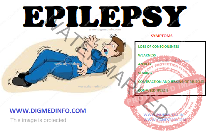
COMMON EPILEPTIC SYNDROMES
INFANTILE
SPASMS
Syn: Salaam
spasm (hypsarrhythmia):
This
condition is seen in infants below 1 year of age. This is characterized by
brief sudden jerky flexion or less commonly extension movements of both arms,
neck and torso. Usually these jerks or spasms occur in clusters, precipitated
by sudden noise or tactile stimulus and may occur several times in a day.
Usually there is evidence of other neurological disorders, secondary cerebral
anoxia or birth injury. As the infant grows, the frequency of the spasms comes
down but other forms of seizures may supervene. The characteristic EEG pattern
is called hypsarrhythmia.
LENNOX-GASTAUT
SYNDROME
This is an
epileptic syndrome with onset between 1 and 6 years of age. It is characterised
by mental retardation, and intractable seizures with mixture of tonic, atonic,
tonicclonic and atypical absence seizures. EEG shows diffuse slow and spike
wave disturbances. The syndrome may occur without any definite cause or may be
associated with a variety of neurodevelopmental or metabolic abnormalities in
which case the prognosis is poor.
BENIGN
ROLANDIC EPILEPSY
This is a
common form of partial epilepsy in childhood with onset between 3 and 11 years.
It is characterised by attacks of hemifacial twitches sometimes with
involuntary vocalizations which may progress to a generalised or unilateral
convulsion. These seizures usually or exclusively occur in sleep. The family
history suggestive of autosomal dominant inheritance may be available. The EEG
abnormality is highly characteristic showing frequent spike discharge in the
rolandic area, especially during non-REM sleep. The prognosis is excellent as
it is not associated with any other neurological, psychiatric or behavioural
disorder and the seizures respond well to anticonvulsant drugs. In some they
may remit spontaneously around puberty.
FEBRILE CONVULSIONS
A febrile
convulsion may be defined as a brief generalized convulsion occurring during
fever in a child in the age group of 6 months to 5 years without pre-existing
or concurrent neurological abnormalities or intracranial infection. About 70%
of febrile convulsions occur in the age of 6 to 18 months. The convulsions
usually last for a few seconds but may extend upto 15 min. At least 5% of children
have one febrile fit before the age of 5 years and nearly 25-50% of them have
recurrent attacks, but the risk progressively diminishes with age. A strong
family history of febrile fits is evident in many cases. The fits usually occur
when the rectal temperature rises above 39°C. Generally, the body temperature
normalises after the fit. About 4 to 20% cases may develop convulsions even without
noticeable fever. Though the risk of developing chronic epilepsy in such
children is only about 2%, it is however higher than in the general population.
The risk of developing epilepsy in later life is higher in the following
groups:
1. When the
first febrile convulsion is complicated, i.e. prolonged (> 30 minutes) or
localised or is followed by multiple seizures in 24 hours.
2. Presence
of neurological abnormalities before or after the onset of convulsion.
In general,
the intellectual performance of children who had febrile convulsions does not
differ from normal’s but prolonged or recurrent frequent febrile convulsions lead
to brain damage and medial temporal sclerosis, which in later life, causes
epilepsy.
PROGRESSIVE MYOCLONIC EPILEPSIES
(PME)
These are
rare, distinctive epileptic disorders with myoclonic seizures, tonic-clonic
seizures and progressive neurologic dysfunction, particularly ataxia and
dementia. Myoclonic seizures are described as sudden, brief, lightning like
jerks that may be generalised or limited to one or more muscle groups. They are
not associated with loss of consciousness and are frequently precipitated by stimuli
such as movement, bright light or stress. Physiological myoclonus may occur in
many normal persons during sleep or in other disease states. This has to be
distinguished from PME. A heterogenous group of disorders may give rise to PME.
a.
Biochemical disorders: Unverricht-Lundborg disease, Lafora body disease,
neuronal ceroid lipofuscinosis, sialidosis, mitochondrial encephalomyopathy,
noninfantile neuronopathic Gaucher’s disease, and others.
b.
Clinically defined groups: May give rise to seizures West’s syndrome,
Ramsay-Hunt syndrome (dyssynergia cerebellaris myoclonica), and others.
Diagnosis of
these groups of disorders depends on the clinical patterns, characteristic
fundal changes and biopsy studies of the skin, skeletal muscle, liver, rectal
mucosa or brain. Study of these diseases is the realm of the specialist.
Management consists of accurate diagnosis, genetic counselling and control of
myoclonus by sodium valproate or clonazepam.
REFLEX EPILEPSY
Epilepsy
precipitated by external stimuli has been designated as reflex epilepsy. The
common stimuli which precipitate reflex epilepsy in susceptible people are hot water
bath of the head, photic stimulation such as flickering light and TV watching,
exposure to sunlight, reading, hearing music, startle and eating. Avoidance of precipitating
factors and prophylactic anticonvulsants serve to prevent the attacks.
EPILEPSY AND
PREGNANCY
One-fourth
to one-third of pregnant women with epilepsy get aggravation of seizure
tendency during pregnancy. Status epilepticus may complicate 1–2% of epileptics
who are in labor. Since drug levels of antiepileptic drugs are lower in many
pregnant women, it is better to monitor free drug levels every month during
pregnancy. Risks of AED during pregnancy include the following;
First
trimester of pregnancy—congenital malformation in child–12.3%
Status
epilepticus: generalized convulsions during labour:
— Risk of
hypoxia and acidosis for mother and fetus is high.
— Increased
rate of neonatal hypoxia, low APGAR scores in the baby.
Pregnant
women with epilepsy have higher risks of hyperemesis gravidarum, pre-eclampsia,
abruptio placentae and premature labour. Serum AED levels rise after delivery
and therefore monitoring is required.
Diagnosis
Diagnosis of
epilepsy is essentially clinical. History is most important in making the
diagnosis. Careful interrogation of witnesses of an attack is essential to
determine the nature of the diagnosis. Epilepsy should be differentiated from
simple faint and syncopal attacks. The epileptic attack can occur during day or
night regardless of the position of the patient. Syncopal attacks usually do
not occur in the recumbent posture. Also the occurrence of pallor at the onset,
gradual loss of consciousness and prompt return of consciousness on adopting
recumbent posture are diagnostic of syncope. In epilepsy loss of consciousness
is abrupt and consciousness returns only slowly. Occurrence of post-ictal
headache and vomiting should suggest the possibility of epilepsy. So also, occurrence
of seizure during deep sleep is a strong point in favour of epilepsy.
Occurrence of injuries such as biting the tongue or due to falls should suggest
seizure disorder since these are practically absent in hysterical convulsions.
Hysterical
attacks should be differentiated by the lack of aura, absence of injuries and
incontinence, presence of peculiar grimacing or squirming movements and the retention
of consciousness during a motor seizure, which involves both sides of the body.
Moreover hysterical convulsions are bizarre and they continue for long periods,
as long as the patient is being observed.
Diagnosis of
the type of epilepsy depends upon the description of the attack and clinical
examination. The epileptic focus and pattern of epilepsy are determined by investigations.
The
electroencephalogram (EEG) is the most useful investigation to establish the
diagnosis of epilepsy. The EEG gives positive records in 60-90% of cases, if
the records are repeated during or after an attack and after 24 hours of sleep
deprivation. Refinements in EEG procedure include the use of special electrodes
such as sphenoidal, nasoethmoidal, nasopharyngeal and in selected cases, intracerebral
electrodes. Photic stimulation, sleep and hyperventilation are measures used to
elicit abnormalities in the EEG.
However,
normal EEG does not exclude the diagnosis of epilepsy. Conversely, a small
number of normal persons may show paroxysmal EEG abnormalities. Thus EEG can be
used only as an additional evidence to the clinical diagnosis of epilepsy. So,
also its role in predicting remission or
selection of antiepileptic drugs is also limited. But in confirmed epileptics
with EEG abnormalities, the decision to stop drug therapy can be based on the
persistence of the abnormality.
In epilepsy
occurring for the first time after 20 years of age, partial epilepsy, presence
of focal neurological deficit and in those where the disease is resistant to conventional
treatment, further investigations to exclude anatomical abnormalities and space
occupying lesions in the brain are indicated. These include CT and MRI scans of
brain.
MANAGEMENT OF EPILEPSY
This
consists of (i) treatment of the acute convulsions, and (ii) prophylactic
management of convulsive and nonconvulsive seizures.
The latter
consists of:
1. Removal
of precipitating or causative factors.
2.
Antiepileptic medication.
3. Social rehabilitation.
EPILEPSY – General Characteristics, Etiology, Pathogenesis, Classification, Partial Seizures, Primary Generalized Epilepsies, Common Epileptic Syndromes, Infantile Spasms, Lennox-Gastaut Syndrome, Benign Rolandic Epilepsy, Febrile Convulsions, Progressive Myoclonic Epilepsies (PME), Reflex Epilepsy, Epilepsy and Pregnancy and Management of Epilepsy
Endemic
fluorosis is caused by chronic fluoride intoxication acquired by ingestion of
water containing high concentration of fluorides. It is characterized by dental
and skeletal changes.
EPIDEMIOLOGY
The disease
is present in many states in India where fluoride content of drinking water
exceeds 2 ppm. Andhra Pradesh, Punjab and North Karnataka show high prevalence.
Endemic areas have also been found in Tamil Nadu, Haryana, Rajasthan, Uttar
Pradesh and Delhi and its surrounding areas. In Kerala three districts are
affected. The disease is present in certain parts of China, Japan, South
Africa, Saudi Arabia and USA. The disease is more prevalent in males who are engaged
in hard manual work because of their higher consumption of water. Total
hardness of drinking water (calcium and magnesium hardness) has a protective
role. Presence of fluoride up to 0.5 to 0.8 ppm in drinking water is considered
safe in India. With higher levels, fluoride accumulates in the skeletal system
and teeth.
Pathophysiology
Ingestion of
fluoride causes reduction of ionized calcium. This hypocalcemia leads on to secondary
hyperparathyroidism and increased osteoclastic activity. Increased levels of
lactic acid and citric acid are produced from the osteoclasts thereby
increasing the hydrogen ion concentration and lysis of lysosomes. Lysosomal
enzymes like protease, collagenase and hyaluronic acid produce disintegration
of hydroxyproline and other ground substances of bone and other calcified tissues
like teeth. This is responsible for the signs and symptoms of fluorosis like
dental hypoplasia with areas of hypocalcification, hypomineralization and
softening. The bones are heavier and irregular. There is excessive subperiosteal
bone formation at the sites of muscular, fascial and tendinous attachments.
Ligaments show various grades of calcification. The most advanced changes are
seen in the spine. There is narrowing of the vertebral canal, which leads to
compression of the spinal cord. Marked changes are seen in the ribs, pelvis,
sternum, mandible, and skull. Over a period of 10-20 years, the subject
develops crippling deformities.
CLINICAL FEATURES
Dental
fluorosis Enamel and dentin of teeth have strong affinity for fluoride during
the formation of teeth. Mottled enamel is an early, sensitive and easily
distinguishable manifestation in children. This has been taken as an index of
endemicity in epidemiological surveys. Dental fluorosis develops only if the
child has lived in the endemic area during dentition. It can be graded
depending on the severity.
Grade-I:
White chalky opacities or patches on enamel without faint yellow lines.
Grade-II:
Distinct brownish discoloration.
Grade-III:
Besides pigmentation there is pitting of enamel surface, sometimes with
chipping of edges
Premature
loss of teeth is not rare. Both permanent and deciduous teeth may be affected.
Skeletal fluorosis
Skeletal fluorosis
is not easily recognizable in the early stages. The initial symptoms are nonspecific
such as pain in the neck and back associated with rigidity, joint pains, and
paresthesia of the limbs.
These cases
may be mistaken for rheumatoid arthritis, ankylosing spondylitis or
osteoarthritis. The physical findings include kyphosis, limitation of movements
of the spine and exostoses. Exostoses can easily be palpated along the anterior
border of the tibia, over the olecranon, and along the medial border of the scapula.
These are diagnostic. In advanced fluorosis, kyphosis, fixed flexion
deformities of hips and knees and paraplegia, may develop.
Non-skeletal manifestations:
These
include neurological manifestations like tingling sensation in fingers and
toes, weakness and stiffness of skeletal muscles, nervousness and depression,
gastrointestinal manifestations like non ulcer dyspepsia, abdominal pain,
diarrhea and/or constipation. The red blood cell membrane becomes more pliable
due to decreased calcium and forms into echinocytes. Early destruction of
echinocytes results in anemia. Fluoride has inhibitory effect on iodine uptake and
so may cause enlargement of thyroid.
Complications
About 8-10%
of cases show compression of spinal cord and the roots by protruding
osteophytes. The vertebral arteries may also be occluded. The clinical picture
may resemble cervical myeloradiculopathy, cervical myelopathy or radiculopathy,
dorsal myelopathy and peripheral neuropathy. Bladder involvement manifests as
precipitancy of micturition or retention of urine.
Occasionally,
peripheral neuropathy manifests as acroparesthesia, but with only minimal
sensory or motor defects. Cranial nerve involvement is rare.
Investigations
Radiologic
and biochemical investigations should be carried out.
Radiology
The classic
features are osteosclerosis, irregular osteophyte formation, and calcification
of ligaments, especially in the vertebral column. In advanced cases, the bones
look chalky white. Irregular subperiosteal new bone formation may be observed
along the muscular, fascial, and tendinous attachments. Interosseous membrane
of the forearm shows calcification and this has been taken as a definite
radiological index of skeletal fluorosis. Skull shows thickening and sclerosis
of the vault.
CT and MRI
CT of the
bones may show prominent cortical thickening and increased density with
irregular contours. Bony excrescences are detected in both the pelvic bones and
the lower extremities. The sacroiliac joints may be narrowed. MRI may show
reduction in the intervertebral disc spaces and multiple disc prolapse. The medullary
canal may be narrowed by bony excrescences and by the ossified posterior
longitudinal ligament.
Biochemistry
Fluoride
content is increased in the blood, urine, and bone ash. Serum fluoride level
varies from 0.05 to 0.8 mg/dL. Serum alkaline phosphatase is moderately raised (15-30 KA
units). Serum calcium, phosphorus and magnesium are normal.
TREATMENT
Endemic
fluorosis is a preventable disease, which can be eradicated by providing
fluoride-free drinking water. Defluoridation of water may be affected by using
bone meal and metasilicate of magnesium (serpentine) but this is not yet widely
used. Vitamins and antioxidants have been tried in many cases. Changing the
dietary habits by restricting use of fluoride rich food is also important. There
is no effective treatment for the established case. Patients with skeletal
fluorosis, when fed with fluoride-free drinking water, seem to improve over the
years. Improvement in the dental changes is reported within weeks. Cases of
spinal compression require laminectomy.
Prevention of Endemic Fluorosis
In endemic areas fluorosis can be prevented by reducing the fluoride content of water to less than 1 ppm. Two methods are available for defluoridation of water, the Nalgonda technology and the activated alumina technology. In Nalgonda technique alum and lime are used in various proportions depending on fluoride content of water. In activated alumina technology activated alumina is used in domestic filters. Encouraging the use of calcium, vitamin C and vitamin E may help to reduce the skeletal changes.
DROWNING – General Considerations, Pathology and Clinical Features, Management, Prognosis and Prevention
GENERAL CONSIDERATIONS
Definition:
Drowning is
the pathological state leading to death resulting from the aspiration of water
into the respiratory tract or due to asphyxia immersion. More than 2 lakh
persons die annually of drowning, 25% in sea and 75% in inland waters. Two
types of drowning have been recognized – dry drowning and wet drowning. In dry drowning
death is due to laryngeal spasm, which proves fatal in 20% of the subjects.
This also prevents the entry of water into the lungs.
In wet
drowning water enters the lungs. The consequences differ between fresh water
and sea water drowning. In fresh water drowning, water is quickly absorbed from
the lungs, leading to hemodilution and hemolysis with release of potassium from
the red blood cells. In addition to hypoxia and ventilatory failure, hyperkalemia
precipitates ventricular arrhythmias, which may prove fatal.
In salt
water drowning the fluid in the lung is hyperosmotic. It absorbs more fluid
into the alveoli causing pulmonary edema and respiratory failure. Hypernatremia
follows later when the salt is absorbed into the circulation. In addition to
the metabolic and local effects, impurities and contaminants give rise to local
infection.
Secondary
Drowning or near-drowning occurs a few hours or few days after the initial
resuscitation due to the secondary changes in the lungs such as pulmonary
edema, pneumonia, pneumothorax, electrolyte disturbances and metabolic or
respiratory acidosis. This accounts for 25% of deaths.
Immersion
syndrome
In this,
sudden death occurs due to cardiac arrest caused by vagal stimulation brought
about by sudden immersion into cold water.
PATHOLOGY AND CLINICAL FEATURES
Lungs:
Pulmonary edema develops. Fresh water interferes with surfactant leading to
formation of hyaline membrane, atelectasis and hypoxemia. Aspiration of foreign
particles worsens the atelectasis. Bacterial infection leads to pneumonia or
lung abscess.
Heart:
Arrhythmias such as ventricular fibrillation and cardiac arrest may occur.
Electrocardiogram may show nonspecific changes due to asphyxia.
Kidneys:
Acute tubular necrosis may develop in neardrowning in fresh water due to
hemolysis and prolonged hypotension.
Central
nervous system: Asphyxia leads to loss of consciousness, cerebral edema and
convulsions. Sequelae of anoxic encephalopathy such as transient hemiparesis, quadriparesis,
choreoathetosis, aphasia and faciobrachial weakness may develop.
MANAGEMENT
First aid:
(1) clear the airway of water and foreign bodies by putting the patient head
low and by suction, (2) institute mouth-to-mouth breathing as early as
possible, (3) closed chest cardiac massage should be instituted if heart sounds
are absent, and (4) all cases must be hospitalized to prevent death from
secondary drowning.
Hospital
treatment: This aims at (1) maintenance of adequate oxygenation, (2) correction
of metabolic and electrolyte imbalance and (3) prevention of secondary effects.
Adequate oxygenation is achieved by the use of controlled ventilation with 100%
oxygen, later to be reduced to 40%. If these measures fail to respond intubation
and application of positive and expiratory pressure (PEEP) respiration should
be resorted to. The PEEP increases the functional residual capacity, thereby minimizing
intrapulmonary shunts and ventilation perfusion abnormalities, and promotes
better oxygenation. If bronchospasm is present, an aerosoal of salbutamol 200 mcg
should be administered. Acidosis is to be corrected with sodium bicarbonate
given intravenously in a dose of 0.7 to 1 mmol/kg bw. Proper correction of
electrolyte imbalance and acidosis should be monitored with laboratory
estimations.
Constant
observation and appropriate management of pulmonary edema, pneumonia and
pneumothorax serves to prevent secondary drowning. Prophylactic antibiotics have
to be used to prevent respiratory infections. Skiagram of the chest is
necessary in all cases to detect complications. Atelectasis has to be managed
with bronchoscopic aspiration. In severe cases of pulmonary edema, dexamethasone
given in a dose of 0.5-1 mg/kg bw in 24 hours IM or IV has been successful. If
signs of intracranial hypertension develop, it is treated with hyperventilation
and IV infusion of 200 mL of 20% mannitol.
Prognosis
This depends
on the extent and duration of hypoxia and the first aid. Patients presenting
with coma and cardiac irregularities have higher mortality and morbidity.
Immersion in
cold water causes death earlier due to rapid cooling, but survivors show less
tendency to develop neurological sequelae. Residual complications of near drowning
include convulsive disorders, intellectual impairment, cardiac neurosis and
pulmonary atelectasis leading to bronchiectasis.
Prevention:
Education of the public on the hazards in water and first aid measures to save drowning victims should be available in places of water sports, holiday resorts and beaches. Trained lifeguards should be available at public swimming places.
DROWNING – General Considerations, Pathology and Clinical Features, Management, Prognosis and Prevention
Diet is the
corner stone in the management of diabetes. The objective of dietary therapy is
the optimization of glycemic control and to provide a nutritious and balanced diet.
In type 1 DM patients the total energy input has to be relatively higher in
order to regain ideal weight and growth. In type 2 patients the calories need
to be restricted in order to avoid obesity. Dietary articles such as saturated fats,
excess salt and cholesterol which promote vascular complications have to be
avoided.
Goals of Medical Nutrition Therapy
1. To
achieve and maintain near normal glycemia(euglycemia).
2. To
achieve and maintain optimal lipid profile. (total cholesterol < 150,
triglyceride ± 120, HDL>50 and LDL<100/mg/dL).
3. To
achieve and maintain normal blood pressure levels around 120 to 130 / 80 to 85
mmHg.
4. To adjust
the nutrient intake to restore and maintain ideal body weight to avoid
dyslipidemia, cardiovascular disease, hypertension and nephropathy. During
childhood and pregnancy adjustment for growth also should be provided.
5. For
elderly patients, provision for proper nutrition and psychosocial needs.
In type 2
diabetic patients the first step would be dietary therapy alone along with
exercise. They should be given a trial of dietary therapy for 4-8 weeks. About 50%
of the patients come under control with diet alone. Proper patient education
helps to improve adherence to treatment.
The
following points have to be considered while prescribing a diet for a diabetic-
1. The type
of diabetes- type 1 or type 2
2. The
weight of the individual in comparison with his ideal body weight (BMI)
3. His
occupation and activities and to assess his caloric requirements
4. The
presence of any complications
An appropriate
assessment of the caloric needs of each diabetic individual should be done.
Total Caloric Intake
This is the
most important step while prescribing a diet. Total caloric intake depends on
the patient’s body weight, degree of physical activity and the presence of any
other comorbid illnesses. Obesity is an important factor in terms of target
cell resistance to insulin action.
The body
mass index (BMI) will help to determine the total caloric requirement.
BMI = Weight
(in Kg) / Height in m2. It is desirable to keep the BMI between 22 and 25.
Ideal body
weight can be readily calculated by the formula: ideal body weight = height in
cm – 100.
The
recommended daily allowance is 30 Kcal/kg of desirable body weight to patients
with average activity except in the case of obesity where appropriate changes have
to be made.
Having
calculated the number of calories required, they are distributed into at least
three principal meals and one or two small snacks. These calories are derived
from three principal sources—carbohydrates, protein and fats. Each fraction has
its own importance and should provide 60% of the calories from carbohydrates
and 20% each from proteins and fats.
When
instructing on the diet regime the following points should be stressed.
1. It is not
a reduction in the diet; on the other hand it is a modulation to suit the
particular need of the individual. This concept will help to reduce the psychological
resistance in accepting the diet.
2. The
patient and his spouse should be counseled together, so that the latter will
understand the principles and help to provide the diet appropriately.
3. Whenever
possible, the dietary articles should be prescribed in terms of weight so that
at least on a few occasions the actual quantities would be determined and
adopted.
4. It is
better to prescribe the diet in terms of the primary food articles such as
rice, meat, fish and others so that the patient can determine the various items
of the menu in relation to the allowed foodstuff.
5. Many
patients are under the wrong impression that reducing the food further than
what is prescribed may be beneficial and this should be avoided.
6. Both the
quantity to diet and its timing are important since other aspects of management
such as medication and exercise are timed in relation to the diet. As far as
possible the diet should conform to the cultural habits and socioeconomic
condition of the patient.
7. Vast
majority of treatment failure can be avoided by proper dietary instructions.
8. At all
follow-up visits enquiry on the diet should be made and the need for strict
adherence stressed.
9. As far as
possible the patient should be involved in the formulation of the diet and such
a participatory instruction assures better compliance.
Carbohydrates
The amount
of carbohydrates to be permitted in diet of diabetic patients has been an area
of controversy. Till recently most people recommended restriction of carbohydrates
in the diet to provide only 30-40% of the calories. Our diets in India are
cereal based and have a high carbohydrate component (about 70%).
The American
Diabetic Association and the European Diabetic Association study groups have
also altered their dietary recommendations. In an attempt to reduce cardiovascular
morbidity and mortality, they now recommend a liberalized use of carbohydrates
in the diet up to 50-60% of the calories. This also helps to reduce the intake
of saturated fats. Modification in the type of carbohydrate can be achieved by
increasing the intake of legumes and pulses, green leafy vegetables and other vegetables,
which will increase the content of complex carbohydrates and fiber.
Fats
The fat
content of diet should be 20-25% of the total calories. The distribution of the
type of fat should be equal, i.e. saturated fats and mono and polyunsaturated fats
should be equally distributed to make up the total fat intake. The dietary
cholesterol should be less than 300 mg/day. Invisible fat is derived in a fair
amount from cereals, legumes and seeds and contributes to 5-10% of the total
energy intake. Milk and Milk products contribute approximately to 40-45% of the
total fat content in vegetarian diets. Milk fat is a saturated fat.
Proteins
Protein
intake has been recommended as 0.8 g/kg body weight and should contribute to
12–20% of the total caloric intake. Vegetable proteins derived from cereals and
lentils, do not contain cholesterol. They have high fiber content. Animal
protein is rich in saturated fats and tends to increase cholesterol and
triglycerides. Lean meat and fish are to be preferred to fatty meat in order to
minimize the risk. When renal failure occurs, strict-protein restriction is
instituted.
The dietary
salt should be less than 6 g/day. In the presence of hypertension or renal
failure it should be reduced to around 3 g/day. Alcohol should be avoided as
far as possible. Alcohol intake will increase the risk of hypoglycemia by
inhibiting gluconeogenesis. It may induce ketoacidosis, lactic acidosis and may
contribute to peripheral neuropathy. Alcohol
also induces hypertriglyceridemia and hyperuricemia. Alcohol is an additional
source of calories, without any further nutritional values each mL providing 7
calories. If consumed it should be taken only in moderate quantities (1-2
drinks/day i.e. 20-40 mL/day).
EXERCISE
In type 2
diabetes, regular exercise forms an important component of therapy along with
dietary regulation and oral hypoglycemic agents. However a careful assessment of
the expected benefits and associated risks of exercise in individual patients
should be made while incorporating an exercise program in the treatment.
Appropriate monitoring should be done to avoid complications.
Exercise
should not be recommended indiscriminately in all type 1 diabetic patients but
efforts should be made, to make it possible for those who want to exercise to
do so as safely as possible.
Endocrine/Physiological Responses during
Exercise
1.
Suppression of insulin release – directly as well as through epinephrine.
2.
Sympathetic system activation – which inhibits insulin release (by
alpha-receptor stimulation) and stimulates lipolysis.
3.
Non-insulin dependent glucose uptake in the periphery. In both type 1 and type
2 diabetic patients who are under good control the response to exercise will be
normal. In untreated type 1 patients there is an increased production of FFA
from adipocyte lipolysis, which leads to decreased glucose uptake in the
periphery. This can even precipitate diabetic ketoacidosis in some patients.
In the
well-controlled patients this does not occur While prescribing exercise, it
should suit the social and economic condition of the patient and his work schedule.
Regular exercise as part of the therapeutic intervention should be taken by all
diabetics, irrespective of the physical activity entailed in their regular
occupation.
Maximum
benefit is achieved by exercises such as brisk walking (4-5 km/hour), swimming,
cycling and such other aerobic exercises. At least five sessions a week should
be performed in order to achieve optimal benefit. While starting the exercise
program in persons above the age of 35 years clinical assessment of their
cardiovascular status should be done and introduction of the exercise regimen should
be gradual, so as not to precipitate any acute cardiovascular events.
For the
average middle-aged Indian diabetic the following exercise regimen is adequate.
• Walk 3 km
on level ground over a period of 45 minutes
• Swim for
30 minutes at average speed without cardiovascular distress.
• Cycle on
level ground at 8 km/hour for 30 minutes.
Regular
sports and games activities can be undertaken and should be encouraged by those
who desire to do them, with special provision made for the diet and
medications. Since such sport activities are likely to be intermittent rather
than regular, in practice walking or cycling is more regularly available.
Before an
exercise program is initiated, a fair control of blood glucose is to be ensured
and a thorough clinical evaluation of the patient should be made particularly
in regard to complications of diabetes such as hypertension, coronary artery
disease, peripheral vascular disease, retinopathy and nephropathy.
Yoga exercises
Recently
several well-planned studies have demonstrated the beneficial effects of yogic
practices in diabetics. Patients with diabetes demonstrated a significant fall
in fasting and postprandial blood glucose values and HbA1c, with reduction in
the requirements of OHA and insulin. Type 1 diabetic patients with brittle diabetes
showed marked improvement with the practice of yoga. There was a salutary
effect on the lipid profile, with a fall in serum cholesterol, triglycerides
and an increase in HDL- cholesterol fraction. Certain Asanas (specific postural
manipulations which have to be learnt under supervision) have been identified
as useful in the control of diabetes. Thus yogic practices have a useful role
in the control of diabetes and prevention of its longterm complications.
ORAL HYPOGLYCEMIC AGENTS (OHAs)
The oral
hypoglycemic agents are indicated in type 2 diabetes when diet and exercise
fail to achieve euglycemia. The two major groups of drugs in use are a) Insulin
secretogogues (sulfony1 urea compounds) and b) insulin sensitizers
(glitazones).
Sulphonylurea (SU) Compounds
The SU
compounds stimulate the beta cells of the pancreas to release insulin. Therapy
should be initiated with the smallest dose, taken 15-30 minutes before
breakfast and small increments should be made at weekly intervals till the optimum
dose is reached. The sulfonylureas are similar in effectiveness in equipotent
doses and in the absence of any specific reason such as adverse side effects,
cost or nonavailability, there is no need to change the medication in patients
who are adequately controlled. So also there is no advantage in combining two
or more sulfonylurea drugs.
INSULIN THERAPY
Therapeutics of Insulin
Insulin therapy aims at providing ideal physiological insulin profiles with peaks at meal times and maintenance of basal levels between meals and at night. This can be achieved by administering soluble insulin before each meal and adding a dose of intermediate acting insulin at bed time. The alternative regimes would be; (i) a split dose of soluble and intermediate acting insulin before breakfast and dinner, (ii). a single dose of soluble and intermediate acting insulin before breakfast, (iii) a single dose of intermediate acting insulin before breakfast. With the introduction of premixed insulin it is easier for the patients to adopt these regimes. As in the case of other hypoglycemic agents, diet control and exercise should be instituted along with insulin therapy.
CYANOCOBALAMIN (VITAMIN B12) – Absorption of B12, Etiology of Vitamin B12 Deficiency, Effects of B12 Deficiency and Treatment
Deficiency
of vitamin B12 or folates leads to abnormality in DNA synthesis,
characterized by megaloblastic erythropoiesis and similar changes in many
tissues in the body. Due to its fatal outcome in pre-vitamin B12
days, it was called pernicious anemia.
Absorption of B12
The dietary
vitamin B12 which is bound to proteins has to be liberated from them
to enable absorption. Cooking converts a part of these into dialysable form.
Low pH achieved in the stomach helps further liberation of this vitamin. After
liberation, cobalamin is bound to the intrinsic factor (IF) which is a glycoprotein
with a molecular weight of 44,000 present in the gastric juice. Proteolytic
enzymes of the pancreas play a part in this process.
The vitamin
B12-IF complex is taken up by receptor sites present in the
microvilli of the ileum by passive absorption. In the plasma cyanocobalamin
remains bound to a polypeptide of molecular weight 38,000 known as transcobalamin
II (TC II). This complex passes into cells and its B12 is liberated
by lysosomal enzymes. Liver can store up to 2 mg of vitamin B12
which is adequate for several years. The daily requirement of B12 is
1-2 mcg.
Meat, liver,
eggs, dairy products, and yeast contain adequate amounts of this vitamin.
Purely vegetable sources are deficient in vitamin B12. Therefore,
vegans (persons who do not take any form of animal foods or dairy products)
suffer from nutritional deficiency. Normal levels of vitamin B12 in
serum ranges from 120-900 pg/mL in Indian subjects.
Vitamin B12
deficiency is less common than folate deficiency.
Etiology of Vitamin B12 Deficiency
1.
Malabsorption states—diseases of the ileum.
2. Dietary
inadequacy—vegans.
3. Intrinsic
factor deficiency—pernicious anemia. This
is usually
acquired, rarely this may be congenital.
4. Chronic
disorders destroying the gastric mucosa and partial or total gastrectomy.
5. Blind
loop syndromes-with colonization of the small intestine by bacteria.
6.
Pancreatic insufficiency.
7. Familial
deficiency of transcobalamin II.
8. Inherited
disorders of vitamin B12 metabolism.
9.
Interference with absorption of vitamin B12 by drugs, e.g. PAS,
colchicine, phenformin, neomycin.
Effects of B12 Deficiency
Megaloblastic
anemia develops owing to diminished red cell production and dyserythropoiesis.
In addition, cells from other organs with rapid cell turnover such as the gastrointestinal
tract and cervicovaginal mucosa also show similar abnormalities. The central
nervous system and peripheral nerves are also affected. The neurological lesions
include subacute combined degeneration of the spinal cord, optic neuritis, and
demyelination of the cerebral white matter and peripheral nerves.
Neurological
damage occurs due to impaired DNA synthesis and myelin formation. The
occurrence of neuropathy does not bear any direct relationship to the severity
of anemia.
Treatment
Dietary
deficiency can be corrected by giving 1-2 μg of vitamin B12 orally. If
megaloblastic anemia has developed, larger doses are required (100 μg/day)
oral, if absorption is reliable. Otherwise 1000 μg of hydroxocobalamin is given
intramuscularly once a week for 3-4 weeks, and thereafter the daily dietary
supplementation of 1-2 μg is continued.
There is evidence that vitamin B12 can be absorbed from the buccal mucosa when applied sublingually. This route can be made use of for therapy in mild deficiency states.
CYANOCOBALAMIN (VITAMIN B12) – Absorption of B12, Etiology of Vitamin B12 Deficiency, Effects of B12 Deficiency and Treatment
COPD (Chronic Obstructive Pulmonary Disease) CHRONIC BRONCHITIS – General Characteristics, Pathogenesis, Pathology, Clinical Features, Diagnosis and Management
General Characteristics
Chronic
bronchitis is defined as a disease characterized by hypersecretion of mucus
sufficient to cause cough and sputum on most days for at least three months in
a year for two or more consecutive years. This happens in the absence of any
other specific respiratory or cardiovascular disease.
In the
initial stages the inflammation of the bronchi is intermittent and recurrent,
later it becomes established. The larger air passages are affected during the
early part of the disease, later obstructive features set in when the smaller airways
are also affected. Infection leads to periodic aggravation of the symptoms and
the sputum, which is mucoid, becomes purulent during these episodes. As the airways
obstruction progresses, emphysema sets in. These two processes become
established in the majority of cases so that the condition is termed chronic
bronchitis emphysema syndrome (CBES). The disease is more common in damp, cold,
and dusty regions. Atmospheric pollution is accompanied by a higher incidence
of CBES.
COPD (Chronic Obstructive Pulmonary
Disease)
Chronic
Obstructive Pulmonary Disease (COPD) is a major cause of chronic morbidity and
mortality throughout the world. Chronic Obstructive Pulmonary Disease (COPD) is
a disease state characterized by airflow limitation that is not fully
reversible. The airflow limitation is usually both progressive and associated
with an abnormal inflammatory response of the lungs to noxious particles or
gases.
The most
important risk factor for COPD is cigarette smoking. Pipe, cigar and other
types of tobacco smoking popular in many countries are also risk factors for
COPD. Passive exposure to cigarette smoke also contributes to respiratory
symptoms and COPD.
Other
documented causes of COPD include occupational dusts and chemicals (vapours,
irritants, and fumes) when the exposures are sufficiently intense or prolonged.
Indoor air pollution from biomass fuel used for cooking and heating in poorly
ventilated dwellings. Outdoor air pollution, which adds to the lungs total
burden of inhaled particles, although its specific role in causing COPD isnot
well understood.
Symptoms of COPD include:
Cough
Sputum
production
Dyspnea on
exertion
Episodes of
acute worsening of these symptoms often occur. Chronic cough and sputum
production often precede the development of airflow limitation by many years;
although not all individuals with cough and sputum production go on to develop
COPD.
Pathogenesis
Chronic
obstructive pulmonary disease (COPD) is a descriptive term given to the
syndrome seen mostly in the elderly, who have airflow obstruction, not completely
relieved by therapy. At least three distinct pathological processes may occur
concurrently or separately in different subjects lead to COPD.
These are:
1. Emphysema
which is due to destruction of alveolar walls
2. Chronic
bronchitis with hypersecretion of mucus, and 3. Asthma with airway remodeling.
COPD
encompases chronic obstructive bronchitis with obstruction of small airways and
emphysema with enlargement of air spaces, destruction of lung parenchyma, loss
of lung elasticity and closure of small airways. The inflammatory process in
COPD differs from that in asthma in several ways. The type of inflammatory
cells, inflammatory mediators, final outcome and response to treatment are
different. In COPD the inflammation affects the peripheral airways-the
bronchioles. The cells are macrophages, CD8 lymphocytes and
neutrophils. The lung parenchyma is affected. Unlike as in asthma there is no preponderance
of eosinophils.
Oxidative
stress also plays a significant role in the pathogenesis. Even though the lungs
bear the main brunt of the disease, systemic effects also occur. Muscle weakness
and wasting may develop as part of the systemic disorder.
Pathology:
The
bronchial mucosa shows hypertrophy and increase of the mucous glands and goblet
cells with consequent overproduction of viscid mucus. The distal airways show
narrowing of lumen caused by increased height of the epithelium and increased
thickness of the muscle and connective tissue. The mucosa becomes ulcerated and
when the ulcers heal, fibrosis occurs resulting in distortion of the lumen with
stenosis and dilatation.
Distortion
of the airways leads to permanent obstruction. Secondary infection occurs in
the later stages. The ciliary movement is further impaired by the abnormally
viscid mucus. This aggravates infection and a vicious cycle is established. Severe
recurrent infections cause the development of microabscesses in the bronchial
wall. These heal with fibrosis. Squamous metaplasia occurs. Distortion and obstruction
of the bronchial lumen result in air trapping and emphysema of the alveoli,
some show collapse and fibrosis.
The main
pathological process can be summarized as follows:
1.
Inflammation of the bronchi with enlargement of mucous glands and smooth muscle
hyperplasia, all leading to wall thickening
2. Acinar
distension due to the destruction of lung parenchyma probably mediated by
imbalance of protease-antiprotease (alpha-1 antitrypsin) enzymes causing loss
of support of small airways
3. Fibrosis
and narrowing of the airways leading to increase in airway resistance.
The
capillary bed is distorted and truncated and this aggravates the progression of
pulmonary arterial hypertension. The pulmonary arteries become distended and
atheromatous. Pulmonary hypertension gives rise to right ventricular
hypertrophy and dilatation. Chronic cor pulmonale supervenes as time passes.
CLINICAL FEATURES
The clinical
picture is varied depending on the severity and duration. The most frequent
early symptom is cough recurring year after year, especially so in winter
months. Later the cough becomes constant. Expectoration is mucoid and the
sputum is tenacious, especially on waking up in the morning. Main complaint is
the feeling of tightness of the chest. Physical examination reveals mild wheeze
which disappears as the patient clears the bronchi by expectoration. Variable
degrees of bilateral rhonchi and coarse crepitations are heard as adventitious
sounds.
Initially
acute infections give rise to fever and purulent sputum. As the infection
becomes established, fever and other general symptoms come down. At this stage
the quantity and character of the sputum are more reliable indicators of
infection. The sputum becomes copious in amount when bronchiectatic changes
develop. With the development of emphysema the chest assumes the inspiratory
position and the respiratory excursions are considerably diminished. At this
stage dyspnea is far out of proportion to the physical findings in the chest.
Key Indicators for Considering the
Diagnosis of COPD
Chronic cough: Present intermittently or every day.
Often present throughout the day; seldom only nocturnal
Chronic sputum production: Any pattern of chronic sputum production may
indicate COPD
Acute bronchitis: Repeated episodes
Dyspnea that
is: Progressive (worsens over time), Persistent (present every day), Worse on
exercise, Worse during respiratory infections
History of exposure to risk factors: Tobacco smoke (including popular
local preparations), Occupational dusts and chemicals and smoke from home
cooking and heating fuel
DIAGNOSIS
Chronic
bronchitis should be diagnosed from the history of recurrent cough extending
over several years, mucopurulent sputum and the physical findings of bronchial
obstruction and emphysema. X-ray is normal in the early stages but the features
of emphysema may be evident later. X-ray may be helpful in identifying precipitating
conditions like pneumonia, pneumothorax during the time of an exacerbation.
HRCT is useful in quantifying the severity of emphysematous changes and locating
areas with bronchiectatic changes.
Lung
function tests show reduction in vital capacity, increase in the closing volume
and features of airway obstruction. Based on the spirometry values severity of
obstruction can be categorised as mild, moderate severe and very severe.
Differential diagnosis:
Chronic
bronchitis has to be distinguished from asthma. Differentiation is easy in the early
stages but there is considerable overlap of symptoms and signs in the advanced
stages, and, therefore, the clinical assessment is difficult.
Other
conditions like pulmonary tuberculosis, bronchiectasis, heart failure and
bronchogenic carcinoma, obliterative bronchiolitis, diffuse pan bronchiolitis
have to be ruled out in atypical cases.
Course and prognosis: Established chronic bronchitis is incurable.
Over several years the condition progresses to produce complications and death.
Each infective exacerbation leads to further deterioration in lung function and
precipitates the development of respiratory failure and cor pulmonale.
Exacerbation can occur as a result of bacterial infection of the lower
respiratory passages, viral infections of the respiratory tract or due to
non-infective causes such as environmental allergens and pollutants.
Complications: These include:
1. Frequent
respiratory infections,
2.
Respiratory failure, and
3.
Right-sided heart failure (cor pulmonale).
MANAGEMENT
General measures: Most effective single step to
prevent deterioration is to stop smoking. This single measure itself affords
considerable relief of symptoms. Environmental allergens and pollutants must be
avoided by the patient.
Other
general measures include improvement in general health, regular exercise,
deep-breathing exercises, adequate sleep, treatment of obesity, and eradication
of foci of sepsis in the throat, nose and paranasal sinuses. If these measures
are started during the early phase of the disease, further progression can be
arrested.
Drugs: Apart from the general measures, no
active treatment is indicated in the early stages.
Treatment
Treatment of
infective episodes: A broad spectrum antibiotic should be employed for 7-10
days during an infective episode. Tetracycline or ampicillin may be started initially.
Depending on the microbiological tests, the antibiotic may have to be changed.
Bronchospasm
has to be relieved by the use of bronchodilators such as aminophylline,
salbutamol or any other β-agonist. Ipratropium bromide is an anticholinergic muscarine
receptor blocker, which blocks vagal reflexes responsible for
bronchoconstriction. It is indicated when bronchospasm is troublesome.
Ipratropium
bromide delivered by a metered dose inhaler in a dose of 40-80 μg helps to
relieve bronchospasm without appreciable side effects. In COPD response of the
airways to ipratropium is excellent. Tiotropium is a newer analogue which is
equally effective.
Inhibitors of inflammatory response: Since inflammation plays a major
role in the pathogenesis of COPD newer pharmacological agents are under trial.
These include mediator antagonists which are capable of counteracting the
effects of leukotrienes, lipoxygenases, interleukin B, tumor necrosis factor
and the like. Protease inhibitors which inhibit neutrophil elastases, cathepsin
and anti-inflammatory agents such as phosphodiesterase inhibitors are all in
the process of development.
Several
newer drugs especially phosphodiesterase-4 inhibitors are under trial.
Roflumilast is a drug of this class given in a dose on 250 or 500 mg orally
daily for 24 weeks. Results are encouraging.
Use of
N-acetyl cysteine or bromhexine hydrochloride 8 mg thrice daily orally helps to
liquefy the sputum. Steam inhalations help to improve vital capacity, relieve
emphysema and open up the airways by expectorating the sputum.
Nonpharmacological measures: Noninvasive positive pressure
ventilation and oxygen therapy at home using a simple nasal mask which
eliminates the need for endotracheal intubation is beneficial. Long-term oxygen
therapy has shown improved survival with better quality of life.
Pulmonary rehabilitation: Structured programme of education
and exercises is very effective. Blowing into an air pillow repeatedly for
10-15 times twice a day and bending over a pillow held firmly on to the
abdomen, in order to push the diaphragm up during expiration are simple
maneuvers which can be practised at home.
Management of chronic respiratory
failure: When
hypoxemia becomes severe, i.e. PaO2 below 55 mm Hg or SaO2
below 80% at rest, continuous oxygen inhalation may have to be instituted for
at least 18 hours a day. Oxygen therapy helps to reduce pulmonary arterial
tension and allay cor pulmonale. In intractable cases lung transplantation may have
to be considered.
Lung volume reduction surgery (LVRS): This has been found to be useful in selected
cases. The rationale for this technique is to reduce the volume of over
inflated emphysematous lung by 20-30%, in order to improve the elastic recoil
of the lungs, to improve the configuration of the diaphragm, chest wall
mechanics and gas exchange.
Lung transplantation: This procedure is in vogue for more
than a decade in advanced countries.
Transplantation
of a single lung or both heart and lungs as a whole is possible.
Transplantation should be considered if the recipient is below 55 years of age,
and is free from underlying conditions such as advanced diabetes, malignancy,
hepatic or renal failure and conditions which impair mechanics of the chest
wall. The donor should be ABO and HLA compatible with normal lungs, preferably
between 12 and 50 years of age and with normal cardiopulmonary anatomy. Usually
cadaver lungs are used for transplantation. Complications may occur as in the
case of any other major organ transplantation.
Indications
1. Incurable
respiratory failure—chronic or acute due to pulmonary causes
2. Irreversible structural and functional abnormalities in the lung such as fibrosis, extensive bronchiectasis, cystic disease, emphysema and others.
COPD (Chronic Obstructive Pulmonary Disease) CHRONIC BRONCHITIS – General Characteristics, Pathogenesis, Pathology, Clinical Features, Diagnosis and Management
CHRONIC KIDNEY DISEASE – DEFINITION AND STAGING, ETIOLOGY, NATURAL HISTORY AND PROGRESSION, CLINICAL MANIFESTATIONS, DIAGNOSIS AND MANAGEMENT
GENERAL CHARACTERISTICS
Chronic
kidney disease (CKD) is an important public health problem affecting more than 10%
of the general population. CKD is an important risk factor progressing to end
stage renal disease (ESRD), cardiovascular disease and premature mortality.
Management of ESRD is expensive and beyond the reach of many sections of the population
in developing countries. CKD is a silent disease and if not detected and
treated early, may progress to ESRD. Hence, it is important to identify CKD
early and institute measures to retard its progression.
DEFINITION AND STAGING
Chronic
kidney disease is defined as either kidney damage or GFR <60mL/min/1.73m2
for >3 months. Kidney damage is defined as pathologic abnormalities or
markers of damage, including abnormalities in blood or urine tests or imaging
studies.
CKD is
characterized by kidney damage characterized by structural, functional or
pathological abnormalities in the kidney associated with abnormalities in the composition
of urine and/or blood of more than 3 months duration. Anomalies in the imaging
tests and decrease in the GFR may be demonstrable. If the GFR is less than 60 mL/min/1.73
m2 body surface, persisting for more than 3 months, this by itself can
constitute CKD.
STAGES OF CHRONIC KIDNEY DISEASE
Stage Description GFR (mL/min/1.73m2)
1. Kidney damage with
normal
> 90
or increased GFR
2. Kidney damage with
mild
60-89
decrease in GFR
3. Moderate decrease in
GFR 30-59
4. Severe decrease in GFR 15-29
5. Kidney failure
< 15 or dialysis
Staging
Stage 1 CKD
represents kidney damage when GFR is normal or high. This includes patients
with albuminuria or those with abnormal imaging studies.
In stage 2
CKD, there is evidence of kidney damage with mild decrease in GFR (60- 89 mL/min/1.73
m2). In the absence of kidney damage, this level of GFR per se, does
not constitute CKD.
Stage 3 CKD
includes patients with moderate decline in GFR (59-30 mL/ min/ 1.73 m2).
All patients with GFR of less than 60 mL/min/1.73 m2 are classified
as having CKD irrespective of whether kidney damage is present or not. This is
the stage where serum creatinine starts to rise. Majority of patients still
remain asymptomatic. Nocturia and polyuria are early symptoms that may appear
at this stage.
In stage 4
CKD, the fall in GFR is severe (29-15 mL/min/1.73 m2). Overt uremic
symptoms like loss of appetite, nausea, anemia and recurrent infections occur during
this stage.
Stage 5 CKD
is the stage when GFR is less than 15 mL/min/ 1.73 m2. At this stage
the patient usually requires dialysis or other forms of renal replacement
therapy.
GFR is the
best measure of overall kidney function in health and disease. GFR can be
affected by chronic kidney disease which reduces the number of nephrons. Hemodynamic
factors affect single nephron GFR and they result in fall in the GFR. The
normal GFR of a young adult is around 120-130 mL/min/1.73 m2. Normal
values in women are 8% lower from those of men at all ages. Above the age of 30
years, GFR declines at the rate of approximately 1 mL/min/1.73 m2
per year.
ETIOLOGY
The causes
of CKD are broadly classified as given below.
Glomerular
Diseases
Primary—Focal
segmental glomerulosclerosis (FSGS), IgA nephropathy, membranoproliferative
glomerulonephritis (MPGN)
Cystic
diseases—Autosomal dominant and recessive polycystic kidney disease (ADPKD and
ARPKD)
Heredofamilial—Alport’s
syndrome
Diseases in
the transplant -Chronic allograft nephropathy
Age,
ethnicity and distribution of renal diseases may be different in various
geographical regions. Diabetes and hypertension are the two most important
causes of CKD worldwide. In the developing countries chronic glomerulonephritis
and chronic interstitial nephritis are relatively more common than in the
developed countries.
FSGS, IgA
nephropathy, reflux nephropathy and congenital anomalies of the urinary tract
lead to CRF usually before the age of 40 years. Diabetes, hypertensive nephrosclerosis,
ischemic nephropathy, multiple myeloma, analgesic nephropathy and systemic
vasculitis are more common above the age of 55 years.
NATURAL HISTORY ANDPROGRESSION OF CKD
Many
diseases can intiate kidney injury and lead to progression. Reduction in the
number of functioning nephrons lead to hypertrophy and increased glomerular filtration
by the surviving nephrons. Glomerular hyperfiltration initially maintains GFR
but later, the overworked nephrons succumb to the increased workload and lead
to proteinuria, which causes further damage to the glomeruli and the
tubulo-interstium. The extent of tubulo-interstitial damage is an important
factor determining disease progression. Hypertension which is present in over
90% of cases of advanced CKD leads to further progression of the disease, if
not controlled. Hyperglycemia also causes structural and functional changes in
the glomeruli leading to progressive renal failure. Progressive destruction of
nephrons leads to several adaptive mechanisms which enable the remaining nephrons
to maintain body homeostasis. Though these adaptive changes are beneficial,
later, they lead to maladaptive consequences. For example, early in the course
of renal failure, loss of nephrons result in reduction of phosphate excretion
and lead to hyperphosphatemia.
This causes
a fall in serum calcium due to its binding to the excess phosphorus.
Hypocalcemia stimulates PTH which results in reduction of tubular reabsorption
of phosphate and increased phosphorus excretion. This helps to normalize serum
phosphate levels. With progressive loss of nephrons, higher and more sustained
levels PTH develop leading to secondary hyperparathyroidism with all its
adverse consequences. The nature of the primary kidney disease and its activity
also dictates the progression. The rate of progression of glomerular diseases
is faster than that of tubulo-interstitial diseases.
CLINICAL MANIFESTATIONS OF CKD
Renal
impairment is associated with a variety of signs and symptoms that are
collectively referred to as the uremic state. The symptomatology may involve
any system of the body. However, there is no correlation between the development
of symptoms and the severity of renal disease. The early stages of CKD are
usually asymptomatic and are detected only on investigations.
Edema
Most of the
glomerular diseases are associated with edema. In chronic glomerular disorders,
development of intermittent edema and hematuria indicates disease activity. In
disorders that are not primarily due to glomerular disorders, as in most
tubulointerstitial diseases, edema appears late.
Hypertension
Hypertension
is the most common manifestation and it may appear early during the course of
renal disease (90% in glomerular and 30% in tubulointerstitial diseases). If untreated,
it leads to further damage. Rarely, uncontrolled hypertension can lead to
precipitous and irreversible reduction in GFR. Hypertension is also an
independent risk factor for cardiovascular morbidity.
Cardiovascular Manifestations
Cardiovascular
manifestations include left ventricular hypertrophy (LVH) due to hypertension
and anemia, ischemic heart disease, congestive cardiac failure due to fluid
overload and myocardial dysfunction due to uremia (uremic cardiomyopathy).
Electrolyte disturbances, particularly hyperkalemia can lead to bradycardia, syncope
and cardiac arrythmias. Other manifestations include premature atherosclerosis,
vascular calcification due to secondary hyperparathyroidism and pericarditis.
Pericarditis presents with chest pain. Loud pericardial friction rub is audible
on auscultation. Sometimes, fibrinous pericarditis may lead to pericardial effusion
and cardiac tamponade. CKD is a major risk factor for cardiovascular disease
and even mild impairment of renal function can lead to cardiovascular morbidity
and mortality. Conversely, compromise in cardiac function worsens renal
function too.
Gastrointestinal Manifestations
The GI
manifestations like anorexia, nausea, vomiting, dyspeptic symptoms,
constipation or diarrhea usually appear in Stage 4 CKD (GFR between 15 and 30
mL/min). The symptoms are mainly due to gastrointestinal mucosal ulcerations.
Uremic stomatitis with dry mucous membranes, multiple small oral ulcers and
parotitis are seen in advanced uremia. Rarely, gastrointestinal hemorrhage may occur.
‘Uremic fetor’ describes the ammoniacal odor occurring in patients with
advanced renal failure and it is due to the hydrolysis of urea in saliva by
bacterial urease. Intractable hiccoughs may occur in advanced uremia. Severe
abdominal pain and paralytic ileus may occur as a result of hypokalemia.
Ascites may occur in advanced renal failure.
Neuropsychiatric Manifestations
Paresthesias,
sensory or motor peripheral neuropathy, pruritus, restless legs syndrome and
bladder dysfunction are the neurologic manifestations. Subtle to gross behavioral
abnormalities such as anxiety, depression, personality changes and disturbances
of sleep are the psychiatric manifestations.
Flapping
tremor (asterixis) and myoclonic jerks are features of uncontrolled uremia. Rarely,
convulsions and coma may occur. When a patient is initiated on dialysis, abrupt
and rapid removal of urea from the blood may lead to higher concentration of
urea in the brain since it does not cross the blood-brain barrier rapidly. This
leads to cerebral edema and transient neuropsychiatric manifestations,
collectively called ‘dialysis disequillibrium syndrome’. This can be prevented
by adjusting the initial few sessions of dialysis so as to achieve very gradual
fall in blood urea. Patients on long term dialysis may develop features of
dementia
and this is
attributed to aluminum intoxication which may occur due to intake of aluminum
containing antacids or through the water used for dialysis. This is no longer a
major problem at present because of water treatment which involves removal of
contaminants like aluminium in dialysis water and withdrawal of use of aluminum
hydroxide as a phosphate binder.
Cutaneous Manifestations
The
characteristic sallow complexion in renal failure is due to pallor and the
deposition of yellowish brown urochrome pigment. Recurrent skin infections, dry
scaly skin with severe itching, rashes, erythema, vesicles and ulcerations are
common. Cutaneous calcification in association with secondary hyperparathyroidism
contributes to the severe itching. Pruritic hyperkeratotic papular eruptions or
Kyrle’s disease occurs in diabetics. In stage V CKD, precipitation of urea on
the surface of the skin gives rise to ‘uremic frost’. Bleeding into the skin
and mucosa may occur as a result of platelet dysfunction. Nail changes include
pitting, burrowing and ‘half and half’ nails. In half and half nail, the distal
half of the nail is pink or brown and the proximal half is white or pale. The
conjunctival deposition of calcium leads to redness and gritty feeling in the
eye also called the ‘uremic red eye’ while deposition of calcium as a band in
the lamina propria of the cornea leads to ‘band keratopathy.’
Hematological Manifestations
Anemia is
common in chronic kidney disease. It appears when the GFR is below 50 mL/min
(Stage 3 CKD) and progressively worsens as GFR declines further. Symptoms
appear much later, (stage 4 CKD). Usually the anemia is normocytic
normochromic. The most important cause is decreased secretion of erythropoietin
(EPO). Other factors, apart from EPO deficiency which contribute to renal
anemia include the following.
1.
Circulating uremic toxins – bone marrow resistance to the effect of EPO.
2. Reduced
RBC survival (120 days to 80 days) probably due to mild hemolysis.
3. Platelet
dysfunction – Bleeding, including occult gastrointestinal blood loss.
4. Iron
deficiency.
5.
Hyperparathyroidism – bone marrow suppression or fibrosis.
6. Folic
acid deficiency.
7. Chronic
inflammation.
8. Aluminum
toxicity (rare).
Prolonged
anemia can lead to worsening of renal function and lead to left ventricular
hypertrophy and increased myocardial oxygen demand. Disturbances in the coagulation
system and platelet dysfunction are common in advanced uremia. Platelet
dysfunction occurs due to factors like retention of uremic toxins, nitric oxide
and hyperparathyroidism.
Skeletal Abnormalities – Renal
Osteodystrophy
As renal
failure progresses, hyperphosphatemia, hypokalemia and secondary
hyperparathyroidism develop leading to skeletal abnormalities. Renal tubular
defects and altered vitamin D metabolism also contributes to skeletal changes
of renal osteodystrophy. Stunting of growth, bone deformities and rickets occur
due to end organ resistance to hormones as a result of circulating uremic
toxins. Adults with advanced CKD manifest with high turnover bone disease (due
to hyperparathyroidism), low turnover bone disease (due to vitamin D deficiency
or aluminum toxicity) or adynamic bone disease (due to excessive suppression of
PTH). Skeletal deformities are more pronounced especially in children. These
manifest even before there is significant reduction of GFR. This is called non-uremic
renal osteodystrophy. This is caused by renal tubular acidosis and disturbances
in Vitamin D metabolism. With advancing renal failure, bone disease and growth
retardation become more evident. Short stature is an important feature of
chronic kidney disease of childhood. Uremic toxins and end-organ resistance to growth
hormone/insulin like growth factor have been implicated. Prolonged use of
corticosteroids may also contribute to growth retardation.
Children
with advanced chronic renal failure caused by chronic tubulointerstitial and
glomerular diseases also develop severe rickets with deformities (Uremic renal
osteodystrophy). In adults, the most common skeletal disturbance is
hyperparathyroid bone disease (osteitis fibrosa) characterized by increased osteoclastic
bone resorption. This is known as high turnover bone disease. Typical
radiographic features include subperiosteal resorption in the phalanges, ‘salt
and pepper” pattern in the skull, vascular calcifications in peripheral
arteries like radial and femoral arteries and osteosclerosis (rugger jersey
spine). Large osteoclastic tumours may be seen (brown tumours) in the skeleton around
weight bearing areas. A rare and unusual syndrome in patients with severe
osteitis is calciphylaxis. This is due to extra-osseous calcium deposition in
soft tissues. It manifests as painful violaceous mottling of the skin followed
by progressive gangrenous ulcerations at the fingers, toes and ankles.
Osteomalacia is the second pattern of bone disease seen in chronic kidney
disease. Vitamin D deficiency and/or aluminum intoxication occurring in those
receiving long-term hemodialysis aggravate this condition. It is characterized
by severe bone pain, recurring fractures and proximal myopathy. In some cases,
a mixed pattern of hyperparathyroidism and osteomalacia may be found. In
addition, adynamic bone disease is being increasingly recognized. It is a
histological diagnosis showing lack of bone formation and resorption. It is
probably due to excessive suppression of PTH by calcium supplements and Vitamin
D therapy.
Respiratory Manifestations
Patients
with advanced kidney disease may develop dyspnea due to pulmonary edema,
pleural effusion or severe metabolic acidosis. Flash pulmonary edema occurs in
patients with renovascular diseases due to accelerated hypertension. ‘Uremic
lung’ may be seen radiologically as a butterfly shadow in the area of the hilum
and this is due to noncardiogenic pulmonary edema associated with increased
pulmonary capillary permeability and exudation of proteinaceous fluid into the
alveoli. Uremic serositis may present as pleurisy with associated pleural
friction rub or underlying hemorrhagic pleural effusion.
DIAGNOSIS
Often CKD
may go unnoticed until renal failure is advanced. Strong clinical suspicion and
appropriate investigations are essential for early diagnosis. The diagnosis is
established by demonstrating decreased GFR or markers of kidney damage.
Estimation of serum creatinine and a creatinine clearance are usually used for this
purpose.
Markers of
kidney damage include proteinuria, hematuria or abnormalities of urinary
sediment. Since elevation of serum creatinine occurs only when GFR is below 50%
of normal, early detection of CKD depends on the other markers.
Detection of
more than 5 erythrocytes per high power field in a freshly voided specimen of
urine indicates significant microscopic hematuria if present on repeated examinations.
Presence of dysmorphic RBCs and acanthocytes in urine usually indicates
pathology in the glomerulus. Coexistence of proteinuria and cellular casts points
to renal parenchymal disease. Urinary specific gravity and osmolality may be
relatively fixed at around 1010 and 290 respectively, signifying the inability
of the kidneys to concentrate or dilute urine. Low serum calcium, high serum
phosphorus with high serum alkaline phosphatase is often seen in advanced
kidney disease. Serum bicarbonate is often reduced as a result of metabolic
acidosis. High anion gap is due to decreased ammonium ion production and decreased
excretion of hydrogen ions. Serum levels of sodium are usually normal until
renal failure is very advanced. The ability of the kidney to maintain serum potassium
homeostasis is preserved till the patient is in stage IV CKD. Hyperkalemia may
set in early in patients consuming excessive potassium in diet and those taking
potassium sparing diuretics, ACE inhibitors or angiotensin receptor blockers
(ARBs).
X-ray of the
skeleton may show features of renal rickets in children or osteomalacia,
osteitis fibrosa or osteosclerosis in adults. Presence of smaller kidneys
detected by ultrasound imaging suggests long standing kidney disease. The exceptions
to this rule include diabetes mellitus, multiple myeloma, polycystic kidney
disease and obstructive uropathy. Demonstration of scarring of the kidneys may be
the earliest indicator of parenchymal damage in diseases like reflux
nephropathy. Asymmetry of the kidneys on ultrasound examination may be an
indicator of underlying renovascular disease. Calcification and stone disease
may be demonstrated by plain X-ray KUB or by ultrasound. Doppler imaging of the
kidneys and renal blood vessels helps to detect renovascular disease early.
MANAGEMENT OF CHRONIC KIDNEY DISEASE
All patients
require conservative management in the early stages. When the patient
approaches Stage V CKD, renal replacement therapy has to be introduced.
The general
management consists of:
1. Treatment
of reversible causes.
2.
Preventing or slowing the progression of the renal disease.
3. Treatment
of the complications of renal dysfunction.
4. Identification and adequate preparation for renal replacement therapy.
CHRONIC KIDNEY DISEASE – DEFINITION AND STAGING, ETIOLOGY, NATURAL HISTORY AND PROGRESSION, CLINICAL MANIFESTATIONS, DIAGNOSIS AND MANAGEMENT
CHLAMYDIA (Psittacosis/Parrot Fever) – General Characteristics, Life cycle, Definition, Epidemiology, Pathology, Clinical Features, Diagnosis, Treatment and Control
General Characteristics
Chlamydiae
are small gram-ve organisms which remain intracellular. They have both DNA and
RNA. They possess cell wall and divide by binary fission. They have a unique biphasic
life cycle with dimorphic forms which are functionally and morphologically
distinct.
Life Cycle
The
elementary body (EB) is an infectious but metabolically inactive form which
attaches to the epithelial cell surface and enters the cell by endocytosis.
Once endocytosed it differentiates into a larger pleomorphic form called
reticular body (RB) and remains inside a membrane bound vacuole thereby evading
phagolysosomal fusion.
Once inside
the cell they interrupt the metabolic processes of the host cells and utilize
them for their own metabolism and replication. These processes are not yet
fully understood. Antigenically they are complex, possessing antigens of genus,
species and serotype specificity. The human pathogens include the species
Chlamydia trachomatis, Ch. pneumoniae and Ch. psittaci.
PSITTACOSIS (PARROT FEVER)
Definition
Psittacosis
is an infectious disease caused by the organism Chlamydia psittaci which is a
primary pathogen of birds. Chlamydia psittaci is a gram-negative obligate
intracellular parasite.
Epidemiology
The disease
is present all over the world. Psittacine birds (parrots, parakeets,
cockattoos) are commonly affected, but all types of birds may suffer. Infection
in man results from close contact with birds. The organism is present in the
feces of infected birds. Humans acquire the infection by inhalation of bird’s
feces or handling the tissues of infected birds.
Pathology
Chlamydia
psittaci reaches the pulmonary alveoli and the reticuloendothelial cells of the
spleen and the liver. In the lung patchy consolidation develops. Liver and
spleen are enlarged due to congestion. The heart, meninges, and the brain also
show congestion. The alveolar exudate is mainly mononuclear.
Clinical Features
The average
incubation period is about ten days. The onset is sudden with fever, malaise,
sore throat, and intense photophobia. Temperature ranges from 37-39°C. There is
relative bradycardia. Tachypnea may occur. There may be cough with scanty
mucopurulent sputum. Signs of pulmonary consolidation may be evident. After
about 10 days of fever the temperature comes down by lysis. Mortality may reach
20%. Respiratory failure and toxemia are the usual causes of death.
Diagnosis
Psittacosis
has to be differentiated from viral pneumonia, typhoid fever, influenza and Q
fever. History of contact with birds is the most helpful clue to diagnosis. In
the first few days of the illness the organisms can be isolated from the sputum
and blood by inoculation into mice or into the yolk sac of embryonated eggs or
into irradiated cell cultures. Diagnosis can be established by demonstrating
antibodies by ELISA or microimmunofluorescence.
Treatment
Tetracycline
is very effective. It is given in a dose of 4 g/day for 2 days and then 2 g
daily till recovery is complete. Doxycycline 100 po bd up to 3 weeks is a
convenient alternative.
Control
Quarantine of birds imported from other countries should be insisted upon and the infected birds should be killed. Carrier state among the birds is reduced by incorporating tetracycline in bird feeds
CHLAMYDIA (Psittacosis/Parrot Fever) – General Characteristics, Life cycle, Definition, Epidemiology, Pathology, Clinical Features, Diagnosis, Treatment and Control