ADVANCED CARDIAC LIFE SUPPORT – Airway and Ventilatory Support, Electrocardiographic Monitoring, Correction of Acidosis and Fluid Replacement, Termination of Cardiopulmonary Resuscitation and Drugs in Advanced Cardiac Life Support
The provision of basic life support-airway maintenance, ventilatory assistance, and external chest compression is the first step toward facilitating survival for the victim of cardiopulmonary arrest.
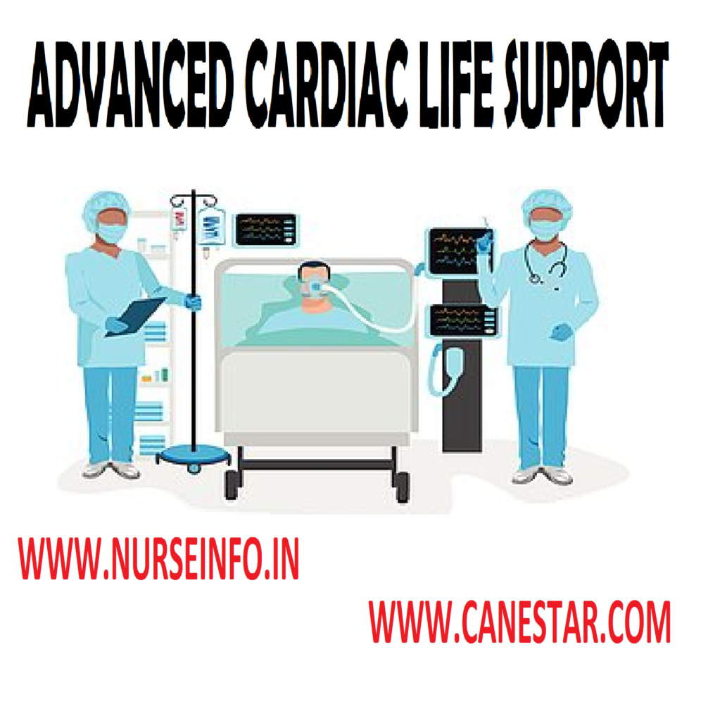
AIRWAY AND VENTILATORY SUPPORT
- Oxygenation: essential aspect of resuscitation is to achieve optimal ventilation and oxygenation. Although artificial ventilation provides a normal partial pressure of oxygen, there is arterial hypoxemia because of diminished cardiac output, intrapulmonary shunting and ventilation perfusion mismatch which can be corrected with supplementary oxygen
- Airway adjuncts: there is a possibility of continued soft tissue obstruction which can be relieved by oropharyngeal and nasopharyngeal airway. Oral airway must be of proper size and positioned so that the patients tongue is completely encircled by the airway.
- Masks: a well-fitting mask can be used to supply higher concentration of oxygen through an oxygen insufflation inlet.
- Ventilation circuits: if the patient’s spontaneous ventilation is inadequate assistance is provided using manual ventilating unit. There are two types of manual resuscitators which are self-refilling units are referred as bag mark devices or Ambu bags. The nonself-refilling Magill circuit with 1 piece circuit
- Endotracheal intubation: in most of the cardiopulmonary arrests ventilation can be achieved by simple airway restoration. Endotracheal intubation should be attempted by experienced person when all the equipment is ready. After placing the endotracheal tube in place good airway control is possible, with better regulation of FiO2, airway pressure and ventilatory pattern is offered. Ventilation and tube function is to be monitored closely as there is danger of blocked or malfunctioning endotracheal tube.
- Support of circulation: chest compressions during advanced cardiac life support are performed in the same manner as Basic Life Support.
ELECTROCARDIOGRAPHIC MONITORING
- ECG monitoring is essential during resuscitation. Many defibrillators have the built in ECG monitoring circuit and quick look paddles. Such units sense, the patient ECG pattern from the defibrillator paddle upon application and display. For continuous monitoring, standard ECG machine or monitoring unit with display screen is used
- Defibrillation: in ventricular fibrillation, a precordial thumb is employed by giving a sternum to a height of 8-12 inches. If this reverts, the rhythm to sinus bolus lidocaine is given. If ventricular fibrillation persists proceed to Basic Life Support and defibrillation
- Electrical defibrillation: electrical defibrillation involves passing an electrical current through a fibrillating heart allowing for uniform depolarization and organized cardiac electromechanical activity. To use the defibrillator effectively apply conductive jelly to the paddles and apply to the chest to determine rhythm. Select the energy level marked in Joules (100-300). Place the paddles on the chest in appropriate locations. Make sure that personnel are not in contact with patient or the cot. Evaluate the effectiveness and administer epinephrine and sodium bicarbonate device.
CORRECTION OF ACIDOSIS AND FLUID REPLACEMENT
It is necessary to have a proper venous access for correction of acidosis administration of other drugs and fluid replacement. Peripheral veins are conveniently used during arrest but in case of vasoconstriction and venous collapse, central venous cannulation should be attempted making sure that it does not interfere with resuscitative efforts.
Correction of acidosis should be attempted if the arrest is continued for few minutes. Acidosis can be respiratory acidosis which results from failure of carbon dioxide elimination. Carbon dioxide production is continuous but the gas cannot be removed because of pulmonary and cardiac failure. PaCO2 rises and PO2 lowers. Metabolic acidosis develops with tissue hypoperfusion occurs. Blood gas shows an acid pH. It is due to reduction in the (HCO3) which is associated with an equivalent rise in chloride leaving the anion gap unchanged or the fall in (HCO3) which is accompanied by equivalent rise in anion gap.
By ensuring adequate alveolar ventilation carbon dioxide removal can be achieved and residual metabolic acidosis corrected by the administration of sodium bicarbonate. The actual amount of bicarbonate required in each case is determined on the basis of blood gas results that are the patient’s base deficit. Sodium bicarbonate is used sparingly as against the earlier practice because major part of metabolic acidosis can be corrected by adequate alveolar ventilation. The initial dose of sodium bicarbonate is 1mEq/kg which is given slowly. Further administration should be guided by blood gas determination. Formula could be used – 0.3 multiply wt (kg) multiply based deficit. If blood gas result is not available half of the initial dose every 10-15 minutes is appropriate
Volume replacement: isotonic crystalloid is the best for rapid expansion of circulatory blood volume. Collapsed jugular and peripheral veins, dryness of mucous membranes, absence of normal secretions, and peripheral vasoconstriction with appropriate history suggest dehydration and volume deficit.
Volume replacement should be attempted and restored until cardiac function is restored. It can be initiated with ringer lactate or normal saline. If the quantity of fluid is required in excess of 1-2 liters for adults, colloids is added. Volume infusion can be guided by central venous pressure and pulmonary capillary wedge pressure.
DRUGS IN ADVANCED CARDIAC LIFE SUPPORT
Drugs
- Oxygen
- Adrenaline
Pharmacological Effects: correction of hypoxia. 1. Elevates blood pressure. 2. Stimulates cardiac contraction
Indications for use: respiratory and cardiac arrest from any cause. 1. Ventricular fibrillation 2. Asystole 3. Electromagnetic dissociation
- Sodium bicarbonate (NaHCO3)
- Calcium chloride/calcium gluconate
Pharmacological effects: for correction of metabolic acidosis. Stimulates spontaneous contraction
Indications for use: prolonged cardiac arrest (10-15 minutes) 1. Asystole 2. Electromechanical transfusion 3. Massive blood transfusion
- Xylocaine
Pharmacological Effects: suppressive ventricular arrhythmias
Indications for use: 1. Ventricular tachycardia 2. Recurrent/refractory ventricular fibrillation 3. Prophylaxis in patients of myocardial infarction
- Bretylium tosylate
Pharmacological Effects: suppresses ventricular arrhythmias
- Atropine
Pharmacological Effects: accelerates cardiac rate
- Isoproterenol
Pharmacological Effects: 1. Stimulates spontaneous contraction 2. Accelerates cardiac rate
Indications for use: 1. Third degree heart block/Asystole 2. Electromechanical dissociation
- Potassium chloride
Pharmacological Effects: correction of hypokalemia
Indications for use: refractory cardiac arrest/ventricular arrhythmia due to hypokalemia
- Deriphyllin
Pharmacological Effects: bronchial dilatation
Indications for use: Bronchospasm due to secretion or bronchial asthma/COPD
- Hydrocortisone
Pharmacological Effects: 1. Suppresses anoxic tissue damage 2. Membrane stabilizer 3. Protects against circulating toxins 4. Suppression of harmful inflammation
Indications for use: 1. shock-hypovolemic, anaphylactic 2. Bronchial asthma (acute) 3. Hemolysis 4. Decreases intracranial pressure 5. Acute adrenal insufficiency
TERMINATION OF CARDIOPULMONARY RESUSCITATION
It is difficult to decide to terminate unsuccessful resuscitative efforts. Inability to restore adequate cardiovascular function is the basis of the decision. Most definitive signs which act as a guideline are absence of reactive pupils, lack of spontaneous activity and response to deep pain and absent brainstem reflexes. Family of the patient should receive high priority in making the decisions of terminating the life support.
Post-resuscitation support: in case of a successful resuscitation, post-resuscitation support plays an important role in deciding the final outcome. Transition from emergency service to critical care unit need to be carried out smoothly. A thorough assessment and examination should be carried out. Diagnostic studies required further should be completed. Ventilatory support is continued at optimal level. Cardiac support with minimal cardiac work is maintained with appropriate drugs.
Transportation is arranged only after proper stabilization and critical care unit is ready to receive the patient. Patient should be accompanied by a nurse and a physician with adequate equipment. Portable ventilators are available which can be used while transferring the patient from emergency unit to critical care unit. If it is not available Ambu bag with an oxygen source can be used during shifting to provide artificial ventilator.