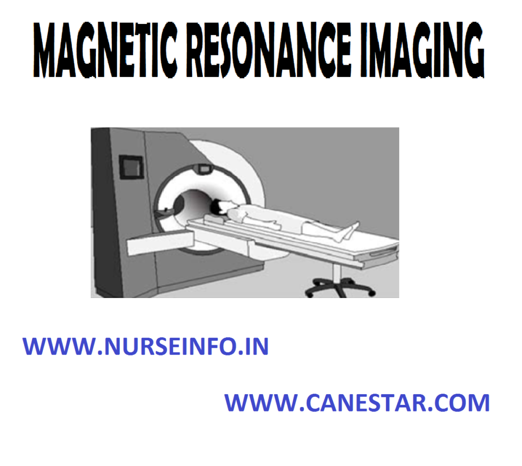
MAGNETIC RESONANCE IMAGING – Definition, Purpose, Principle, Instruction, Preparation of the Client, MRI Equipment, Procedure, After Care, Advantages of MRI, Disadvantages and Contraindications
Magnetic resonance imaging is also called as nuclear magnetic resonance; this technique was independently discovered by Felix Bloch and Purcell in 1952. He explained that when the nuclear energy is exposed to a magnetic field, it behaves like a magnet. This nuclear magnetism with its magnetic field helps the nuclear energy to act as a small magnet
DEFINITION
- Magnetic resonance imaging (MRI) is a noninvasive diagnostic test with a powerful magnetic field to obtain images of different areas of the body
- Magnetic resonance imaging or MRI uses a powerful magnetic field and radiofrequency waves to produce computerized images of internal organs and tissues
PURPOSE
- To produce tissue analysis and images not readily seen on standard X-ray
- To detect tiny lesions of multiple sclerosis on brain and spinal cord
- To detect slipped disc in the spinal cord
- To get a clear image of internal structures in response to the magnetic field, created by harmless, low energy radiowaves
- To detect, localize and stage malignancies of the CNS, spine, head and neck and musculoskeletal system
PRINCIPLE
- MRI does not employ ionizing radiations, so it is free from radiations where as CT scan is by X-ray
- The picture from an MRI are opposite of the CT scan. In MRI the bones appear black whereas in CT scan bones appear white
- MRI is used to study the tissue metabolism by spectroscopy where as CT not
- MRI is used to obtain sectional views in any plane unlike CT scan which is more or less restricted to cross-sectional imaging
- MRI detects water because it focuses on the behavior of hydrogen atom in water molecule. This allows MRI to distinguish between water proof and water rich tissues
- MRI gives early warning of myocardial infarction or stroke with the help of sodium or phosphorus ions
INSTRUCTIONS
- The client informed that it is painless noninvasive procedure and he will hear a lot of noises during the procedure
- All jewelry, eye glasses and hair pins/clips or any other metal objects should be removed
- Carefully question and screen for the presence of any metal implantation
- Consent for contrast and general anesthesia to be taken
- Patient should wear only cotton dress
- No dietary restriction for MRI even for contrast, unless anesthesia is planned
- Extra blanket may be provided as the client is in the air conditioned for more than 45 minutes
PREPARATION OF THE CLIENT
- Explain the procedure to the patient in a simple language
- Remove all metal objects, clips and jewelry from the patient’s body
- Give information about actual procedure, staff involved, duration and sensation to be experienced and probable outcome
- The patient is assured the investigation is safe and painless
- Psychological support and assistance to be given for claustrophobia
MRI EQUIPMENT
- Magnet
- Radiofrequency (RF) coils (transmitter/receiver)
- Gradient coils
- Computer
- Display unit
- Digital storage facilities
PROCEDURE
- After removing all metal objects, the client lies on a padded stretcher that slides into tunnel like chamber
- Place the head in a plastic helmet like structure
- Place the arms at the side of the X-ray table is the rolled several feet into the scanner
- The patient is placed in a strong magnetic field up to 40,000 times stronger than the earth’s magnetic field and is then subjected to precise, computer programmed bursts of radiofrequency waves
- The client feels nothing and hears only loud noises caused by the pulsating radiofrequency waves the resemble a Jackhammer or drill which lasts about a few minutes
AFTER CARE
- Ask the patient to get up slowly, it the client may feel dizzy provide bed rest
- Check the vital signs and record it
- Assess the allergic reactions if dye administered
- For clients who had MRI under general anesthesia kept fasting for 3-4 hours and IV fluids to be given
ADVANTAGES OF MRI
- Does not expose the client to radiation because it is non-ionizing radiation
- Results are obtained rapidly
- Multisectional imaging
- It is safe even contrast dye is used
- Cost affordable when comparing with other invasive surgical procedures
- Provides tissue characterization and blood flow
- Provides clear images of moving organs
- Helps to detect disorders that cause loss of myelin from nerve such as multiple sclerosis
DISADVANTAGES
- Long imaging time
- Many protocol options
- Correct choice of machine parameters essential
- Poor bone and calcium detail
- Not available in all areas
- Difficult to manage and monitor patients who are critically ill
CONTRAINDICATIONS
- Clients with pacemaker are contraindicated
- Cannot use in clients who are extremely obese
- Cannot use in clients with metal implants


