
DEEP VEIN THROMBOSIS – Etiology, Risk Factors, Pathophysiology, Clinical Manifestation, Diagnostic Evaluation and Management (Surgical and Nursing)
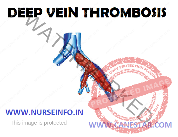
Deep vein thrombosis or deep venous thrombosis (DVT) is a blood clot in a deep vein. A clot inside a blood vessel is called thrombus. Deep vein thrombosis (DVT) is a condition in which a blood clot forms in one or more of the deep veins in the body, usually in a calf or thigh muscle of legs.
ETIOLOGY
- Combination of venous stasis
- Hypercoagulability
- Physical damage or endothelial activation
- Genetic factors: deficiencies in antithrombin, protein C
RISK FACTORS
- Acquired
Older age
Major surgery and orthopedic surgery
Cancers especially pancreatic
Immobilization, as in orthopedic casts the sitting position, and travel, particularly by air
Pregnancy and the postpartum period
Antiphospholipid syndrome (such as lupus anticoagulant)
Trauma and minor leg injury
Previous oral contraceptives
Hormonal replacement therapy
Central venous catheters
Inflammatory diseases and some autoimmune diseases
Nephritic syndrome
Obesity
Infection
HIV
Polycythemia vera
Chemotherapy
- Inherited
Antithrombin deficiency
Protein C deficiency
Protein S deficiency (type I)
Factor V Leiden
Prothrombin
Dysfibrinogenemia
Non-O-blood type
- Mixed
Low free protein S
Activated protein C resistance
High factor VIII levels
Hyperhomocysteinemia
High fibrinogen levels
High factor IX levels
High factor XI levels
PATHOPHYSIOLOGY
When legs are inactivated —- ineffective blood pools by gravity in the veins —- thrombus develops in local process —- platelets adhere to endothelium —- adenosine diphosphate is released by dead tissue —- this leads to platelet plug form —- risk of embolization
CLINCIAL MANIFESTATION
- Swelling in one or both legs
- Pain or tenderness in one or both legs, which may occur only while standing or walking
- Warmth in the skin of the affected leg
- Red or discolored skin in the affected leg
- Visible surface veins
- Leg fatigue
DIAGNOSTIC EVALUATION
- Physical examination: swelling in the leg from fluid can result in ‘pitting’ after pressure is applied
- CT scan: an abdominal CT scan shows a common iliac vein thrombosis
- D-dimer test: a type of blood test that detects pieces of blood clots that have broken down and are loose in the bloodstream
- Duplex ultrasound: during this test, high-frequency sound waves bounce off the inside of body, producing images of blood vessels. An ultrasound image demonstrates a blood clot in the left common femoral vein
MANAGEMENT
- Anticoagulation: anticoagulation, which prevents further coagulation but does not act on existing clots, is the standard treatment for DVT. Parenteral anticoagulant (such as fondaparinux, or heparin) for at least five days and a vitamin K antagonist an oral anticoagulant
- Graduated compression stockings and walking: in addition to anticoagulation treatment, the graduated compression stockings, which apply higher pressure (30 to 40 mm Hg) at the ankles and a lower pressure around the knees is suggested. Walking is also suggested over bed rest for those without severe pain or edema
SURGICAL MANAGEMENT
- Inferior vena cava filters: inferior vena cava filters (IVC filters) are used on the presumption that they reduce PE. They are only recommended in some high-risk scenarios
- Thrombolysis: thrombolysis, which acts to break up clots, can be systemic or catheter-directed, patients may choose thrombolysis, if it concerns over the complexity, bleeding risk, and cost of the procedure
- Mechanical thrombectomy: a mechanical thrombectomy device can remove a thrombosis
Nursing Management
Nursing Diagnosis: Risk for hemorrhage related to graft procedure
Interventions
- Monitor pulse rate
- Monitor central venous pressure
- Provide sterile dressing on wound
- Give vitamin K as per doctor’s advice
Nursing Diagnosis: Pain related to disease condition as evidenced by verbal communication.
Interventions
- Assess for the presence of pain, the scale and intensity of pain
- Teach the client about pain management and relaxation with distraction
- Secure the chest tube to restrict movement and avoid irritation
- Assess pain-reduction measures
- Provide analgesics as indicated
Nursing Diagnosis: risk for impaired gas exchange related to cough and pain from incision
Interventions
- Airway management
Open the airway with headtilt, chinlift, jaw thrust
Set the position to maximize ventilation
Use tools airway
Perform chest physiotherapy
Teach breathing deeply and coughing effectively
Perform suction
Auscultation of breath sounds
Give bronchodilators
- Oxygenation therapy
Provide humidification system of oxygen equipment
Monitor the flow of oxygen and the amount given
Monitor signs of oxygen toxicity
DEEP VEIN THROMBOSIS – Etiology, Risk Factors, Pathophysiology, Clinical Manifestation, Diagnostic Evaluation and Management (Surgical and Nursing)
