CHEST X-RAY (Purpose, Indications, Normal Findings, Standard Positions Used, Portable Chest X-ray, Lateral View, Lateral Decubitus Position, Oblique Position, Lordotic Position and Nursing Considerations)
Chest X-ray can help in the diagnosis of the large variety of pulmonary problems, including pneumonia, lung cancer, emphysema, pulmonary edema, and many others. When performed in a radiology department, usually two view are ordered; a posterior to anterior view and lateral view. This gives a radiologist a more three-dimension perspective of the chest X-ray may also be taken of the sinuses in cases of sinusitis
PURPOSE
- Chest X-ray studies done as part of routine screening procedures
- To detect or identify the pulmonary diseases
- To monitor the status of respiratory disorders and abnormalities (e.g. pleural diffusion, atelectasis and tuberculosis cavities lesions)
- To confirm endotracheal or tracheotomy tube placement
- To detect traumatic chest injuries after any major accidents
INDICATIONS
- Chest X-ray is used to identify abnormalities in chest structures and lung tissue for diagnosis and injuries of the lungs and to monitor treatment
- Chest films may reveal abnormalities when there are no physical signs or symptoms of pulmonary disease
- Chest films show the bony structures (e.g. ribs, sternum, clavicles, scapulae, and upper portion of the humerus)
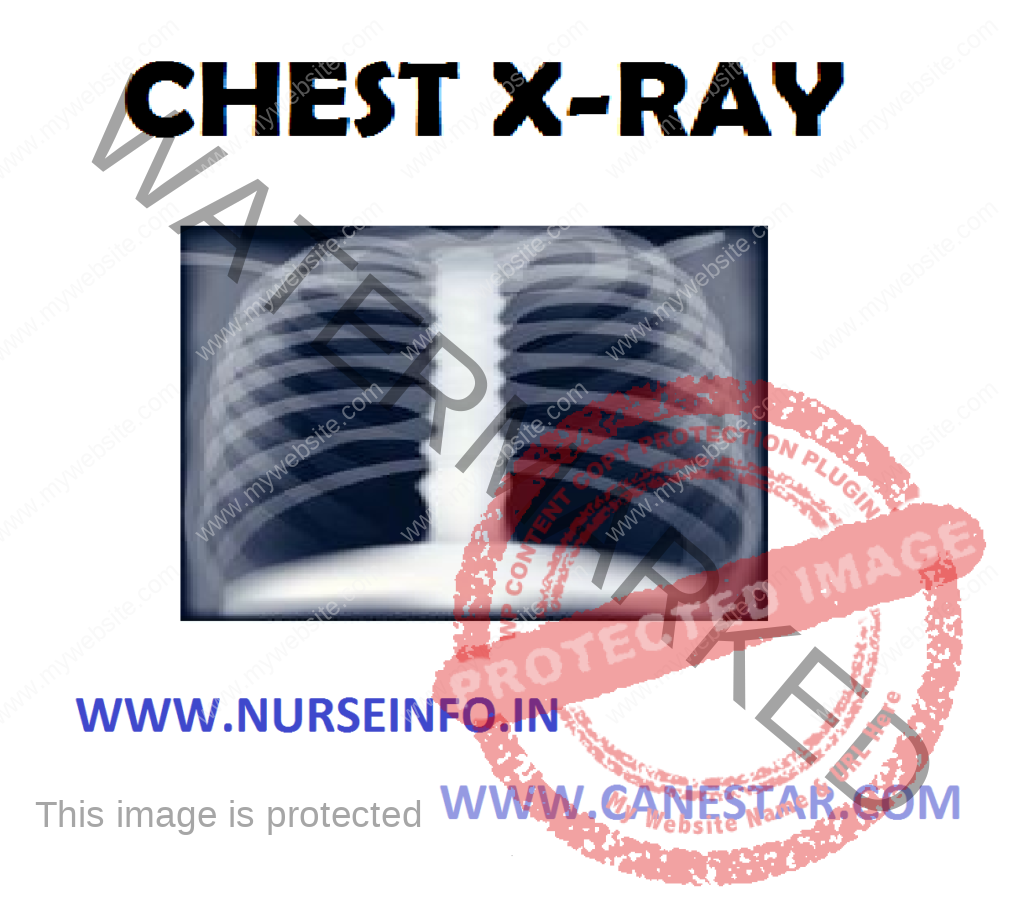
NORMAL FINDINGS
- The vertebral column is visible vertically through the middle of the thorax. The two hemidiaphragm normally appear rounded, smooth and sharply defined, with the right hemidiaphragm slightly elevated above the left
- The junction of the rib cage and the diaphragm, called the costophrenic angle is normally clear visible and angled
- Heart tissue is dense and appears white but less intensely white then bony structures. The heart shadow is normally clearly outlined and extends primarily onto the left side of the thorax and occupies no more than one third of the chest width
- Close observation shows the trachea in the upper middle chest almost superimposed over the cervical and thoracic vertebrae. The trachea bifurcates at the level of the forth thoracic vertebra into the right and left main stem bronchi
- The pulmonary blood vessels, bronchi and lymph nodes are located in the hilum on both the right and left sides of the midthorax
- Lung tissue appears black on X-ray film. Vascular lung structures are visible as white, thin, wispy strings fanning out from the hilum
STANDARD POSITIONS USED
- Adult chest X-ray studies are taken with the clients standing or sitting facing the X-ray film, with the chest and shoulder in direct contact with the film cassette
- The shoulders are rotated forward to pull the scapulae away from the lung field
- The X-ray cathode penetrates from the posterior. This position is called posteroanterior (PA) position
- The radiograph is usually taken at pull inspiration, which causes the diaphragm to move downwards
- Radiographs taken on expiration are sometimes requested for demonstrating the degree of diaphragm movement or for assisting in the assessment and diagnosis of pneumothorax
PORTABLE CHEST X-RAY
- For clients unable to be transported to the radiology department, portable chest radiography may be taken
- Portable radiographs are usually taken with the film placed behind the client, and the X-ray beam penetrates from the front of the chest anterioposterior (AP) position
- Because the X-ray beam enters from the anterior chest, the heart will appear larger than it reality is and larger than on a PA view
LATERAL VIEW
- It usually accompanies a standard PA view. It is taken from either the right or left side of the chest
- The arms are raised above the head, and the side of the chest is placed against the film
- The lateral view allows better visualization of the heart and diaphragm dome
- When used in conjunction with a PA film, a lateral position gives a three-dimension view, allowing more specific identification of an abnormality’s location
LATERAL DECUBITUS POSITION
- This position may be used when it is necessary to determine whether opaque areas on the pleura are due to solid or liquid media
- This view is taken with the client lying on either the right or left side, depending on which side of the chest is being assessed
- In a left lateral decubitus position, the client is lying on the left side. The term decubitus refers to a lying down position
OBLIQUE POSITION
- This position is used to see behind and around underlying structures. The shoulders are rotated either to the right or left of the film
- By turning the client, the angle at which the X-ray beam passes through the chest is shifted
- In a right oblique position, the right side is closest to the film. The view may be taken from either an anterior or posterior position
LORDOTIC POSITION
- This position is useful if clearer visualization of the upper lung fields is needed
- This angle of the X-ray cathode is lowered and the beam directed at an upward angle
- This angle removes the clavicles and first and second ribs from the field of vision
NURSING CONSIDERATION
- Explain to the client that this test detects or monitor the progress of respiratory distress
- Explain the client, who will perform the procedure, where and when it will be done, and that it takes only minutes
- Inform that the client must wear a gown without snaps but may keep his pants, socks and shoes on
- Instruct the client, to remove all jewelry from his neck and chest
- Inform the client, that this procedure is performed in radiology department; the client will stand or sit in front of a machine
- Inform that the client is asked to take a deep breath and to hold it for a few seconds while the X-ray is taken
- Reassure him that the amount of radiation exposure is minimal. Faculty personnel will leave the area when the technician takes the X-ray because they are potentially exposed to radiation many times a day
- If the client is incubated, make sure that no one disconnects the tubes during the procedure
- Care should be taken for female clients of childbearing age