BRONCHIECTASIS – Etiology and Types, Signs and Symptoms, Diagnostic Evaluations and Management
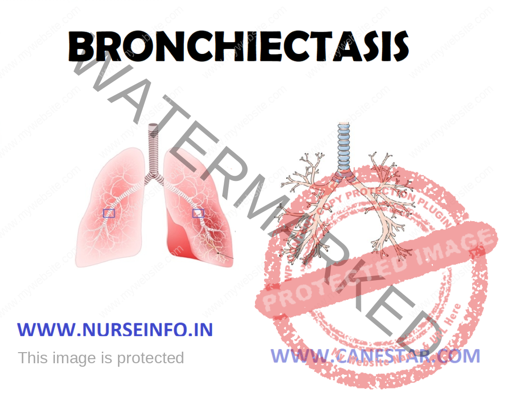
Bronchiectasis is a condition in which some of the bronchi have become scarred and permanently enlarged along with the loosening of integrity of bronchial wall. During the disease process, the cilia are damaged so that they are unable to effectively sweep away the mucus. As a result, mucus accumulates in parts of the lung that are affected and the risk of developing lung infections is increased.
ETIOLOGY AND TYPES
Currently bronchiectasis usually occurs as the result of an illness, such as pneumonia (approximately 25% of all cases). Other causes include:
- Cystic fibrosis
- Immune deficiency
- Recurrent aspiration of fluid into the lungs (as occurs with gastroesophageal reflux)
- Inhalation of a foreign object into the lungs
- Inhalation of harmful chemicals, e.g. ammonia
- In rare cases, it may be congenital
TYPES
- Cylindrical bronchiectasis: bronchi are enlarged and cylindrical
- Varicose bronchiectasis: bronchi are irregular with areas of dilatation and constriction
- Saccular or cystic bronchiectasis: dilated bronchi form clusters of cysts. This is the most severe form of bronchiectasis and is often found in patients with cystic fibrosis
SIGNS AND SYMPTOMS
The main symptom of bronchiectasis is a mucus-producing cough. The cough is usually worse in the mornings and is often brought on by changes in posture. The mucus may be yellow-green in color and foul-smelling, indicating the presence of infection.
OTHER SYMPTOMS
- Coughing up blood (more common in adults)
- Bad breath
- Wheezing chest: a characteristic crackling sound may be heard when listening with a stethoscope
- Recurring lung infections
- A decline in general health
- In advanced bronchiectasis, breathlessness can occur
DIAGNOSTIC EVALUATIONS
An initial diagnosis of bronchiectasis is based on the patient’s symptoms, their medical history and a physical examination. Further diagnostic tests may include:
- Chest X-ray
- CT (computerized tomography) scan
- Blood tests
- Testing of the mucus to identify any bacteria present
- Checking oxygen levels in the blood
- Lung function tests (spirometry)
MANAGEMENT
Bronchiectasis is a chronic (long-term) condition that requires lifelong maintenance. Good management of the condition is vital to prevent ongoing damage to the lungs and worsening of the condition.
The ultimate goal of treatment is to clear mucus from the chest and prevent further damage to the lungs. The two main types of treatments used are:
Medications
Some or all of the following groups of medications may be used:
- Antibiotics are used to treat acute infections. Where the infections is severe, hospitalization and treatment with intravenous antibiotics may be required.
- Bronchodilators (as used in people with asthma) to improve the flow of air to the lungs
- Corticosteroids to reduce inflammation in the lungs
- Occasionally, medications to thin the mucus may be used
- Vaccination against flu and pneumococcus
Physiotherapy and Exercise
- Chest physiotherapy and postural drainage are used to remove secretions from the lungs
- Other factors important in managing the condition include avoiding dust, smoke and other respiratory irritants, and maintaining a balanced nutritious diet
PREVENTION
The ministry of health recommends the following measures to help prevent bronchiectasis in children:
- Not smoking during pregnancy and having a smoke free home
- Breastfeeding your children
- Eating a healthy balanced diet
- Early detection and treatment of chest infections
- Making sure homes are warm and dry (making chest infections less likely)
- Immunization for diseases like measles and whooping cough
NURSING MANAGEMENT
Nursing Intervention
- Improve gas exchange
Administer oxygen to maintain PaO2 of 60 mm Hg, using devices that provide increased oxygen concentration
Monitor fluid balance by intake and output measurement, urine-specific gravity, daily weight measurement
Provide measures to prevent atelectasis and promote chest extension and secretion clearance as per order, spirometer
Elevate head level to 30 degrees
Monitor adequacy of alveolar ventilation by frequent measurement of respiratory system
Administer antibiotic, cardiac medication and diuretics as prescribed by doctor
- Maintain airway clearance
Administer medication to increase alveolar function
Perform chest physiotherapy to remove mucus
Administer IV fluids
Suction patient as needed to assist with removal of secretions
- Relieving pain
Watch patient for signs of discomfort and pain
Position the head elevated
Give prescribed morphine and monitor for pain-relieving sign
- Reducing anxiety
Correct dyspnea and relive physical discomfort
Speak calmly and slowly
Explain diagnostic procedure


