Home Blog Page 64

MYASTHENIA GRAVIS

MYASTHENIA GRAVIS – Etiology, Signs and Symptoms, Diagnostic Evaluation and Management (Medical, Surgical and Nursing)

Myasthenia gravis is a chronic inflammation neuromuscular disease characterized by varying degrees of weakness of the skeletal (voluntary) muscles of the body. The name myasthenia gravis, which is Latin and Greek in origin, literally means “grave muscle weakness.”

ETIOLOGY

Autoimmunity

In myasthenia gravis, the immune system produces antibodies that block or destroy muscles receptor sites for a neurotransmitter called acetylcholine. Antibodies may also block the function of a protein called a muscle-specific receptor tyrosine kinase. This protein is involved in forming the nerve-muscular junction. When antibodies block the function of this protein, it may lead to myasthenia gravis.

Thymus Gland

Tumors of the thymus (thymomas). Usually, thymomas are not cancerous. In some people, myasthenia gravis is not caused by antibodies blocking acetylcholine or the muscle-specific receptor tyrosine kinase. This type of myasthenia gravis is called antibody-negative myasthenia gravis. Antibodies against another protein, called lipoprotein-related protein 4, may play a part in the development of this condition.

Genetic Factors

Rarely, mothers with myasthenia gravis have children who are born with myasthenia gravis (neonatal myasthenia gravis). If treated promptly, children generally recover within two months after birth.

Factors that can worsen Myasthenia Gravis

  • Fatigue
  • Illness
  • Stress
  • Extreme heat
  • Some medications – such as beta blockers, quinidine gluconate, quinidine sulfate, quinine (qualaquin), phenytoin (Dilantin), certain anesthetics and some antibodies.

SIGNS AND SYMPTOMS

Eye Muscles

In more than half the people who develop myasthenia gravis, their first signs and symptoms involve eye problems, such as:

  • Drooping of one or both eyelids (ptosis)
  • Double vision (diplopia), which maybe horizontal or vertical, and improves or resolves when one eye is closed.

Face and Throat Muscles

In about 15 percent of people with myasthenia gravis, the first symptoms involve face and throat muscles, which can cause:

  • Altered speaking
  • Difficulty swallowing
  • Problems chewing
  • Limited facial expressions

Neck and Limb Muscles

Myasthenia gravis can cause weakness in neck, arms, and legs, but this usually happens along with muscle weakness in other parts of body, such as eyes, face or throat.

The disorder usually affects arms more often than legs. However, if it affects legs, patient may waddle when walk. If neck is weak, it may be hard to hold up head.

Patient may have difficulty in:

  • Breathing
  • Swallowing
  • Chewing
  • Walking
  • Using arms or hands
  • Holding up head

DIAGNOSTIC EVALUATION

Diagnosis may be made on the basis of neurological health by testing:

  • Reflexes
  • Muscle strength
  • Muscle tone
  • Senses of touch and sight
  • Coordination
  • Balance

The key sign that points to the possibility of myasthenia gravis is muscle weakness that improves with rest. Tests to help confirm the diagnosis may include:

  • Edrophonium test: Injection of the chemical edrophonium chloride (Tensilon) may result in a sudden, although temporary, improvement in muscle strength. This is an indication that the patient may have myasthenia gravis. Edrophonium chloride blocks an enzyme that breaks down acetylcholine, the chemical that transmits signals from nerve endings to muscle receptor sites.
  • Ice Pack Test: in this test, a bag filled with ice is placed on eyelid. After two minutes, doctor removes the bag and analyzes droopy eyelid for signs of improvement.
  • Blood Analysis: a blood test may reveal the presence of abnormal antibodies that disrupt the receptor sites where nerve impulses signal muscles to move.
  • Repetitive nerve stimulation: in this nerve conduction study, electrode is attached to skin over the muscles to be tested.
  • Single-fiber electromyography (EMG): electromyography (EMG) measures the electrical activity traveling between brain and muscle. It involves inserting a fine wire electrode through skin and into a muscle.
  • Imaging scans: CT scan or an MRI to check if there is a tumor or other abnormality in thymus.
  • Pulmonary function tests: to evaluate whether condition is affecting breathing.

MANAGEMENT

Medical Management

  • Cholinesterase inhibitors: medications such as pyridostigmine enhance communication between nerves and muscles. These medications do not cure the underlying condition, but they may improve muscle contraction and muscle strength.
  • Corticosteroids: corticosteroids such as prednisone inhibit the immune system, limiting antibody production. Prolonged use of corticosteroids, however, can lead to serious side effects, such as bone thinning, weight gain, diabetes, and increased risk of some infections.
  • Immunosuppressants: such as azathioprine, mycophenolate mofetil, cyclosporine or tacrolimus. Side effects of immunosuppressants can be serious and may include nausea, vomiting, gastrointestinal upset, increased risk of infection, liver damage, and kidney damage.
  • Plasmapheresis: this procedure uses a filtering process similar to dialysis. Blood is routed through a machine that removes the antibodies that block transmission of signals from nerve endings to muscles’ receptor sites. Other risks associated with plasmapheresis include a drop in blood pressure, bleeding, heart rhythm problems or muscle cramps. Some people may also develop an allergic reaction to the solutions used to replace the plasma.
  • Intravenous immunoglobulin (IVIg): this therapy provides normal antibodies, which alters the immune system response. IVIg has a lower risk of side effects than do plasmapheresis and immune-suppressing therapy. Side effects, which usually are mild, may include chills, dizziness, headaches, and fluid retention.

Surgical Management

About 15 percent of the people with myasthenia gravis have a tumor in their thymus gland, a gland under the breastbone that is involved with the immune system. If patient is having a tumor, called a thymoma, thymectomy maybe performed as an open surgery or as a minimally invasive surgery

Minimally invasive thymectomy may include:

Video-assisted thymectomy: in one form of this surgery, surgeons make a small incision in neck and use a long thin camera (video endoscope) and small instruments to visualize and remove the thymus gland through neck.

Robot-assisted thymectomy: in a robot-assisted thymectomy, surgeons make several small incisions in the side of chest. Surgeons conduct the procedure to remove the thymus gland using a robotic system, which includes a camera arm and mechanical arm.

Nursing Management

Nursing Diagnosis

  • Ineffective breathing pattern related to respiratory muscle weakness
  • Impaired physical mobility related to weakness of voluntary muscles
  • Risk for aspiration related to the weakness of bulbar muscles.
  • Self-Care deficit related to muscle weakness, general fatigue.
  • Imbalanced Nutrition: less than body requirements related to dysphagia, intubation, or muscle paralysis.

Interventions

  • Assess the breathing pattern
  • Administer oxygen in case of emergency arrest
  • Encourage deep breathing exercise to strengthen the respiratory muscle tone
  • Install grab bars or railings in places
  • Keep floors clean, and move any loose rugs out of areas.
  • Use electric appliances and power tools
  • Try using an electric toothbrush, electric can openers and other electrical tools to perform tasks when possible to save the energy
  • Wearing an eye patch if have double vision, as this can help relieve the problem
  • Try wearing the eye patch while you write, read or watch television. Periodically switch the eye patch to the other eye to help reduce eyestrain
  • Encourage to eat when patient have good muscle strength
  • Take time chewing the food, and take a break between bites of food
  • Encourage to eat small meals several times a day may be easier to handle
  • Encourage to eat mainly soft foods and avoid foods that require more chewing, such as raw fruits or vegetables.
MYASTHENIA GRAVIS – Etiology, Signs and Symptoms, Diagnostic Evaluation and Management (Medical, Surgical and Nursing)

MYASTHENIA GRAVIS – Etiology, Signs and Symptoms, Diagnostic Evaluation and Management (Medical, Surgical and Nursing)
PARKINSONISM

PARKINSONISMEtiology and Risk Factors, Signs and Symptoms, Disease Stages, Pathophysiology, Diagnostic Evaluation, Nursing Management

Parkinson’s disease is a neurodegenerative disorder which leads to progressive deterioration of motor function due to loss of dopamine- producing brain cells and is characterized by progressive loss of muscle control, which leads to trembling of the limbs and head while at rest, stiffness, slowness, and impaired balance.

ETIOLOGY AND RISK FACTORS

  • Age is the largest risk factor for the development and progression of Parkinson’s disease. Most people who develop Parkinson’s disease are older than 60 years years of age.
  • Men are affected about 1.5 to 2 times more often than women.
  • A small number of individuals are at increased risk because of a family history of the disorder.
  • Head trauma, illness, or exposure to environmental toxins such as pesticides and herbicides maybe a risk factor.

SIGNS AND SYMPTOMS

  • Tremors: Trembling in fingers, hands, arms, feet, legs, jaw or head. Tremors most often occur while the individual is resting, but not while involved in a task. Tremors may worsen when an individual is excited, tired, or stressed.
  • Rigidity: Stiffness of the limbs and trunk, which may increase during movement. Rigidity may produce muscle aches and pain. Loss of fine hand movements can lead to cramped handwriting (micrographia) and may make eating difficult.
  • Bradykinesia: Slowness of voluntary movement. Over time, it may become difficult to initiate movement and to complete movement. Bradykinesia together with stiffness can also affect the facial muscles and result in an expressionless, ‘mask-like’ appearance.
  • Postural instability: Impaired or lost reflexes can make it difficult to adjust posture to maintain balance. Postural instability may lead to falls.
  • Parkinsonian gait: Individuals with more progressive Parkinson’s disease develop a distinctive shuffling walk with a stooped position and a diminished or absent arm swing. It may become difficult to start walking and to make turns. Individuals may freeze in mid-strike and appear to fall forward while walking.

SECONDARY SYMPTOMS OF PARKINSON’S DISEASE

While the main symptoms of Parkinson’s disease are movement- related, progressive loss of muscle control and continued damage to the brain can lead to secondary symptoms. Some of the secondary symptoms include:

  • Anxiety , insecurity and stress
  • Confusion , memory loss, and dementia
  • Constipation
  • Depression
  • Difficulty swallowing and excessive salivation
  • Diminished sense of smell
  • Increased sweating
  • Male erectile dysfunction
  • Skin problems
  • Slowed, quieter speech, and monotone voice
  • Urinary frequency/urgency
  • Slow blinking
  • Stooped position

PARKINSON’S DISEASE STAGES

  • Stage one: During this initial phase of the disease, a patient usually experiences mild symptoms. These symptoms may inconvenience the day- to-day tasks the patient would otherwise complete with ease. Typically these symptoms will include the presence of tremors or experiencing shaking in one of the limbs.
  • Stage two: In the second stage of Parkinson’s disease, the patient’s symptoms are bilateral, affecting both limbs and both sides of the body. The patient usually encounters problems walking or maintaining balance and the inability to complete normal physical tasks becomes more apparent.
  • Stage three: Stage three symptoms of Parkinson’s disease can be rather severe and include the inability to walk straight or to stand. There is a noticeable slowing of physical movements in stage three.
  • Stage four: This stage of the disease is accompanied by severe symptoms of Parkinson’s. Walking may still occur, but it is often limited and rigidity and bradykinesia are often visible. During this stage, most patients are unable to complete day-to-day tasks, and usually cannot live on their own. The tremors or shakiness that takes over during the earlier stages however, may lessen or become nonexistent for unknown reasons during this time.
  • Stage five: The last or final stage of Parkinson’s disease usually takes over the patients physical movements. The patient is usually unable to take care of himself or herself and may not be able to stand or walk during this stage. A patient at stage five usually requires constant one-on-one nursing care.

PATHOPHYSIOLOGY

  • A substance called dopamine acts as a messenger between two brain areas – the substantia nigra and the corpus striatum – to produce smooth, controlled movements.
  • Most of the movement related symptoms of Parkinson’s disease are caused by a lack of dopamine due to the loss of dopamine – producing cells in the substantia nigra.
  • When the amount of dopamine is too low, communication between the substantia nigra and corpus striatum becomes ineffective, and movement becomes impaired.
  • The greater the loss of dopamine, the worse the movement- related symptoms. Other cells in the brain also degenerate to some degree and may contribute to non-movement related symptoms of Parkinson’s disease.

DIAGNOSTIC EVALUATION

  • At least two of the three major symptoms are present (tremor at rest, muscle rigidity, and slowness).
  • The onset of symptoms started on one side of the body.
  • Symptoms are not due to secondary causes such as medication or strokes in the area controlling movement.
  • Symptoms are significantly improved with levodopa.

MANAGEMENT

  • Medical management
  • Levodopa, Sinemet, levodopa and Carbidopa
  • Pramipexole, Ropinirole, Bromocriptine
  • Selegiline, Rasagiline
  • Amantadine or anticholinergic medications to reduce early or mild tremors
  • Entacapone

Other medications may include

  • Memantine, rivastigmine, galantamine for cognitive difficulties
  • Antidepressants for mood disorders
  • Gabapentin, duloxetine for pain
  • Fludrocortisone, Midrodrine, Botox, Sidenafil for autonomic dysfunction
  • Armodafinil, clonazepam, Zolpidem for sleep disorders.

NURSING MANAGEMENT

Nursing Diagnosis

  • Impaired Physical Mobility related to stiffness and muscle weakness
  • Self-care deficits related to neuromuscular weakness, decreased strength, loss of muscle control/coordination
  • Impaired bowel elimination: Constipation related to medication and decreased activity
  • Imbalanced nutrition: Less than body requirements related to tremor, slowing the process of eating, difficulty chewing and swallowing
  • Impaired verbal communication related to the decrease in the volume of speech, delayed speech, inability to move facial muscles.
  • Ineffective individual coping related to depression and dysfunction due to disease progression.
  • Knowledge deficit related to information resources inadequate maintenance procedures.

Interventions

  • Examine existing mobility and observation of an increase in damage
  • Do exercise program increases muscle strength
  • Encourage bath and massage the muscle
  • Help clients perform ROM exercises, self-care according to tolerance
  • Collaboration physiotherapists for physical exercise
  • Assess the ability and the rate of decline and the scale of 0-4 to perform ADL
  • Avoid what not to do the client and help if needed
  • Collaborative provision of laxatives and consult a doctor of occupational therapy
  • Teach and support the client during the client’s activities
  • Environmental modifications
  • Refer to speech therapy
  • Teach clients to use facial exercises and breathing methods to correct the words, volume, and intonation
  • Breathe deeply before speaking to increase the volume and number of words in sentences of each breath

Practice speaking in short sentences, reading aloud in front of the glass or into a voice recorder (tape recorder) to monitor progress.

PARKINSONISM
PARKINSONISM

BELL’S PALSY

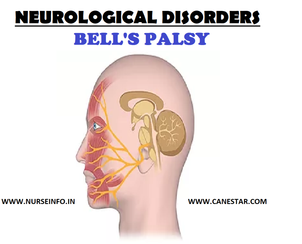
NEUROLOGICAL DISORDERS – BELL’S PALSY

BELL’S PALSY – Etiology, Signs and Symptoms and Management (medical, surgical and nursing)

Bell’s palsy is a paralysis or weakness of the muscles on one side of face. Damage to the facial nerve that controls muscles on one side of the face causes that side of face to droop. The nerve damage may also affect sense of taste. The weakness usually affects one side of the face. Rarely, both sides are affected.

ETIOLOGY

It is thought that inflammation develops around the facial nerve as its passes through the skull from the brain. The inflammation may squash (compress) the nerve as it passes through the skull. The nerve then partly, or fully, stops working until the inflammation goes. If the nerve stops working, the muscles that the nerve supplies also stop working.

  • Cold sore (herpes simplex) virus
  • Chickenpox (varicella-zoster) virus

SIGNS AND SYMPTOMS

  • Weakness of the face which is usually one-sided. The weakness normally develops quickly.

Face may droop to one side

Food may get trapped between gum and cheek. Drinks and saliva may escape from the side of mouth.

Difficult to close the eye, this may causes a watery or dry eye.

Difficult able to wrinkle forehead, whistle or blow out check

Difficulty with speech

  • Painless or cause just a mild ache
  • Loud sounds maybe uncomfortable and normal noises may sound louder than usual. This is because a tiny muscle in the ear may stop working
  • Lose of sense of taste on the side of the tongue that is affected.

MANAGEMENT

  • Anti-inflammatory drugs

The steroid tablet most commonly used is called prednisolone. Steroids help to reduce inflammation, which is probably the reason they help.

  • Antiviral drugs
  • Eye protection
  • An eye pad or goggles to protect the eye.
  • Eye drops  to lubricate the eye during the day
  • Eye ointment to lubricate the eye overnight
  • Tape the upper and lower lid together when you are asleep. Other procedures are sometimes done to keep the eye shut until the eyelids recover
  • Physiotherapy; a treatment called, ‘facial retraining’ with facial exercises may help
  • Injections of botulism toxin (Botox) may help it spasm develops in the facial muscles
  • Various surgical techniques can help with the cosmetic appearance

Complications

  • Corneal ulcers
  • Blindness
  • Impaired nutrition

Medical Management

The objectives of management are to maintain facial tone and to prevent or minimize complication with the help of the following:

  • Corticosteroid therapy (prednisone) maybe initiated to reduce inflammation and edema, which reduces vascular compression and permits restoration of blood circulation to the nerve.
  • Early administration of corticosteroids appears to diminish severity, relieve pain, and minimize denervation.
  • Facial pain is controlled with analgesic agents or heat applied to the face to prevent muscle atrophy, or surgical exploration of the facial nerve.
  • Surgery may be performed if a tumor is suspected, for surgical decompression of the facial nerve, and for surgical rehabilitation of a paralyzed face.

Surgical Management

  1. Facial Nerve Decompression

The surgeon decides if the maxillary segment should be decompressed externally or if the labyrinthine segment and geniculate ganglion should be decompressed with a middle fossa craniotomy.

  • Subocularis Oculi Fat Lift (SOOF)

The SOOF is deep to the orbicularis oculi muscle and superficial to the periosteum below the inferior orbital rim. An SOOF lift is designed to lift and suspend the midfacial musculature. The procedure may also elevate the upper lip and the angle of the mouth to improve facial symmetry.

  • Lateral Tarsal Strip Procedure

An SOOF lift is commonly performed in conjunction with a lateral tarsal strip procedure to correct horizontal lower-lid laxity and to improve apposition of the lid to the globe. first, lateral canthotomy and cantholysis is performed. Then, the anterior lamella is removed and the lateral tarsal strip is shortened and attached to the periosteum at the lateral orbital rim.

  • Implants in Eyelid

Implantable devices have been used to restore dynamic lid closure in cases of severe, symptomatic lagophthalmos. These procedures are best for patients with poor Bell phenomenon and decreased corneal sensation. Gold or platinum weights, a weight-adjustable magnet, or palpebral springs can be inserted into the eyelids. Pretarsal gold-weight implantation is most commonly performed. The implants are easily removed if nerve function returns.

  • Tarsorrhaphy

Tarsorrhaphy decreases horizontal lid opening by fusing the eyelid margins together, increasing support of the precorneal lake of tears and improving coverage of the eye during sleep. The procedure can be done in the office and is particularly suitable for patients who are unable or unwilling to undergo other surgery. It can be completed as either a temporary or a permanent measure.

Permanent tarsorrhaphy is performed if nerve recovery is not expected. Tarsorrhaphy can be performed laterally, centrally, or medially. The lateral procedure is the most common; however, it can restrict the monocular temporal visual field. Central tarsorrhaphy offers good corneal protection, but it occludes vision and  can be cosmetically unacceptable. Medial or paracentral tarsorrhaphy is performed lateral to the lacrimal puncta and can offer good lid closure without substantially affecting the visual field.

Other surgeries

Muscle Transportation, Nerve Grafting, and Brow Lift.

  1. Transposition of temporalis

Transposition of the temporalis muscle can be used to reanimate the face and to provide lid closure by using the fifth cranial nerve. Strips from the muscle and fascia are placed in the upper and lower lids as an encircling sling. Patients initiate movement by chewing or clenching their teeth.

  • Facial nerve grafting or hypoglossal-facial nerve anastomosis

Reinnervation of the facial nerve by means of facial nerve grafting or hypoglossal-facial nerve anastomosis can be used in cases of clinically significant permanent paralysis to help restore relatively normal function to the orbicularis oculi muscle or eyelids.

  • Direct brow lift

Brow ptosis is repaired with a direct brow lift. Care should be taken in the presence of corneal decompression because lifting the brow can cause worsening of lagophthalmos, especially if lid closure is poor. A gold-weight implant can be placed or lower-lid resuspension can be performed simultaneously to prevent this complication.

Nursing Management

Teaching patients with Bell’s palsy to care for them at home is an important nursing priority.

Teaching Eye care

Because the eye usually does not close completely, the blink reflex is diminished, so the eye is vulnerable to injury from dust and foreign particles. Corneal irritation and ulceration may occur. Distortion of the lower lid alters the proper drainage of tears. Encourage the client for the following:

  • Cover the eye with a protective shield at night
  • Apply eye ointment to keep eyelids closed during sleep
  • Close the paralyzed eyelid manually before going to sleep
  • Wear wraparound sunglasses or goggles to decrease normal evaporation from the eye.

Teaching about Maintaining Muscle Tone

  • Show patient how to perform facial massage with gentle
  • Upward motion several times daily when the patient can tolerate the massage
  • Demonstrate facial exercises, such as wrinkling the forehead
  • Blowing out the cheeks, and whistling, in an effort to prevent muscle atrophy
  • Instruct patient to avoid exposing the face to cold and drafts

Diet and Nutrition

  • Instruct patient to chew on the unaffected side of his mouth
  • Provide soft and nutritionally balanced diet. Eliminate hot fluids and foods
  • Give frequent mouth care, being particularly careful to remove residues of food that collects between the cheeks and gums.
NEUROLOGICAL DISORDERS - BELL’S PALSY 
BELL’S PALSY – Etiology, Signs and Symptoms and Management (medical, surgical and nursing)
NEUROLOGICAL DISORDERS – BELL’S PALSY
BELL’S PALSY – Etiology, Signs and Symptoms and Management (medical, surgical and nursing)

CARDIOVASCULAR EMERGENCIES 2

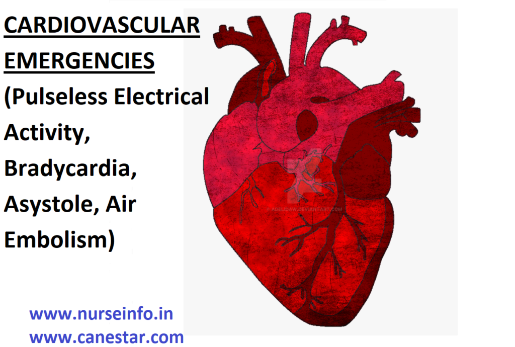
CARDIOVASCULAR EMERGENCIES (Pulseless Electrical Activity, Bradycardia, Asystole, Air Embolism)

Cardiovascular emergencies include pulseless electrical activity, bradycardia, asystole and air embolism.

Pulseless Electrical Activity

  • Assess the patient and conduct a primary ABCD survey
  • Review for the most frequent causes of pulseless electrical activity, the five Hs and five and hypothermia and tablets (drug overdose, accidents), tamponade (cardiac), tension pneumothorax, thrombosis (coronary), and thrombosis (pulmonary embolism)
  • Administer epinephrine (1 mg IV push repeated every 3 to 5 minutes) or atropine (1 mg IV if the heart rate is slow, repeated every 3 to 5 minutes as needed, to a total dose of 0.04 mg/kg).
  • Conduct a secondary ABCD survey

BRADYCARDIA

  • Determine whether the bradycardia is slow (heart rate less than 60 beats/min or relatively slow
  • Conduct a primary ABCD survey
  • Check for serious signs or symptoms caused by the bradycardia
  • If no serious signs or symptoms are present, evaluate for a type II second-degree atrioventricular block or third-degree atrioventricularblock
  • If neither of these types of heart block is present, observe
  • If one of these types of heart block is present, prepare for transvenous pacing, if symptoms develop, use a transcutaneous pacemaker until the transvenous pacer is placed
  • If serious signs or symptoms are present, begin the following intervention sequence:

Atropine, 0.5 up to a total of 3 mg IV

Transcutaneous pacing, if available

Dopamine, 5 to 20 mcg/kg/min

Epinephrine, 2 to 10 mcg/min

Isoproterenol, 2 to 10 mcg/min

Conduct a secondary ABCD survey

ASYSTOLE

  • Conduct a primary ABCD survey
  • Perform transcutaneous pacing immediately if needed. Consider transvenous pacing if transcutaneous pacing fails to capture
  • Administer epinephrine ( 1 mg IV push, repeated every 3 to 5 minutes) or atropine (1 mg IV repeated every 3 to 5 minutes, up to a total of 3 mg)
  • Conduct a secondary ABCD survey
  • If asystole persists, consider withholding or ceasing resuscitative efforts

HYPERTENSIVE EMERGENCY

A hypertensive emergency is an acute, severe elevation in blood pressure accompanied by end-organ compromise. In newly hypertensive patients, a hypertensive emergency is usually associated with a diastolic blood pressure higher than 120 mm/Hg

Etiology

  • Essential hypertension
  • Renal causes
  • Renal artery stenosis
  • Glomerulonephritis
  • Vascular causes
  • Vasculitis
  • Hemolytic-uremic syndrome
  • Thrombotic thrombocytopenia purpura
  • Pregnancy-related causes
  • Preeclampsia
  • Eclampsia
  • Pharmacologic causes
  • Sympathomimetics
  • Clonidine withdrawal, beta blocker withdrawal
  • Cocaine
  • Amphetamines
  • Endocrine causes
  • Cushing’s syndrome
  • Pheochromocytoma
  • Renin-secreting adenomas
  • Thyrotoxicosis
  • Neurologic causes
  • Central nervous system trauma
  • Intracranial mass
  • Autoimmune cause
  • Scleroderma renal crisis

Signs and Symptoms

Symptoms of end-organ involvement include:

  • Headache
  • Blurry vision
  • Confusion
  • Chest pain
  • Shortness of breath
  • Back pain (e.g., aortic dissection)
  • If severe, seizures and altered consciousness

Treatment

  • Nitroprusside
  • Labetalol
  • Fenoldopam
  • Enalaprilat

AIR EMBOLISM

An air embolism, or more generally gas embolism, is a pathological condition caused by a gas bubble, or bubbles, in a vascular system.

An air embolism, also called a gas embolism is when an air bubble or air bubbles enter a vein or artery and block it. When the embolism enters a vein, it is called a venous air embolism. When the air enters an artery, it is called an arterial air embolism.

These air bubbles can travel to brain, heart, or lungs and cause a heart attack, stroke, or respiratory failure.

Etiology

  • An air embolism can occur when veins or arteries are exposed and pressure allows air to travel into them. This can happen in several ways, such as:

Injections and surgical procedures

  • A syringe or IV can accidentally inject air into veins. Air can also enter veins or arteries through a catheter that is inserted into them
  • Air can enter veins and arteries during surgical procedures. This is most common during brain surgeries (lung trauma and scuba diving)

This is possible if a person hold his breath, for too long when under water. These actions can cause the air sacs in lungs, called alveoli, to rupture. When the alveoli rupture, air may move to arteries, resulting in an air embolism. (Explosion and blast injuries) (Air into the vagina)

In this case, the air embolism can occur if there is a tear or injury in the vagina or uterus. The risk is higher in pregnant women, who may have a tear in their placenta.

Signs and Symptoms

  • Loss of consciousness
  • Cessation of breathing
  • Vertigo
  • Convulsions
  • Tremors
  • Loss of coordination
  • Loss of control of bodily functions
  • Numbness
  • Paralysis
  • Extreme fatigue
  • Weakness in the extremities
  • Areas of abnormal sensation
  • Visual abnormalities
  • Hearing abnormalities
  • Personality changes
  • Cognitive impairment
  • Nausea or vomiting
  • Bloody sputum

Diagnostic Evaluation

  • Ultrasound
  • CT scan
  • X- ray

Treatment

  • A large bubble of air in the heart (as can follow certain traumas in which air freely gains access to large veins) will present with a constant “machinery” murmur.
  • It is important to promptly place the patient in Trendelenburg Position
  • The Trendelenburg position keeps a left-ventricular air bubble away from the coronary artery ostia so that air bubbles do not enter and occlude the coronary arteries.
  • Left lateral decubitus positioning helps to trap air in the nondependent segment of the right ventricle (where it is more likely to remain instead of progressing into the pulmonary artery and occluding it).
  • Administration of high percentage oxygen is recommended for both venous and arterial air embolism. This is intended to counteract ischemia and accelerate bubble size reduction
  • For venous air embolism the trendelenburg or left lateral positioning of a patient with an air-lock obstruction of the right ventricle may move the air bubble in the ventricle and allow blood flow under the bubble.
  • Hyperbaric therapy with 100% oxygen is recommended for patients presenting clinical features of arterial air embolism, as it accelerates removal of nitrogen from the bubbles by solution and improves tissue oxygenation. This is recommended particularly for cases of cardiopulmonary or neurological involvement. Early treatment has greatest benefits, but it can be effective as late as 30 hours after the injury.
CARDIOVASCULAR EMERGENCIES (Pulseless Electrical Activity, Bradycardia, Asystole, Air Embolism)

CARDIOVASCULAR EMERGENCIES 1

CARDIOVASCULAR EMERGENCIES (Ventricular Tachycardia or Ventricular Fibrillation, Hypertensive Emergency and Cardiac Temponade)

VENTRICULAR TACHYCARDIA or Ventricular Fibrillation

  • Conduct a primary ABCD survey (airway, breathing, circulation, differential diagnosis). Place airway device as soon as possible. Confirm placement, secure device, and confirm oxygenation and ventilation. Establish IV access, identify rhythm and administer drugs appropriate for rhythm and condition. Search for and treat identified reversible causes, with focus on basic CPR and early defibrillation.
  • On arrival to an unwitnessed cardiac arrest or downtime longer than 4 minutes, five cycles (approximately 2 minutes) of CPR are to be initiated before evaluation or rhythm. If the cardiac arrest is witnessed or downtime is shorter than 4 minutes, one shock may be administered immediately if the patient is in ventricular fibrillation or pulseless ventricular tachycardia.
  • If the patient is in ventricular fibrillation or pulseless ventricular tachycardia, shock the patient once using 200 J on biphasic (or equivalent monophasic, 360 J)
  • Resume CPR immediately after attempted defibrillation, beginning with chest compressions. Rescuers should not interrupt chest compression to check circulation (e.g., evaluate rhythm or pulse) until five cycles or 2 minutes of CPR have been completed.
  • If there is persistent or recurrent ventricular tachycardia or ventricular fibrillation despite several shocks and cycles of CPR, perform a secondary ABCD survey with a focus on more advanced assessments and pharmacologic therapy. Pharmacologic therapy should include epinephrine (1 mg IV push, repeated every 3 to 5 minutes) or vasopressin (a single dose of 40 U IV, one time only)
  • Consider using antiarrhythmics for persistent or recurrent pulseless ventricular tachycardia or ventricular fibrillation. These include amiodarone, lidocaine, magnesium and procainamide.
  • Resume CPR and attempts to defibrillate

HYPERTENSIVE EMERGENCY

A hypertensive emergency is an acute, severe elevation in blood pressure accompanied by end-organ compromise. In newly hypertensive patients, a hypertensive emergency is usually associated with a diastolic blood pressure higher than 120 mm Hg.

Etiology

  • Essential hypertension
  • Renal causes
  • Renal artery stenosis
  • Glomerulonephritis
  • Vascular causes
  • Vasculitis
  • Hemolytic-uremic syndrome
  • Thrombotic thrombocytopenia purpura
  • Pregnancy-related causes
  • Preeclampsia
  • Eclampsia
  • Pharmacologic causes
  • Sympathomimetics
  • Clonidine withdrawal, beta blocker withdrawal
  • Cocaine
  • Amphetamines
  • Endocrine causes
  • Cushing’s syndrome
  • Pheochromocytoma
  • Renin-secreting adenomas
  • Thyrotoxicosis
  • Neurologic causes
  • Central nervous system trauma
  • Intracranial mass
  • Autoimmune cause
  • Scleroderma renal crisis

Signs and Symptoms

Symptoms of end-organ involvement include:

  • Headache
  • Blurry vision
  • Confusion
  • Chest pain
  • Shortness of breath
  • Back pain (e.g. aortic dissection)
  • If severe, seizures and altered consciousness

Treatment

  • Nitroprusside
  • Labetalol
  • Fenoldopam
  • Enalaprilat

CARDIAC TEMPONADE

Cardiac tamponade is pressure on the heart that occurs when blood or fluid builds up in the space between the heart muscle (myocardium) and the outer covering sac of the heart (pericardium)

Etiology

Cardiac tamponade can occur due to:

  • Dissecting aortic aneurysm (thoracic)
  • End-stage lung cancer
  • Heart attack (acute MI)
  • Heart surgery
  • Pericarditis caused by bacteria or virus
  • Wounds to the heart

Other possible causes include:

  • Heart tumors
  • Hypothyroidism
  • Kidney failure
  • Leukemia
  • Placement of central lines
  • Radiation therapy to the chest
  • Recent invasive heart procedures
  • Recent open heart surgery
  • Systemic lupus erythematosus

Signs and Symptoms

  • Anxiety, restlessness
  • Chest pain (radiating to the neck, shoulder, back or abdomen), (sharp, stabbing), (worsened by deep breathing or coughing)
  • Difficulty breathing
  • Discomfort, sometimes relieved by sitting upright or leaning forward
  • Fainting, light-headedness
  • Pale, gray, or blue skin
  • Palpitations
  • Rapid breathing
  • Swelling of the abdomen or other areas

Other symptoms that may occur with this disorder:

  • Dizziness
  • Drowsiness
  • Weak or absent pulse

Diagnostic Evaluations

  • Chest CT or MRI of chest
  • Chest X-ray
  • Coronary angiography
  • ECG

Treatment

  • The fluid around the heart must be drained as quickly as possible
  • Pericardiocentesis is a procedure that uses a needle to remove fluid from the pericardial sac, the tissue that surrounds the heart
  • A procedure to cut and remove part of the pericardium (surgical pericardiectomy or pericardial window) may also be done
  • Fluids are given to maintain normal blood pressure until pericardiocentesis can be performed
  • Medications that increase blood pressure may also help sustain the patient’s life until the fluid is drained
  • The patient may be given oxygen. This reduces the workload on the heart by decreasing tissue demands for blood flow
  • The cause of tamponade must be identified and treated
CARDIOVASCULAR EMERGENCIES (Ventricular Tachycardia or Ventricular Fibrillation, Hypertensive Emergency and Cardiac Temponade)

Cardiopulmonary Arrest

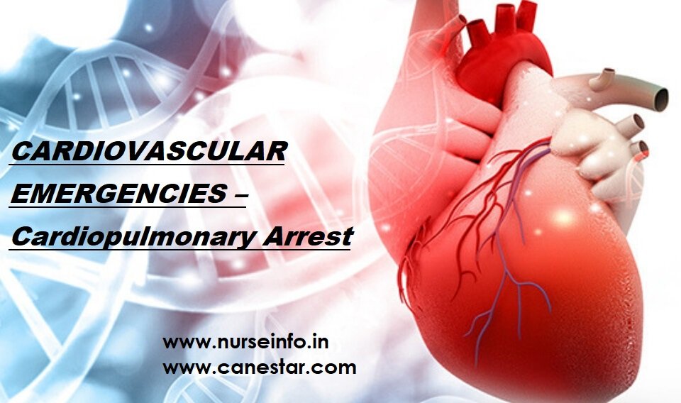
CARDIOVASCULAR EMERGENCIES – Cardiopulmonary Arrest (Types, Diagnosis and Treatment)

Cardiovascular emergencies are life-threatening disorders that must be diagnosed quickly to avoid delay in treatment and to minimize morbidity and mortality.

Cardiac emergencies includes

  • Cardiopulmonary arrest
  • Hypertensive emergency
  • Aortic aneurysm (ruptured)
  • Aortic dissection
  • Air embolism (arterial)
  • Cardiac tamponade
  • Cardiac arrhythmia
  • Ventricular fibrillation
  • Myocardial infarction (heart attack)

CARDIOPULMONARY ARREST

Cardiac arrest, also known as cardiopulmonary arrest or circulatory arrest, is the cessation of functional circulation of the blood due to failure of the heart to contract effectively.

A cardiac arrest is different from (but may be caused by) a heart attack, where blood flow to the muscle of the heart is impaired. It is different from congestive heart failure, where circulation is substandard, but the heart is still pumping sufficient blood to sustain life.

Cardiac arrest is a medical emergency that, in certain situations, is potentially reversible if treated early. Unexpected cardiac arrest can lead to death within minutes: this is called sudden cardiac arrest (SCD).

CLASSIFICATION OF CARDIOPULMONARY ARREST

Cardiac arrest is classified into two types:

  1. Shockable
  2. Nonshockable

SHOCKABLE

  • In this type the rhythm is present but abnormal
  • The two “shockable” rhythm are ventricular fibrillation and pulseless ventricular tachycardia
  • Ventricular fibrillation: It is a condition in which there is uncoordinated contraction of the cardiac muscle of the ventricles in the heart, making them quiver rather than contract properly.
  • Ventricular fibrillation is a medical emergency that requires prompt Advanced Life Support Interventions. If this arrhythmia continues for more than a few seconds, it will likely degenerate further into asystole (‘flatline’). This condition results in cardiogenic shock and cessation of effective blood circulation. As a consequence, sudden cardiac death (SCD) will result in a matter of minutes. If the patient is not revived after a sufficient period (within roughly 5 minutes at room temperature), the patient could sustain irreversible brain damage and possibly become brain dead due to the effects of cerebral hypoxia. On the other hand,  death often occurs if sinus rhythm is not restored within 90 seconds of the onset of VF, especially if it has degenerated further into asystole.
  • Ventricular tachycardia: it is a type of tachycardia, or a rapid heartbeat that starts in the bottom chambers of the heart called ventricles. The ventricles are the main pumping chambers of the heart. This is a potentially life-threatening arrhythmia because it may lead to ventricular fibrillation, asystole, and sudden death.

NONSHOCKABLE

  • When a patient’s ECG shows flatlines it is called an asystole. It is not a shockable rhythm because there is no signal left to reset. This means the electrical impulse is gone and cannot be recovered.
  • ‘Nonshockable’ rhythms are asystole and pulseless electrical activity
  • Asystole: a life threatening cardiac condition characterized by the absence of electrical and mechanical activity in the heart. Clinical signs include apnea and lack of pulse. Without cardiac monitoring, asystole cannot be distinguished from ventricular fibrillation
  • Pulseless electrical activity or PEA refers to a cardiac arrest situation in which a heart rhythm is observed on the electrocardiogram that should be producing a pulse, but is not.

DIAGNOSIS AND TREATMENT

The American Heart Association, in collaboration with the International Liaison Committee on Resuscitation, has established guidelines for resuscitation of cardiac arrest patients. In each resuscitation scenario, four concepts should always apply:

  1. Activate EMS or the designated code team
  2. Perform basic life support (CPR)
  3. Evaluate heart rhythm and perform early defibrillation as indicated
  4. Deliver advanced life support (e.g. intubation, intravenous (IV) access, and transfer to a medical center or intensive care unit).
CARDIOVASCULAR EMERGENCIES – Cardiopulmonary Arrest (Types, Diagnosis and Treatment)

CARDIOVASCULAR EMERGENCIES – Cardiopulmonary Arrest (Types, Diagnosis and Treatment)

AMLODIPINE – NORVASC

AMLODIPINE – NORVASC

Classification, uses, dosages, common side effects, interaction, generic and brand name

CLASSIFICATION: Calcium Channel Blockers

USES:

It is used for treating high blood pressure (hypertension) and coronary heart disease. Not recommended for heart failure without doctor’s guidance.

It can be taken or given orally.

DOSAGES:

High blood pressure:

Adults (18 years to 59 years) – starting with 5 mg once daily, upon doctor’s advice maximum dose can go upto 10 mg daily

Senior people more than 60 years age: 2.5 mg daily once

According to the patient’s blood pressure value dosage may vary.

Vasospastic Angina:

Adults (18 years to 59 years):  5 mg to 10 mg daily once

Senior people more than 60 years age: 5 mg daily once

Chronic Artery Disease:

Adults (18 years to 59 years):  5 mg to 10 mg daily once

Senior people more than 60 years age: 5 mg daily once

Top of Form

NOTE: Dosages can be given or taken as per physician’s instructions and guidance that depends upon the patient’s health condition

COMMON SIDE EFFECTS

Mild side effects are

  • Headache
  • Fatigue
  • Nausea
  • Abdominal pain
  • Drowsiness

Serious Side effects (rarely occurs) are

  • Edema
  • Dizziness
  • Flushing

INTERACTIONS:

Drugs(to name a few) that may interact with amlodipine include:

Check with your doctor for better understanding about the interactions with these drugs before taking them along with amlodipine

  • Diltiazem
  • Ritonavir
  • Itraconazole
  • Ketoconazole
  • Cyclosporine
  • Simvastatin

GENERIC NAME: amlodipine

BRAND NAME: Norvasc, katerzia, tekamlo, amturnide, caduet, lotrel, amlobenz, consensi, tribenzor, azor, olmesartan, prestalia, twynsta, exforge, valsartan

AMLODIPINE - NORVASC Classification, uses, dosages, common side effects, interaction, generic and brand name
AMLODIPINE – NORVASC Classification, uses, dosages, common side effects, interaction, generic and brand name

AMOXICILLIN

AMOXICILLIN

Classification, uses, dosages, common side effects, interaction, generic and brand name

CLASSIFICATION: Beta-lactam (β-lactam) antibiotic , penicillin antibiotic

USES:

It is used for treating bacterial infections that comprises middle ear infection, pneumonia, Urinary tract infections, skin infections, salmonella infections, Chlamydia infections, streptococcal infections, lyme disease, acute otitis media. 

It can be taken or given orally.

DOSAGES:

For Ears, Nose, and Throat Infections

Immediate release and chewable tablet dosage:

Adult (18 years to 64 years)

Typical dosage: 250 mg every 8 hours or 500 mg every 12 hours.

Pediatric (3 months to 17 years) (Doctor’s advice on dosages is must)

If kid weighs less than 40kgs then the typical dosage: 25 mg/kg/day in divided doses every 12 hours, or 20 mg/kg/day in divided doses every 8 hours.

If kid weighs more than 40kgs can be dosed adult dosage.

Before providing any medicine to children doctor recommendation is must

Extended Release tablets for adult dosage

18 years to 64 years

Typical dosage is 775 mg once per day for 10 days.

Seniors aged more than 65 years and pediatric dosage for extended release tablets can be discussed with your doctor.

Urinary tract infection, Skin Infection Dosage

Adult – 18 years to 64 years

Typical dosage: 250 mg every 8 hours or 500 mg every 12 hours.

Lower Respiratory tract Infection

Adult – 18 years to 64 years

Typical dosage: 500 mg every 8 hours or 875 mg every 12 hours.

Top of Form

NOTE: Dosages can be given or taken as per physician’s instructions and guidance that depends upon the patient’s health condition

COMMON SIDE EFFECTS

Mild side effects are

  • Abnormal taste sense
  • Headache
  • Nausea
  • Skin  rash
  • Diarrhea
  • Vaginal yeast infection
  • Stomach upset
  • Vomiting

Serious Side effects (rarely occurs) are

  • Dizziness
  • Seizures
  • Breathing problems
  • Hives
  • Trouble passing urine
  • Dark urine
  • Jaundice
  • Tiredness
  • Blistering
  • Liver damage

INTERACTIONS:

Drugs(to name a few) that may interact with amoxicillin include:

Check with your doctor for better understanding about the interactions with these drugs before taking them along with amoxicillin

  • Chloramphenicol
  • Erythromycin
  • Azithromycin
  • Sulfamethoxazole
  • Oral contraceptives
  • Doxycycline
  • Tetracycline
  • Allupurinol
  • Probenecid

GENERIC NAME: amoxicillin

BRAND NAME: amoxil, trimox, moxatag, sumox, moxillin, larotid, trumox

AMOXICILLIN - Classification, uses, dosages, common side effects, interaction, generic and brand name
AMOXICILLIN – Classification, uses, dosages, common side effects, interaction, generic and brand name

INSULIN (Regular) or HUMAN INSULIN

INSULIN  (Regular) or HUMAN INSULIN

Classification, uses, dosages, common side effects, interaction, generic and brand name

CLASSIFICATION: Protein 

USES:

It is used for treating type-1 diabetes, type-2 diabetes, gestational diabetes, diabetic ketoacidosis, hyperosmolar hyperglycemic states.  High blood potassium levels are also treated using insulin along with glucose.

It can be taken or given through injection under skin. In some cases some forms of insulin are used by injection into a vein or muscle.

DOSAGES:

Dosage calculation formula:

Insulin Dosage Calculation Formula


  • TR = target rate (measured as mg/dL or mmoI/L)
  • CF = corrective factor
  • KF = carbohydrate factor
  • Blood glucose (measured as mg/dL or mmoI/L)

TR, CF, KF should be set by a physician or clinical pharmacist.

Regular human insulin is available in two forms or concentrations:

  • 100 units of insulin per mL (U-100)
  • 500 units of insulin per mL (U-500)

All dosages and units has to be given as per doctor’s advice.

Top of Form

NOTE: Dosages can be given or taken as per physician’s instructions and guidance that depends upon the patient’s health condition

COMMON SIDE EFFECTS

Mild side effects are

  • Arms and legs swelling
  • Dizziness
  • Fast heart rate
  • Weight gain
  • Blurred vision
  • Anxiety
  • Mood swings
  • Hunger
  • Redness
  • Itching

Serious Side effects (rarely occurs) are

  • Hypoglycemia
  • Delirium
  • Fatigue
  • Lack of coordination
  • Impaired vision
  • Seizures
  • Loss of consciousness
  • Numbness in tongue or lips

INTERACTIONS:

Drugs(to name a few) that may interact with insulin (regular) include:

Check with your doctor for better understanding about the interactions with these drugs before taking them along with insulin (regular)

  • Rosiglitazone
  • Thiazolidinediones
  • Pramlintide
  • MAOIs
  • Fluoxetine
  • Lisinopril
  • Losartan
  • Valsartan
  • Metoprolol
  • Diuretics
  • Propranolol
  • Niacin
  • Disopyramide
  • Aspirin
  • Pentoxifylline
  • Corticosteroidsclozapine
  • Isoniazid
  • Pentamidine
  • Glucagon
  • Somatropin
  • reserpine
  • Hormones – estrogens, progesterone
  • Ritonavir

GENERIC NAME: insulin/NPH insulin  

BRAND NAME: Humulin R, Novolin R, Humulin R U-500 (Concentrated), Myxredlin, human mixtard, human mixact, human mixtard 50

INSULIN  (Regular) or HUMAN INSULIN Classification, uses, dosages, common side effects, interaction, generic and brand name
INSULIN (Regular) or HUMAN INSULIN Classification, uses, dosages, common side effects, interaction, generic and brand name

METFORMIN

METFORMINClassification, uses, dosages, common side effects, interaction, generic and brand name

CLASSIFICATION: biguanide 

USES:

It is used for treating type-2 diabetes and PCOS (polycystic ovary syndrome)

It can be taken or given orally.

DOSAGES:

Adult dosage (18 years and above)

Starting dosage: 500 mg, twice per day, or 850 mg, once per day as per doctor’s advice. Take your doses with meals. Dosage may vary per person according to their medical needs. Dosage can be taken or given as per physician’s advice.

Pediatric dosage: as per physician’s guidance. Normally kids above 10 years age 500 mg can be given daily after meal, but has to be discussed with the doctor according to their medical condition. Metformin is not advisable for kids below 10 years has there is no study or research on this.

Senior citizen (80 years and above): People aged 80 years and above should not take metformin unless they have normal kidney function. People at these ages have a higher risk of lactic acidosis.

Top of Form

NOTE: Dosages can be given or taken as per physician’s instructions and guidance that depends upon the patient’s health condition

COMMON SIDE EFFECTS

Mild side effects are

  • Diarrhea
  • Stomach pain
  • Heart burn
  • Gastric
  • Nausea

Serious Side effects (rarely occurs) are

  • Lactic acidosis
  • Low blood sugar level

INTERACTIONS:

Drugs(to name a few) that may interact with metformin include:

Most drugs results to low blood sugar levels if taken along with metformin, some drugs will react slowly to lower the blood glucose level. Check with your doctor for better understanding about the interactions with these drugs before taking them along with metformin

  • Insulin
  • Levothyroxine
  • Liotrix
  • Liothyronine
  • Furosemide
  • Nifedipine
  •  Nicotinic acid
  • Methazolamide
  • Phenytoin
  • Cimetidine
  • Fluphenazine
  • Chlorpromazine
  • Prochlorperazine
  • Birth control pills
  • Estradiol
  • Budesonide
  • Betamethasone
  • Prednisone
  • Fluticasone
  • Isoniazid
  • Brinzolamide
  • Acetazolamide
  • Dorzolamide
  • Topiramate

GENERIC NAME: metformin

BRAND NAME: Fortamet, Glucophage, Glucophage XR, Glumetza, Riomet, ava metformin, Dm2, metformin hydrochloride, glumet tab, gluformin XL, metanorm

 METFORMIN  - Classification, uses, dosages, common side effects, interaction, generic and brand name
METFORMIN – Classification, uses, dosages, common side effects, interaction, generic and brand name
Nurse Info