
NEUROLOGICAL DISORDERS – BELL’S PALSY
BELL’S PALSY – Etiology, Signs and Symptoms and Management (medical, surgical and nursing)
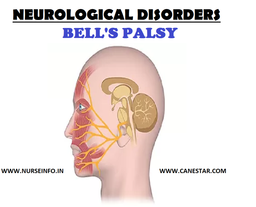
Bell’s palsy is a paralysis or weakness of the muscles on one side of face. Damage to the facial nerve that controls muscles on one side of the face causes that side of face to droop. The nerve damage may also affect sense of taste. The weakness usually affects one side of the face. Rarely, both sides are affected.
ETIOLOGY
It is thought that inflammation develops around the facial nerve as its passes through the skull from the brain. The inflammation may squash (compress) the nerve as it passes through the skull. The nerve then partly, or fully, stops working until the inflammation goes. If the nerve stops working, the muscles that the nerve supplies also stop working.
- Cold sore (herpes simplex) virus
- Chickenpox (varicella-zoster) virus
SIGNS AND SYMPTOMS
- Weakness of the face which is usually one-sided. The weakness normally develops quickly.
Face may droop to one side
Food may get trapped between gum and cheek. Drinks and saliva may escape from the side of mouth.
Difficult to close the eye, this may causes a watery or dry eye.
Difficult able to wrinkle forehead, whistle or blow out check
Difficulty with speech
- Painless or cause just a mild ache
- Loud sounds maybe uncomfortable and normal noises may sound louder than usual. This is because a tiny muscle in the ear may stop working
- Lose of sense of taste on the side of the tongue that is affected.
MANAGEMENT
- Anti-inflammatory drugs
The steroid tablet most commonly used is called prednisolone. Steroids help to reduce inflammation, which is probably the reason they help.
- Antiviral drugs
- Eye protection
- An eye pad or goggles to protect the eye.
- Eye drops to lubricate the eye during the day
- Eye ointment to lubricate the eye overnight
- Tape the upper and lower lid together when you are asleep. Other procedures are sometimes done to keep the eye shut until the eyelids recover
- Physiotherapy; a treatment called, ‘facial retraining’ with facial exercises may help
- Injections of botulism toxin (Botox) may help it spasm develops in the facial muscles
- Various surgical techniques can help with the cosmetic appearance
Complications
- Corneal ulcers
- Blindness
- Impaired nutrition
Medical Management
The objectives of management are to maintain facial tone and to prevent or minimize complication with the help of the following:
- Corticosteroid therapy (prednisone) maybe initiated to reduce inflammation and edema, which reduces vascular compression and permits restoration of blood circulation to the nerve.
- Early administration of corticosteroids appears to diminish severity, relieve pain, and minimize denervation.
- Facial pain is controlled with analgesic agents or heat applied to the face to prevent muscle atrophy, or surgical exploration of the facial nerve.
- Surgery may be performed if a tumor is suspected, for surgical decompression of the facial nerve, and for surgical rehabilitation of a paralyzed face.
Surgical Management
- Facial Nerve Decompression
The surgeon decides if the maxillary segment should be decompressed externally or if the labyrinthine segment and geniculate ganglion should be decompressed with a middle fossa craniotomy.
- Subocularis Oculi Fat Lift (SOOF)
The SOOF is deep to the orbicularis oculi muscle and superficial to the periosteum below the inferior orbital rim. An SOOF lift is designed to lift and suspend the midfacial musculature. The procedure may also elevate the upper lip and the angle of the mouth to improve facial symmetry.
- Lateral Tarsal Strip Procedure
An SOOF lift is commonly performed in conjunction with a lateral tarsal strip procedure to correct horizontal lower-lid laxity and to improve apposition of the lid to the globe. first, lateral canthotomy and cantholysis is performed. Then, the anterior lamella is removed and the lateral tarsal strip is shortened and attached to the periosteum at the lateral orbital rim.
- Implants in Eyelid
Implantable devices have been used to restore dynamic lid closure in cases of severe, symptomatic lagophthalmos. These procedures are best for patients with poor Bell phenomenon and decreased corneal sensation. Gold or platinum weights, a weight-adjustable magnet, or palpebral springs can be inserted into the eyelids. Pretarsal gold-weight implantation is most commonly performed. The implants are easily removed if nerve function returns.
- Tarsorrhaphy
Tarsorrhaphy decreases horizontal lid opening by fusing the eyelid margins together, increasing support of the precorneal lake of tears and improving coverage of the eye during sleep. The procedure can be done in the office and is particularly suitable for patients who are unable or unwilling to undergo other surgery. It can be completed as either a temporary or a permanent measure.
Permanent tarsorrhaphy is performed if nerve recovery is not expected. Tarsorrhaphy can be performed laterally, centrally, or medially. The lateral procedure is the most common; however, it can restrict the monocular temporal visual field. Central tarsorrhaphy offers good corneal protection, but it occludes vision and can be cosmetically unacceptable. Medial or paracentral tarsorrhaphy is performed lateral to the lacrimal puncta and can offer good lid closure without substantially affecting the visual field.
Other surgeries
Muscle Transportation, Nerve Grafting, and Brow Lift.
- Transposition of temporalis
Transposition of the temporalis muscle can be used to reanimate the face and to provide lid closure by using the fifth cranial nerve. Strips from the muscle and fascia are placed in the upper and lower lids as an encircling sling. Patients initiate movement by chewing or clenching their teeth.
- Facial nerve grafting or hypoglossal-facial nerve anastomosis
Reinnervation of the facial nerve by means of facial nerve grafting or hypoglossal-facial nerve anastomosis can be used in cases of clinically significant permanent paralysis to help restore relatively normal function to the orbicularis oculi muscle or eyelids.
- Direct brow lift
Brow ptosis is repaired with a direct brow lift. Care should be taken in the presence of corneal decompression because lifting the brow can cause worsening of lagophthalmos, especially if lid closure is poor. A gold-weight implant can be placed or lower-lid resuspension can be performed simultaneously to prevent this complication.
Nursing Management
Teaching patients with Bell’s palsy to care for them at home is an important nursing priority.
Teaching Eye care
Because the eye usually does not close completely, the blink reflex is diminished, so the eye is vulnerable to injury from dust and foreign particles. Corneal irritation and ulceration may occur. Distortion of the lower lid alters the proper drainage of tears. Encourage the client for the following:
- Cover the eye with a protective shield at night
- Apply eye ointment to keep eyelids closed during sleep
- Close the paralyzed eyelid manually before going to sleep
- Wear wraparound sunglasses or goggles to decrease normal evaporation from the eye.
Teaching about Maintaining Muscle Tone
- Show patient how to perform facial massage with gentle
- Upward motion several times daily when the patient can tolerate the massage
- Demonstrate facial exercises, such as wrinkling the forehead
- Blowing out the cheeks, and whistling, in an effort to prevent muscle atrophy
- Instruct patient to avoid exposing the face to cold and drafts
Diet and Nutrition
- Instruct patient to chew on the unaffected side of his mouth
- Provide soft and nutritionally balanced diet. Eliminate hot fluids and foods
- Give frequent mouth care, being particularly careful to remove residues of food that collects between the cheeks and gums.
BELL’S PALSY – Etiology, Signs and Symptoms and Management (medical, surgical and nursing)
