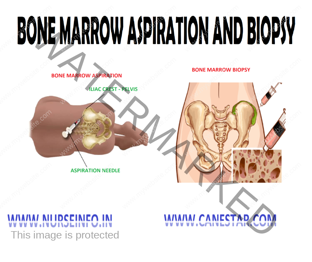
CERVICAL BIOPSY – Indications, Types of Cervical Biopsy, General Instructions, Preliminary Assessment, Preparation of the Patient and Environment, Equipment, Procedure and Post-Procedure Care
A cervical biopsy is a surgical procedure in which a small amount of tissue is removed from the cervix. The cervix is the lower, narrow end of the uterus located at the end of the vagina.
INDICATIONS
- Pap smear, Pap test yielded positive results
- The cervical ectropion is diagnosed (bilateral cervical gap develops after giving birth, especially as a result of multiple micro-breaks)
- The mucosal pathology of the cervix is detected (erosion, polyps, hypertrophy, suspected oncological-cervical tumor)
- Cervical dysplasia stage of II-IV (should be diagnosed and treated in time, it will prevent the development of cancer cells)
- There are serious gaps after childbirth; the cervix is deformed, visible severe scarring on the uterine tissue
TYPES OF CERVICAL BIOPSY
Punch biopsy: one or more small pieces of tissues are removed from the cervix with a punch biopsy forceps
Cervical conization: it is done by taking a cone-shaped section of the cervix with a scalpel or cervitone or by diathermy conization
GENERAL INSTRUCTIONS
- These procedures are frequently performed on an outpatient basis
- The biopsy is usually taken one week after the end of menstruation when the cervix is least vascular
- The patient usually experiences no pain during the cervical biopsy because the cervix does not contain nerve endings for pain
PRELIMINARY ASSESSMENT
- Doctor’s order for any specific instructions
- Written informed consent of the patients or the relatives
- General condition and diagnosis of the patient
- Mental status of the patient to follow instructions
- Articles available in the unit
PREPARATION OF THE PATIENT AND ENVIRONMENT
- The patient is prepared as for routine gynecologic examination
- Shave and clean the perineum
- Explain the procedure to the patient
- Obtain written consent from the patient
- Maintain privacy with screen
- Give lithotomic position to the patient
- There should be good light in the room
- Remain with the patient through the procedure
EQUIPMENT
A sterile tray containing:
- Sponge holder
- Vulsellum
- Biopsy forceps
- Sims vaginal speculum
- Gali pot for lotion
- Gloves, mask and gown
- Leggings
- Specimen bottles with formalin
- Dressing material
An unsterile tray containing:
- Mackintosh and draw sheet
- Kidney tray
- Cautery with its tips sterilized
- Antiseptic for cleaning
PROCEDURE
- Usually, it is done in outpatient department
- The cervix is visualized in a good light and biopsy is taken with a cervical biopsy forceps
- The bleeding from the site is controlled by cauterization
- Patient may have foul smelling discharge for few days
- The patient may be discharged on the same day
POST-PROCEDURE CARE
- To avoid any strenuous activity for the next 24 hours
- To report any bleeding immediately
- To abstain from sexual activities and douching until the doctor gives the permission
- To avoid using tampons until the doctor gives permission. Use clean pads