HEMODIALYSIS – Definition, Indications, Equipment, Preparation of Double Lumen Catheter for Dialysis, Preparation for AV Shunts for Diathesis, Equipment Needed after the Procedure, For AV Fistula, For AV Shunt, Preparation of Equipment, Sites for Hemodialysis, Mechanism of hemodialysis, Types of Dailyzers, Systems Used in Delivering Dialysate, Nursing Care, Role of Nurse in Care of Patient on Hemodialysis with Double Lumen catheter, Post-procedure Care, Nursing Alert and Complications (NURSING PROCEDURE)
DEFINITION
It is defined as the lifesaving procedure in which the toxic waste, removed from the blood through purifying dialyzer and the purified blood return back to the body through arteriovenous fistula
INDICATIONS
- Patients with acute reversible renal failure
- For regular long-term treatment of patient with chronic end-stage renal disease
- Acute poisoning, such as barbiturate or analgesic overdose
EQUIPMENT
Preparation of hemodialysis Machine
- Hemodialysis with appropriate dialyzer
- IV solution, administration set, lines, IV pole
- Dialysate
- Injection heparin, 3 ml syringe with needle, medication label, and hemostats
PREPARATION OF DOUBLE LUMEN CATHETER FOR DIALYSIS
- Povidone-iodine sponges
- Two sterile 4” multiply 4” gauze pads
- Two 3 ml and two 5 ml syringes
- Tape
- Injection heparin bolus syringe
- Clean gloves
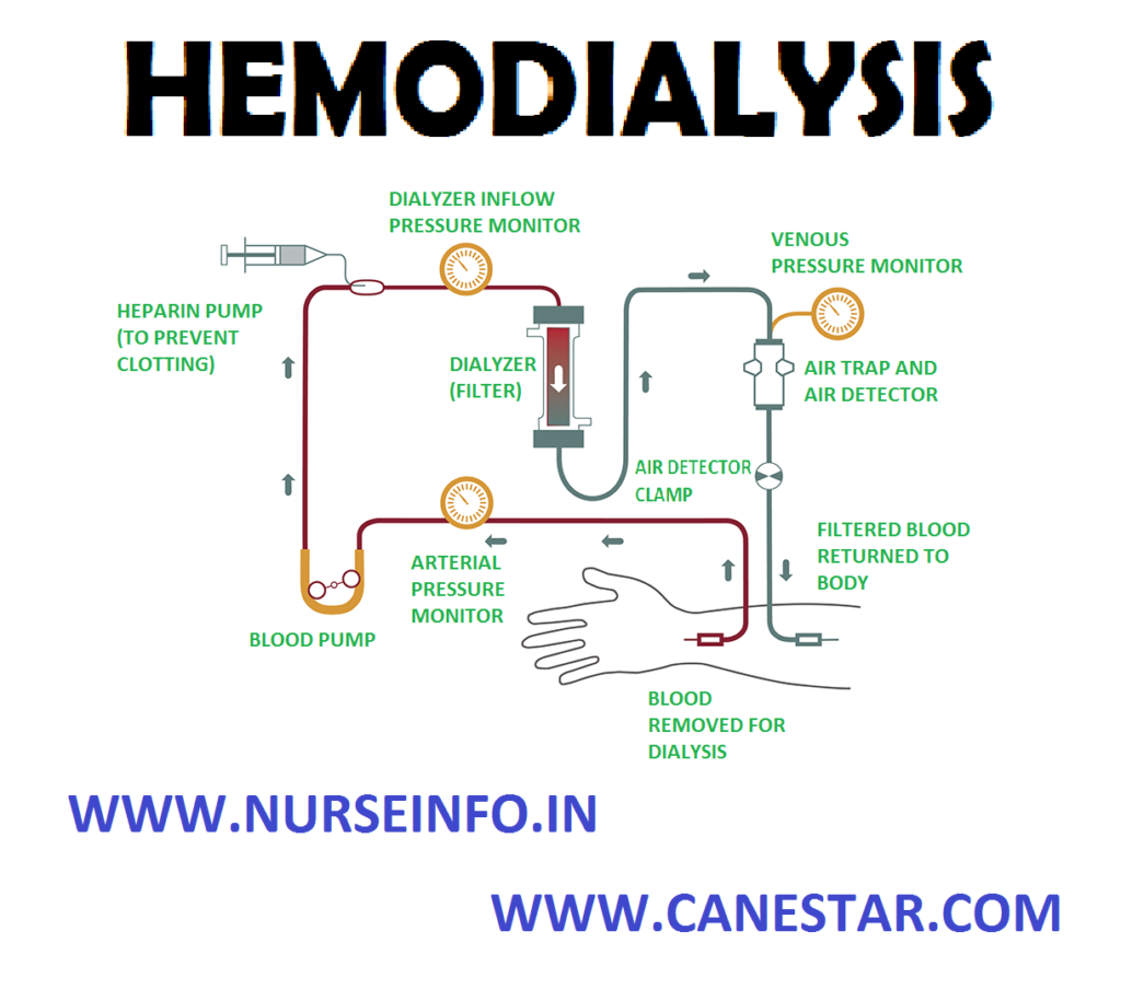
The hemodialysis blood circuit: a dialysis machine pumps blood from the patient, through disposable tubing, through a dialyser, or artificial kidney, and back into the patient. Waste solute, salt and excess fluid is removed from the blood as it passes through the dialyser
PREPARATION FOR AV FISTULA FOR DIALYSIS
- Two winged fistula needles (each attached to 10 ml syringe filled with heparin flush solution)
- Linen-saver pad
- Povidone-iodine sponges
- Sterile 4” multiply 4” gauze pads
- Tourniquet
- Clean gloves
- Adhesive tapes
PREPARATION FOR AV SHUNTS FOR DIATHESIS
- Sterile 4” multiply 4” gauze pad
- Povidone-iodine sponge
- Alcohol sponge
- Sterile gloves
- Two sterile shunt adapters
- Sterile Teflon connectors
- Two bull dog clamps
- Two 10 ml syringes
- Normal saline solution
- Four short strips of adhesive tapes
- Sterile shunt spreader
EQUIPMENT NEEDED AFTER THE PROCEDURE
For Double Lumen Catheter
- Sterile 4” multiply 4” gauze pad
- Povidone-iodine sponges
- Precut gauze dressing
- Clean sterile gloves
- Normal saline solution
- Alcohol sponge
- Heparin flush solution
- Leur-lock injection caps
- Transparent occlusive dressing, tape, skin barrier preparation, materials for culturing drainage
FOR AV FISTULA
- Clean gloves
- Sterile 4” multiply 4” gauze pads
- Two adhesive bandages
- Hemostat
- Sterile absorbable gelatin sponge
FOR AV SHUNT
- Sterile gloves
- Two bull dog clamp
- Two hemostat
- Povidone-iodine solution
- Sterile 4” multiply 4” gauze pads
- Alcohol sponges
- Elastic gauze bandage
PREPARATION OF EQUIPMENT
- Maintain strict aseptic technique to prevent the introduction of infection into the bloodstream
- Test the dialyzer and the dialysis machine for residual disinfectant after rinsing
- Test all the alarms
SITES FOR HEMODIALYSIS
- Subclavian vein catheterization: using the Seldinger technique surgeon introduces the needle into subclavian vein then inserts a guidewire through the introducer needle and then removes the needle
- Using the guide wire thread 5” – 12” (12-30 cm) plastic or Teflon catheter (with a Y hub) into the patient’s vein
- Femoral vein catheterization: using the Seldinger technique surgeon introduces the needle into the left or right femoral vein. Then insert a guide wire through introducer needle and remove the needle. Using the guide wire then thread a 5” -12” plastic or Teflon catheter with a Y tub or two catheters one for inflow and another place about ½” (1.3 cm) distal to the first for outflow
- Arteriovenous fistula: to create fistulas make an incision into the patient’s wrist or lower forearm then a small incision into the side of the artery and another side of a vein, then suture the edges of the incisions together to make a common opening 3-7 mm long
- Arteriovenous shunt: to create a shunt makes a incision in the patient’s wrist, lower forearm, or an ankle. Then insert 6” -10” (15-25 cm) transparent silastic cannula into an artery and another into a vein. Finally, tunnel the cannulas out through a stab wound and join them with a piece of a Teflon tubing
- Arteriovenous graft: to create a graft makes an insertion in the patient’s forearm, upper arm or thigh. Then tunnel a natural or synthetic graft under the skin and suture the distal end to an artery and proximal end to a vein
MECHANISM OF HEMODIALYSIS
- In hemodialysis the blood flows from the patient to an external dialyzer (artificial kidney) through an arterial assess site
- Inside the dialyzer the blood and the dialysate flow counter currently divided by a semipermeable membrane, the composition of the dialysate resembles normal extracellular fluid
- Blood contains excess of specific solutes and dialysate contains electrolytes that may be at abnormal level in the patient’s blood stream
- The dialysate electrolyte composition can be raised or lowered depending on the need
- Excretory function an electrolyte hemostasis area achieved by diffusion, the movement of molecule across the dialyzer’s semipermeable membrane from an area of higher solute concentration to an area of lower concentration
- Water (solvent) crosses the membrane from the blood into a dialysate by ultrafiltration. This process removes excess water, waste products and other metabolites through osmotic pressure and hydrostatic pressure
- Osmotic pressure is the movement of water across the semi permeable membrane from an area of lesser solute concentration to greater solute concentration
- Hydrostatic pressure focus the water from the blood compartment into the dialysate compartment, cleaned from impurities and in excess water the purified blood returns to the body through a venous site
TYPES OF DIALYZERS
There are three types of dialyzers which are as follows:
- Hollow fiber dialyzer: this is most common type contains fine capillaries with semipermeable membrane enclosed in a plastic cylinder. Blood flows through these capillaries as the system pumps dialysate in an opposite direction on the outside of the capillaries
- Flat-plate or parallel flow plate dialyzer: it has two or more layer of semipermeable membrane bound by a semi rigid or rigid structure. Blood ports are located at both ends between the membranes, and dialysate flows in opposite direction along the outside of the membranes
- Coil dialyzer: it consists of one or more semipermeable membrane tubes supported by a mesh and wrapped concentrically around a central core. Blood passes through a coil as the dialysate circulates at a high speed around the coil and the mesh work. Heparin is used to prevent clot formation during dialysis
SYSTEMS USED IN DELIVERING DIALYSATE
Three system types: there are three system types used to deliver the dialysate.
- Batch system: it uses reservoir for recirculating dialysate
- The regenerative system: it uses sorbets to purify and regenerate recirculating dialysate
- The proportioning system: it mixes the concentrate with water to form a dialysate which then circulates through the dialyzer and goes down a drain after a single pass followed by a fresh dialysate
NURSING CARE
- Weigh the patient
- Record the vital signs and blood pressure in sitting and standing position
- Auscultate heart for rate rhythm and abnormalities
- Observe respiratory rate rhythm and quality
- Assess the edema
- Check the mental status and condition of patency in the access site
- Check the last date of dialysis and evaluate the previous lab data
- Place the patient in a comfortable position supine or sitting in a recliner chair with feet elevated. Make sure the site is well supported and resting on a clean drape
- Explain the procedure to the patient if the patient is undergoing the hemodialysis for the first time
- Use standard precaution in all cases to prevent the transmission of infection
- Wash the hand before and after the procedure
ROLE OF NURSE IN CARE OF PATIENT ON HEMODIALYSIS WITH DOUBLE LUMEN CATHETER
- Wash the hands
- Prepare venous access
- Clamp the tubing to prevent the air entry into the catheter
- Clean each catheter extension tube clamp and luer-lock injection cap with povidone-iodine sponge to remove the contaminants
- Place a sterile 4” multiply 4” gauze pad under the extension tubing and place two 5 ml syringe and two sterile gauze pads on the drape
- Prepare the anticoagulant regimen as ordered
- Identify the arterial and venous blood lines and replace them near the drape
- Remove clothes and ensure the catheter patency, remove the catheter cap and attach syringe to each catheter port, open one clamp and aspirate 1.5-3 ml of blood
- Close the clamp and repeat the procedure with other port
- Flush each port with 5 ml of heparin flush solution
- Attach bloodline to patient access, remove the syringe from the arterial port, attach the line to arterial port, and administer heparin according to the protocol which prevents extracorporeal circuit
- Grasp venous bloodline and attach to venous port, open the clamp on extension tubing and secure the tubing to patient’s extremity with a tape to reduce the tension on the tube and minimize the trauma in the insertion site, begin the hemodialysis according to the unit protocol
POST-PROCEDURE CARE
- Wash your hands
- Clamp the extension tubing to prevent air entry
- Clean all connection point on the catheter and bloodlines as well as clamps to reduce the risk of systemic or local infections
- Place a clean drape under the catheter and to sterile 4” multiply 4” gauze pad on the drape beneath the catheter lines, soak the pad with povidone-iodine solution and then prepare the catheter flush solution with normal saline or heparin flush solution as ordered
- Put clean gloves. Grasp each bloodlines with a gauze pad and disconnect each line from the catheter
- Flush each port with a saline solution to clean the extension tubing and the catheter of blood, administer additional heparin flush solution as ordered to ensure catheter patency then attach a luer-lock cap to prevent entry of air or loss of blood
- Clamp the extension tubing
- Hemodialysis is complete redress the catheter in the insertion site also redresses if occluded or soiled
- Position the patient in supine with his face turned away from the insertion site
- Wash your hand and remove the outer occlusive dressing, put on the sterile gloves. Remove the old inner dressing and discard the gloves and the inner dressing
- Set up a sterile field and absorb the site for drainage
- Obtain a drainage sample for culture
- Notify the doctor if the suture is missing
- Put on a sterile glove and clean the insertion site with a alcohol sponge and then clean the site with povidone-iodine sponge and allow to air dry
- Put a precut gauze dressing over the insertion site and under the catheter and place another gauze dressing over the catheter
- Apply a skin barrier preparation to the skin surrounding the gauze dressing, cover the gauze and catheter with a transparent occlusive dressing
- Apply 4 or 5 piece of two inch tape over the cut edge of the dressing to reinforce the over edge
ROLE OF NURSE IN CARE OF PATIENT ON HEMODIALYSIS WITH AN ALL FISTULAS
- Flush the fistula needles using attached syringe containing heparin flush solution
- Place a Mackintosh under the patient’s arm
- Using aseptic technique clean the area of the skin over the fistula with povidone-iodine sponge (3” multiply 10”)
- Discard each pad after one wipe
- Apply the tourniquet above the fistula to distant the vein and facilitate the venous puncture
- Put on clean gloves; perform a venous puncture with a fistula needle
- Remove the needle guard and squeeze the wing tip firmly together
- Insert arterial needle at least one inch above the anastomosis, be careful not to puncture the fistula
- Release the tourniquet and flush the needle with heparin
- Clamp the arterial needle with hemostat and secure the wing tip of the needle with adhesive tape
- Allow the tubing to air dry
- Put on the sterile gloves
- Clamp the arterial side of the shunt with building clamp and also the venous site, when the shunt is open
- Open the shunt by separating its side with your finger
- Both side of the shunt should be exposed
- Adapt the shunt to the lines of the machine. attach a shunt adapter and 10 ml syringe filled with 8 ml of normal saline to the side of the shunt containing Teflon connector
- Attach a new Teflon connector to another side of the shunt with second adapter; attach it also with 10 ml syringe filled with 8 ml of normal saline to the same side
- Next flush the shunt arterial tubing by releasing the clamp and aspirate the saline solution, and then flush the tubes slowly repeat the procedure on the venous site
- Secure the shunt to the adapter with the adhesive tape
- Connect the arterial and venous line to the adapter
POST-PROCEDURE FOR AV
- Wash the hands
- Turn the blood pump on the hemodialysis machine to 50-100 ml per minute
- Put sterile glove and remove the tape from the connection site of the arterial lines
- Clamp the arterial cannula with the bull dog clamp and then disconnect the lines
- Blood in the arterial line will continue to flow toward the dialyzer followed by a column of air, just before the blood reaches the point where the saline solution enters the line clamp the blood line with another hemostat
- Unclamp the saline solution and allow small amount to flow through the line
- Unclamp the hemostat on machine line, just before the last volume of blood enters the patient clamp the venous cannula with bull dog clamp and machines venous lines with hemostat
- Remove the tape from connection site of venous line
- Turn off the blood pump and disconnect the lines
- Reconnect the shunt cannula, remove older two Teflon connectors and discard, connect the shunt and take care that position of the Teflon connector is equal between the two cannula
- Remove the bull dog clamps
- Secure the shunt connection with hypoallergenic tape
- Clean the shunt and the site with gauze pads soaked in povidone-iodine solution
- Make sure the blood flow through the shunt adequately
- Apply the dressing to the shunt site and wrap it securely with elastic gauze bandage
- Attach the bull dog clamps to the outside dressing
- When the hemostat is complete check the vital signs and the mental status
- Compare the findings with the predialysis assessment data
- Document the finding
- Disinfect and rinse the delivery system
NURSING ALERT
- Follow aseptic technique throughout the procedure
- Report immediately any machine malfunction and keep it ready for use at any time
- Avoid unnecessary handling of shunt tubing
- Inspect the shunt for patency, check for any clots, serum, cell separation and temperature of the silastic tubing
- Check the shunt insertion site for signs of infection (purulent drainage, inflammation and tenderness)
- Check for any shunt insertion site is exposed
- Check for any bleeding after removing the AV fistula needle, if bleeding persist soak the sponge and apply thrombin solution
- Monitor patient’s vital signs carefully and blood pressure every 15 minutes
- Check the weight of the patient before and after the procedure
- Check the clotting time of patient’s blood sample and sample from the dialyzer periodically
- Ensure the patient receives light meals during procedure
COMPLICATIONS
- Hyperpyrexia
- Dialysis disequilibrium syndrome (headache, nausea, vomiting, restlessness, hypertension, muscle cramp, backache, and seizures)
- Hypovolemia and hypotension
- Hyperglycemia and hypernatremia
- Cardiac arrhythmias
- Angina
- Reduce toxins, air embolism, chest pain, dyspnea coughing and cyanosis
- Hemolysis (chest pain, dyspnea, cherry red blood, hyperkalemia)


