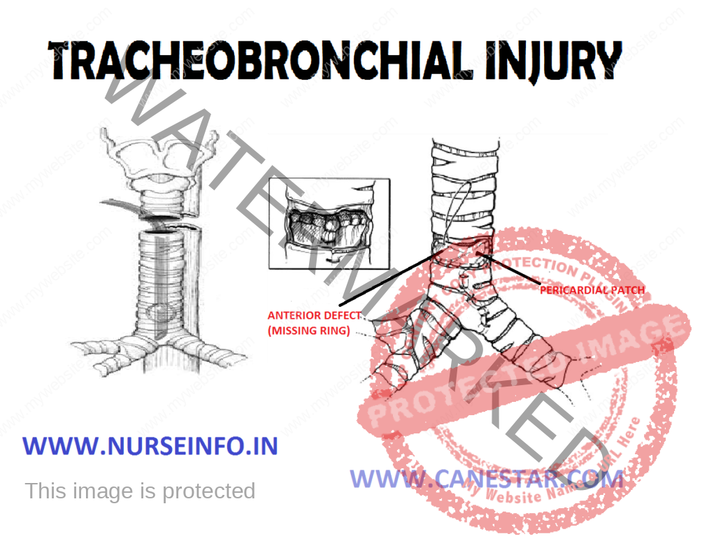
TRACHEOBRONCHIAL INJURY – Etiology, Clinical Manifestations, Diagnostic Evaluations and Management
Tracheobronchial injury (TBI) is damage to the tracheobronchial tree (the airway structure involving the trachea and bronchi). It can result from blunt or penetrating trauma to the neck or chest, inhalation of harmful fumes or smoke, or aspiration of liquids or objects.
ETIOLOGY
- Falls from height
- Motor vehicle accidents
- Explosions are another cause
- Gunshot wounds
- Knife wounds
- Edema (swelling)
CLINICAL MANIFESTATIONS
- Dyspnea
- Respiratory distress
- Coughing
- Hemoptysis
- Stridor (an abnormal, high-pitched breath sound indicating obstruction of the upper airway can also occur)
- Necrosis (death of the tissue)
- Scar formation
- Stenosis
- Due to inhalation of foreign body aspiration
- Medical procedure is uncommon
DIAGNOSTIC EVALUATION
- Chest X-ray: is the initial imaging technique used to diagnose TBI. X-rays may also show accompanying injuries and signs, such as fractures, and subcutaneous emphysema.
- CT scanning detects resulting from blunt trauma. CT are a replacement of bronchoscopy
PREVENTION
Vehicle occupants who wear seatbelts have a lower incidence of TBI after a motor vehicle accident.
TREATMENT
- Endotracheal tube: may be used to bypass a disruption in the airway
- Tracheotomy: an incision can be made in the trachea (tracheotomy)
- Mechanical ventilation: such as positive end-expiratory pressure (PEEP) and ventilation at higher-than-normal pressures may be helpful in maintaining adequate oxygenation.
COMPLICATIONS
- Bronchial stenosis
- Pneumonia
- Bronchiectasis


