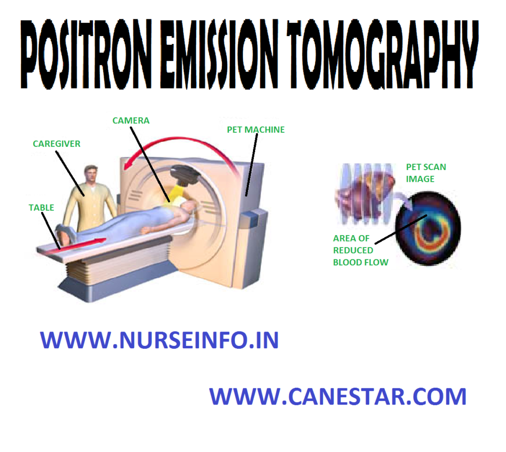
POSITRON EMISSION TOMOGRAPHY – Indications, Client Preparation and During Test (NURSING PROCEDURE)
The positron emission tomography (PET) scanner is a diagnostic imaging tool that allows visualization of regional physiologic function and biochemical changes that often separate normal from diseased myocardium. Cellular metabolic information is obtained by mapping regional myocardial glucose metabolism. Combining information from the perfusion and metabolism images provides a thorough assessment of regional cardiac viability
INDICATIONS
- Detection of coronary artery disease
- Assessment of myocardial viability
- Assessment of progression of coronary artery stenosis
- Documentation of collateral coronary artery circulation
- Differentiation of ischemia from dilated cardiomyopathy
CLIENT PREPARATION
- Ask the client if there is any history to iodine or shellfish or of allergic reaction to dye or contrast material used in a previous X-ray study
- Explain the procedure to the client and obtain written consent from the client or person legally designated to make care decisions for the client
- If contrast medium is to be used, instruct the client to discontinue all food and fluids for 4-8 hours before the test
- Instruct the client to remove all clothes, jewelry and other metal objects. A hospital gown is worn
- Provide appropriate orientation and reassurance so that fear of the unknown is diminished. Many clients feel some degree of apprehension as they enter the enclosed space of the machine. The scan itself is painless
DURING TEST
- Instruct the client to remain motionless while in the scanner and hold his or her breath when instructed to do so
- Keep an emesis basin in a nearby area in case the client vomits


