PNEUMONIA – Types, Pathophysiology, Signs and Symptoms, Diagnostic Evaluation and Management
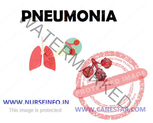
- Pneumonia is an inflammation and consolidation of the lung parenchyma. It is an inflammatory process in lung parenchyma usually associated with marked increase in interstitial and alveoli fluid
- Pneumonitis: it is a more general term that describes an inflammatory process in lung tissues that may predispose a patient to or place a patient at risk for microbial invasions (or)
- Pneumonia is an inflammatory condition of the lung, especially affecting the microscope air sacs (alveoli), associated with fever, chest symptoms, and a lack of air space (consolidation) on a chest X-ray. The inflammation may be caused by infection with viruses, bacteria, or other microorganisms, and less commonly by certain drugs and other conditions.
ETIOLOGY AND RISK FACTORS
The main cause of pneumonia is the:
- Bacteria like Streptococcus pneumoniae, Haemophilus influenza, Staphylococcus aureus
- Viruses like rhinoviruses, coronaviruses, influenza virus
- Fungal agents like Histoplasma capsulatum
- Parasites: a variety of parasites can affect the lungs. These parasites typically enter the body through the skin or the mouth. Once inside the body, they travel to the lungs, usually through the blood. The most common parasites causing pneumonia are Toxoplasma gondii, Strongyloides stercoralis and Ascariasis.
The major risk factors of pneumonia include:
- Advanced age
- A history of smoking
TYPES OF PNEUMONIA
- According to site – Segmental, Lobar and Bilateral
- According to location – broncho or bronchial, interstitial/reticular, alveolar and nacrotizing
- According to organism – Pneumococcal, staphylococcal, influenza, klebsiella, legionnaires disease, mycoplasmal, viral, fungal and parasite
- Other – aspiration
- Upper respiratory infections
- Tracheal intubation
- Prolonged immobility
- Immunosuppressive therapy
- Malnutrition
- Dehydration
- Chronic disease states, like diabetes, heart disease, chronic lung disease, renal disease and cancer
- Exposure to air pollution
- Alcoholism
Pneumonia According to Site
- Segmental pneumonia: it involves one or more segments of the lungs
- Lobar pneumonia: it involves one or more entire lobes
- Bilateral pneumonia: it involves lobes in both the lungs
Pneumonia According to Location and Radiologic Appearance
- Broncho or bronchial pneumonia: it involves terminal bronchials and alveoli
- Interstitial or reticular pneumonia: it involves inflammatory response within the lung tissue surrounding the air space or vascular structure rather than air passage
- Alveolar or acinar pneumonia: it involves fluid accumulation in a lung’s distal air space
- Nacrotizing pneumonia: it causes the death of the lung tissue surroundings by viable tissue. It does not heal and cause permanent loss of functioning parenchyma
Pneumonia According to Organism
- Pneumococcal or streptococcal pneumonia: caused by streptococcus pneumonia
- Staphylococcus pneumoniae: caused by staphylococcus aureus
- Influenza pneumonia: caused by Haemophilus influenza
- Gram-negative bacterial pneumonia: caused by klebsiella pneumoniae
- Legionnaires disease: caused by legionella pneumophilae
- Mycoplasma pneumonia: caused by mycoplasma microorganisms
- Viral pneumonia: caused by influenza A virus and B virus, adenovirus, cytomegalovirus, parainfluenza
- Fungal pneumonia: caused by aspergillus, candidiasis, blastomycosis
- Parasitic pneumonia : caused by protozoa, nematodes
Other Types of Pneumonia
- Aspiration pneumonia: due to aspiration of foreign particles, gastric content or food
- Hypostatic pneumonia: caused by constantly remaining in same position and most common in weak or aged persons
- Ventilator-associated pneumonia (VAP): it is defined as pneumonia occurring in a patient within 48 hours or more after intubation with an endotracheal tube or tracheostomy tube and which was not present before. Early onset VAP occurs within 48 hours and late onset VAP beyond 48 hours of tracheal intubation.
Other Risk Factors
- Previous stroke: people, who have had a stroke, have problems in swallowing, or are bedridden, can easily develop pneumonia
- Age: infants from birth to age too are at risk for pneumonia, as are individuals aged 65 years or older
- Weakened immune system: this includes people who take medications (steroid drugs and anti-cancer drugs) that weaken the immune system and people with HIV, AIDS, or cancer.
- Drug abuse: this includes excessive alcohol consumption and smoking
- Certain medical conditions: asthma, cystic fibrosis, diabetes, and heart failure raise risks for pneumonia
PATHOPHYSIOLOGY OF PNEUMONIA
- Inflammatory pulmonary response to the offending organism or agent —- defense mechanism of lung loses effectiveness —- allow the organism to penetrate the lower airway —- inflammation develops —- inflamed and fluid-filled alveolar sacs cannot exchange oxygen and carbon dioxide effectively —- hypoventilation —- ventilation-perfusion mismatch, alveolar exudates tend to consolidate and become difficult to expectorate
- Acute inflammation occurs that causes excess water, and plasma proteins go to the dependent areas of the lower lobes —- RBCs, fibrin, and polymorphonuclear leukocytes infiltrate the alveoli — containment of the bacteria within the segments of pulmonary lobes by cellular recruitment —- consolidation of leukocytes and fibrin within the affected area —- stage of congestion: engorgement of alveolar spaces with fluid and hemorrhagic exudates —proliferation and rapid spread of organism through the lobe —- stage of red hepatization: coagulation of exudates occurs resulting in the red appearance of the affected lung —- stage of grey hepatization: the decrease in number of RBCs in the exudates is replaced by neutrophils, which infiltrate the alveoli, making the lung tissue to the solid and grayish in color —- pneumonia
SIGNS AND SYMPTOMS
- Productive cough
- Fever accompanied by shaking chills
- Shortness of breath
- Sharp or stabbing chest pain during deep breaths
- Confusion, and an increased respiratory rate
- Malnutrition
- Hemoptysis
- Headache
- Fatigue
- Chest auscultation reveals bronchial breath sounds over the area of consolidation
- Crackling sounds
- Dull sound on percussion
- Unequal chest wall expansion may occur during inspiration
- Pneumococcal pneumonia: sudden onset with a single shaking chill, high fever, stabbing pleuritic chest pain, malaise, weakness, occasional vomiting, tachypnea, dyspnea, elevated WBC count
- Single or multiple lobar consolidations on the X-ray film: cough productive of rusty brown or blood-streaked purulent sputum that turns yellow and mucoid
- Staphylococcus pneumonia: sudden onset with fever, multiple chills, pleuritic pain, dyspnea, decreased breath sounds, elevated WBC counts, and productive cough with purulent golden yellow or blood-streaked sputum
- The chest X-ray may show patchy infiltrates, empyema, abscesses, and pneumothorax
- Influenzal pneumonia: similar to those of pneumococcal pneumonia. Cough productive of apple or lime green purulent sputum, which may be blood-tinged
- Gram-negative: sudden onset with high fever, multiple chills, pleuritic pain, dyspnea, cyanosis and elevated WBC count
- Lobar consolidation on chest X-ray and cough productive of red sputum
- Single or multiple lobe consolidation and small pleural effusion on chest X-ray film, dry cough productive of blood-tinged sputum
- Mycoplasma pneumonia: insidious onset with slowly rising fever, headache, malaise, and normal WBC count
- Viral pneumonia: headache and myalgia followed by high fever, dyspnea, normal breath sounds with occasional wheezing and crackles, elevated WBC count
- Diffused patchy infiltrates on the X-ray film
- Fungal pneumonia: usually asymptomatic
- Parasitic pneumonia: cough, dyspnea, pleuritic chest pain, fever, night sweat, crackles
- Aspiration pneumonia: asymptomatic with minor aspiration
- Major aspiration may lead to tachypnea, apnea, cyanosis, hypotension, lung sounds (crackles, rhonchi, wheezing), hypoxemia, respiratory failure, leukocytosis
DIAGNOSTIC EVALUATION
- Chest computed tomography: A CT scan is similar to an X-ray, but the pictures provided by this method are highly detailed. This painless test provides a clear and precise picture of the chest and lungs
- Sputum test: this test will examine the sputum to determine what type of pneumonia is present
- Pleural fluid test: if there is fluid apparent in the pleural space, a fluid sample can be taken to help determine if the pneumonia is bacterial or viral
- Pulse oximetry: this test measures the level of oxygen blood saturation by attaching a small sensor to finger. Pneumonia can prevent normal oxygenation of blood
- Bronchoscopy: when antibiotics fail, this method is used to view the airways inside the lungs to determine if blocked airways are contributing to pneumonia
MANAGEMENT
Antibiotics are prescribed based on Gram stain results and antibiotic guidelines. Combination therapy may also be used.
- Classifications: antibiotics (aminoglycosides: gentamicin, tobramycin, amoxicillin, erythromycin, penicillin, tetracycline)
- Indications: prevent or treat infections caused by pathogenic microorganisms
- Selected interventions:
Before administering the first dose, assess the client for allergies and determine whether culture has been obtained
After multiple doses, assess the client for superinfection (thrush, yeast infection, diarrhea). Notify the health care provider if superinfection occurs
Assess the insertion site for phlebitis if antibiotics are being administered IV
To assess the effectiveness of antibiotic therapy, monitor the white blood cell count
Monitor peaks and troughs for aminoglycosides
NURSING MANAGEMENT
Nursing Diagnosis
- Ineffective airway clearance related to copious tracheobronchial secretions
Interventions
- Improving airway patency:
Encourage hydration fluid intake (2 to 3 litre/day) to loosen secretions
Provide humidified air using high-humidity face mask
Encourage patient to cough effectively, and provide correct positioning, chest physiotherapy and incentive spirometry
Provide nasotracheal suctioning, if necessary
Provide appropriate method of oxygen therapy
Monitor effectiveness of oxygen therapy
- Activity intolerance related to impaired respiratory function
Interventions
Promoting activity tolerance:
- Counsel patient to rest and to avoid overexertion, which may exacerbate symptoms
- Assist patient into a comfortable position that maximizes breathing (e.g. semi-Fowler’s
- Change position frequently (particularly in elderly patients)
- Risk for fluid volume deficit related to fever and dyspnea
Interventions
- Promoting fluid intake and maintaining nutrition
- Encourage fluids (2 litre/day minimum with electrolytes)
- Administer intravenous fluids and nutrients, if necessary
- Knowledge deficit about treatment regimen and preventive measures
Interventions
Informing patient:
- Instruct on cause of pneumonia and management of symptoms
- Explain treatments in simple manner and using appropriate language
- Repeat instructions and explanations needed
Monitoring and Preventing Complications
- Assess for signs and symptoms of shock and respiratory failure (e.g. evaluate vital signs, pulse oximetry, and hemodynamic monitoring parameters)
- Administer intravenous fluids and medications and respiratory support as ordered
- Initiate preventive measures for atelectasis
- Assess for atelectasis and pleural effusion
- Assist with thoracentesis, and monitor patient for pneumothorax after procedure
- Monitor for superinfection ) rise in temperature, increased cough), and assist in therapy
- Assess for confusion or cognitive changes; assess underlying factors
HEALTH EDUCATION
- Instruct patient to continue taking antibiotics until complete
- Advice patient to increase activities gradually after fever subsides
- Advice patient that fatigue and weakness may linger on
- Encourage breathing exercises to promote lung expansion and clearing
- Encourage follow-up chest radiographs
- Instruct patient to avoid fatigue, sudden changes in temperature, and excessive alcohol intake, which lower resistance to pneumonia
- Review principles of adequate nutrition and rest
- Recommend influenza vaccine and pneumovax to all patients at risk (elderly, cardiac, and pulmonary disease patients)
- Refer patient for home care to facilitate adherence to therapeutic regimen as indicated


