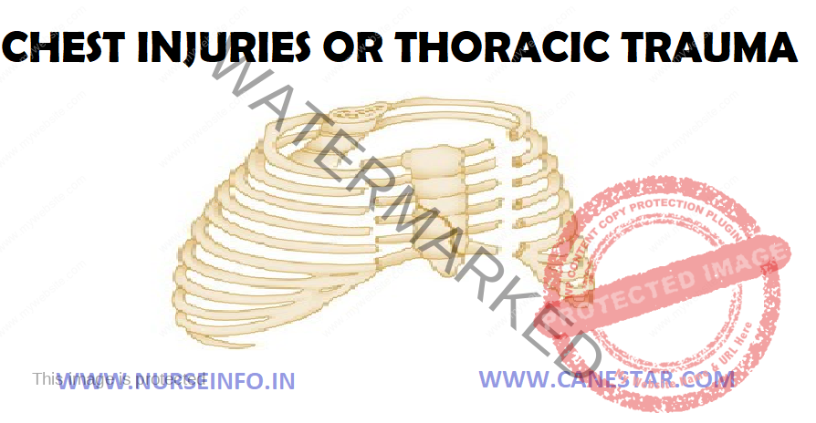
CHEST INJURIES – Classification, Signs and Symptoms, Diagnostic Evaluation and Management
Chest injuries (or thoracic trauma) are a serious injury of the chest. Thoracic trauma is a common cause of significant disability and mortality, the leading cause of death from physical trauma after head and spinal cord injury. Blunt thoracic injuries are the primary or a contributing cause of about a quarter of all trauma-related deaths.
CLASSIFICATION
Chest trauma can be classified as
- Blunt injuries
- Crush injuries
- Penetrating injuries
Blunt Trauma
Mode of injury is important where there has been massive deformity of a car or a history of a fall of 5 meters or more major intrathoracic injuries should always be suspected. The physical nature of chest wall allows for considerable elastic recoil, especially in young patients and, therefore, degree of injury within chest may need to be judged initially by deformity to car rather than appearance of patient.
Penetrating Injuries
- Result in parenchymal damage related to track of missile or stabbing implement and velocity
- More solid structures (e.g. heart and major vessels) suffer greater injury where high-velocity missiles are penetrating weapons
- More lethal complication is hemorrhage
- Often associated with abdominal trauma
Crush Injury
- Occurs where elastic limits of chest and its contents have been exceeded
- Patients usually have AP deformity
- Majority have flail chests with multiple fractures, pneumothorax or hemothorax
- Most have pulmonary contusion
- Injuries of heart, aorta, diaphragm, liver, kidney and spleen are common
Specific Types of Chest Trauma
- Injuries to the chest wall:
Rib fractures, flail chest and sterna fractures
- Pulmonary injury (injury to the lung) and injuries involving the pleural space:
Pulmonary contusion, pulmonary laceration, pneumothorax, hemothorax, hemopneumothorax
- Injury to the airways:
Tracheobronchial tear
- Cardiac Injury
Pericardial tamponade
SIGNS AND SYMPTOMS
- Air hunger, use of accessory muscles, tracheal deviation, cyanosis or distended neck veins (evidence of tension pneumothorax, or tamponade)
- Tracheal deviation (evidence of tension pneumothorax)
- Major defects in the chest (sucking chest wounds)
- Unilaterally diminished breath sounds or hyper-resonance to percussion (evidence of closed pneumothorax or tension pneumothorax)
- Decreased heart sounds (pericardial tamponade)
- Location of foreign bodies
- Location of entry and exit wounds
DIAGNOSTIC EVALUATION
- Chest X-ray
CXR is most useful screening investigation
Look for subcutaneous air, foreign bodies, bony fractures, widening of mediastinum, atelectasis
Inspiratory and expiratory films for checking for pneumothorax
- CT scan:
Valuable tool
Aids in diagnosis and precise location of numerous lesions
Contrast is useful particularly when looking for mediastinal hemorrhage and periaortic hematomas
- Echocardiography: cardiac wall motion abnormalities and valve function and presence of pericardial fluid or blood
- ECG: most common abnormality in thoracic trauma are S-T and T wave changes and findings indicative of bundle branch block
- Angiography: remains the gold standard for defining thoracic vascular injuries
- Bronchoscopy: indications include evaluation of airway injury, hemoptysis, segmental or lobar collapse, and removal of aspirated foreign bodies
MANAGEMENT
Immediate Management
- Assure patient airway, oxygenation and ventilation
- Exclude or treat:
Pneumothorax, hemothorax, cardiac tamponade
- Assess for extrathoracic injuries
- Decompress stomach
- Provide pain relief
- Reconsider endotracheal intubation, ventilation. In particular, take into account gross obesity, significant pre-existing lung disease, severe pulmonary contusion or aspiration, need for surgery for thoracic or extrathoracic injuries
General Management
- Monitoring
Following are danger signs requiring full reassessment:
- Respiratory rate > 20/min
- Heart rate > 100/min
- Systolic BP < 100 mm Hg
- Reduced breath sounds on affected side
- PaO2 < 9 kPa on room air
- PaCO2 > 8 kPa
- Increased size of pneumothorax, hemothorax or increased width of mediastinum on CXR
- Deterioration in any of these signs must be followed by a search for evidence of blood loss, tension pneumothorax, head injury, sepsis or fat embolism. Chest drains should be checked for patency.
- Chest drains: indications for insertion of chest drains in stable patients:
Pneumothorax > 10% in nonventilated patients (i.e. > 1 intercostal space)
Hemothorax > 500 ml (i.e. above neck of 7th rib)
Surgical emphysema
- Antibiotics
Use of prophylactic antibiotics is controversial. Some recommend them for patients treated conservatively in whom a chest drain is inserted
Cefuroxime and metronidazole for patients with perforated viscus (in addition to exploration and drainage)
- Mechanical ventilation
Most enters use PCV or PSV to reduce incidence of barotraumas
PCV and PSV also provide some compensation for air leaks
- Analgesia:
IV opioids in frequent small doses or by continuous infusion
Entonox inhalation during physiotherapy
Intercostal nerve block
Multiple individual nerve blocks
Single large volume (e.g. 20 ml 0.5% bupivicaine) into one intercostals space. Spreads is to block nerves above and below
Intrapleural bupivicaine via intercostal catheters using intermittent injections or continuous infusions
NSAIDs: fully resuscitated patients with normal renal function
Postoperative Intensive Care
- Following tracheobronchial, lung or diaphragmatic repair, high inflation pressures should be avoided
- Tracheal suction must be minimal where there is a tracheobronchial suture line
- Avoid fluid overload
- Prevent gastric distension

CHEST INJURIES – Classification, Signs and Symptoms, Diagnostic Evaluation and Management

