Home Blog Page 57

ABDOMINAL HERNIA

ABDOMINAL HERNIA – Etiology and Risk Factors, Signs and Symptoms, Types, Diagnostic Evaluation and Management

In general, hernia refers to a condition that arises when an organ pushes through a weak area in the muscles or tissue that surrounds and contains it.

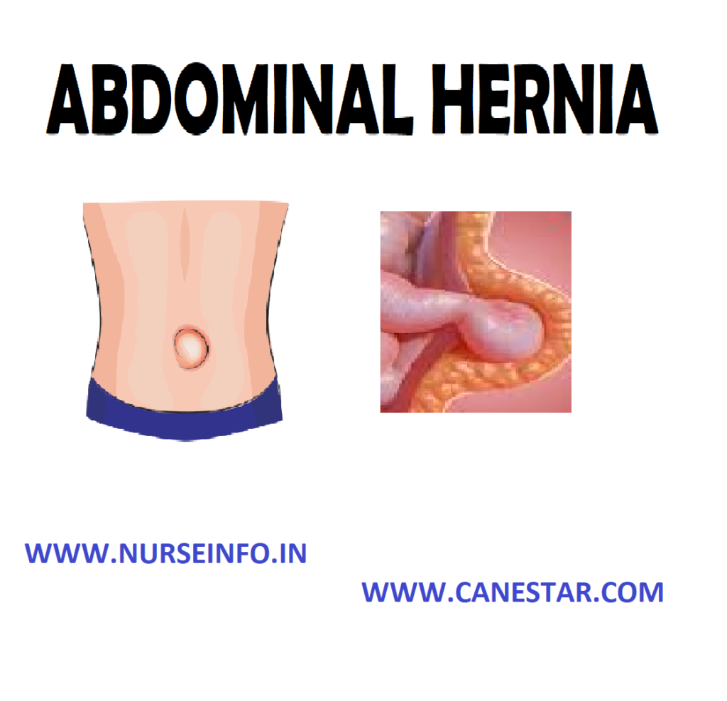
An abdominal hernia occurs when there is a tear in the inner lining of the abdominal wall (the outer layer of muscle, fat and tissue that extends from the bottom of the ribs to the top of the thighs), causing a bulge in the abdominal wall where the organs protrude

The bulge can be either reducible, which means that by applying slight pressure to the area, the organs are being pushed back into the abdominal cavity and the hernia will flatten and disappear, or non-reducible, which means the fat or tissue cannot be pushed back into the abdomen and the hernia will not flatten

ETIOLOGY AND RISK FACTORS

A hernia can develop in anyone, from a newborn baby to a senior citizen. The following may increase risk of developing a hernia by increasing pressure on the abdominal wall:

  • A chronic cough, such as smoker’s cough
  • Obesity
  • Straining during bowel movements or while urinating
  • Pregnancy
  • Straining to lift heavy objects
  • Persistent sneezing, such as that caused by allergies
  • Prolonged seating or standing

The risk of having an abdominal hernia increases with age because the older you get, the weaker your abdominal wall muscles become

SIGNS AND SYMPTOMS

  • The main sign of an abdominal hernia is having a bulge or swelling appear on a part of abdomen. Often, the bulge will disappear in lying down position or push on it and then reappear during standing, cough or sneeze. This is called a reducible hernia
  • Other symptoms are burning, slight discomfort  and a feeling heaviness or aching in abdomen

TYPES OF ABDOMINAL HERNIAS

The following are different types of abdominal hernia:

Inguinal Hernia

When a male’s testicles descend into the scrotum, this causes a naturally weakened area in the wall of the abdomen, called the internal ring. This weakened area makes men more susceptible to a hernia at this location.

It is of two types:

  • Indirect inguinal hernia: an indirect inguinal hernia is the most common type of inguinal hernia. It occurs at the internal ring in the groin area. The intestine drops down into the internal ring and can extend down into the scrotum in men or to the outer folds of the vagina in women. An indirect inguinal hernia can be the result of an inherited weakness at the internal ring or one that occurs later in life. The latter is known as an acquired hernia
  • Direct inguinal hernia: less common than an indirect inguinal hernia, a direct inguinal hernia occurs near the internal ring instead of within it. They are acquired hernias that usually occur after age 40 as a result of aging or injury

Epigastric Hernia

This type of hernia occurs as a result of a weakness in the muscles of the upper-middle abdomen, above the navel. Men are about three times more likely to have an epigastric hernia than women

Umbilical Hernia

They involve protrusion of intra-abdominal contents through a weakness at the site of passage of the umbilical cord through the abdominal wall. These hernias often resolve spontaneously. Umbilical hernias in adults are largely acquired, and are more frequent in obese or pregnant women. Abnormal decussation of fibers at the linea alba may contribute. These hernias can occur in babies, children and adults

Femoral Hernia

Femoral hernias occur just below the inguinal ligament, when abdominal contents pass into the weak area at the posterior wall of the femoral canal. They can be hard to distinguish from the inguinal type. It occurs in the area between the abdomen and the thigh, and appears as a bulge on the upper thigh. This type of hernia is more common in women than men.

Incisional Hernia

An incisional hernia occurs when the defect is the result of an incompletely healed surgical wound. It can occur at the site of an incision from a previous surgery. The fat or tissue pushes through a weakness created by the surgical scar. An incisional hernia can occur months or years after the initial surgery

Diaphragmatic Hernia

Higher in the abdomen, diaphragmatic hernia results when part of the stomach or intestine protrudes into the chest cavity through a defect in the diaphragm. A hiatus hernia is a particular variant of this type, in which the normal passageway through which the esophagus meets the stomach (esophageal hiatus) serves as a functional “defect”, allowing part of the stomach to herniated into the chest.

DIAGNOSIS OF ABDOMINAL HERNIA

  • By taking history
  • Physical examination
  • Examine the bulge or swelling
  • Ultrasound scan
  • X-rays
  • CT scan

MANAGEMENT

Non-Surgical Treatment

Treatment for hernias can be surgical or nonsurgical, depending on their severity. Non-surgical treatments usually help ease pain and discomfort caused by a hernia but do not correct the problem.

Trusses

There are two kinds of trusses for hernias, the spring truss and the umbilical truss. The spring truss is worn around the waist and acts as a support, while the umbilical truss is worn around the midsection. Trusses also come in different sizes and are usually worn under briefs.

Hernia Belts

Hernia belts are lightweight and made in such a way that movement is not restricted while you are recovering. These belts were designed to be worn over briefs, are made with adjustable straps and are lined with foam for extra comfort. The foam compression pads provide gradual pressure and support to the weakened muscles

Bindings

Abdominal bindings are made of elastic and provide uniform compression and support of the abdominal muscles. They can be fastened around the waist.

Hernia Briefs

Hernia briefs look like regular briefs except they are made with spandex and foam pads for extra support and are designed to provide rupture relief. The material is convenient because it is lightweight, cool and washable and is easy to wear. It is important to remember that these nonsurgical treatments for hernias are not meant to correct the problem. They are only meant to provide comfort and pain-relief for those engaged in physically demanding tasks.

Surgical treatment: surgery of hernia can be performed through various approaches;

  • Herniotomy
  • Herniorrhaphy
  • Hernioplasty
  • Laparoscopy
  • Herniotomy (removal of the hernia sac only) alone is adequate for an indirect inguinal hernia in children in whom the abdominal wall muscles are normal.
  • Herniorrhaphy (herniotomy plus repair of the posterior wall of the inguinal canal) may be suitable for a small hernia in a young adult with good abdominal wall musculature.
  • Hernioplasty (reinforcement of the posterior inguinal canal wall with a synthetic mesh) is required for large hernias in the middle-aged and elderly with poor abdominal wall musculature.
  • Laparoscopic hernia repair surgery: during a laparoscopic hernia repair, a surgeon makes small incisions in the abdominal wall, and the abdomen is inflated with carbon dioxide. A laparoscope (a thin, tube-like instrument with a small video camera) and surgical instruments are then inserted through the incisions. While viewing a monitor, the surgeon pushes the herniated intestine back into place and repairs the hernia opening with surgical staples. Mesh is then placed over the defect to reinforce the abdominal wall.

NURSING INTERVENTIONS

  • Find out the recurrent coughing or sneezing causing symptoms and treat them
  • Encourage not to strain too much on the toilet. Encourage to eat enough fruit and vegetables and increase fiber intake
  • Also encourage to drink enough fluids
  • Encourage to maintain a healthy weight
  • Encourage exercising to tone the muscles of the abdomen
  • Provide medical help for chronic constipation, allergies or a chronic cough
  • Encourage not to be heavy work that will increase the intra-abdominal pressure
  • Prepare the patient for diagnostic tests as needed
  • Administer prescribed antacids and other medications
  • To reduce intra-abdominal pressure and prevent aspiration, have the patient sleep in a reverse Trendelenburg position with the head of the bed elevated.
  • Assess the patient’s response to treatment
  • Observe for complications, especially significant bleeding, pulmonary aspiration, or incarceration or strangulation of the herniated stomach portion
  • After endoscopy, watch for signs of perforation such as falling blood pressure, rapid pulse, shock, and sudden pain caused by endoscope
  • To enhance compliance, teach the patient about the disorder. Explain significant symptoms, diagnostic tests, and prescribed treatments.
  • Review prescribed medications, explaining their desired actions and possible adverse effects
  • Teach the patient dietary changes to reduce reflux
  • Encourage the patient to delay lying down for 2 hours after eating
ABDOMINAL HERNIA – Etiology and Risk Factors, Signs and Symptoms, Types, Diagnostic Evaluation and Management

PORTAL HYPERTENSION

PORTAL HYPERTENSION – Etiology, Pathophysiology, Signs and Symptoms, Diagnostic Evaluation and Management

Portal hypertension refers to abnormally high pressure in the hepatic portal vein. It is defined as a portal pressure of 12 mm Hg or more (compared with the normal 5-10 mm Hg)

ETIOLOGY

  • Prehepatic causes (congential atresia or stenosis, portal vein thrombosis, splenic vein thrombosis, extrinsic compression tumors)
  • Hepatic cause (chronic hepatitis, myeloproliferative diseases, idiopathic portal hypertension, granulomata, nodular (nodular regenerative hyperplasia, partial nodular transformation), toxins, fibropolycystic disease (including congenital hepatic fibrosis)
  • Posthepatic – blockage of hepatic veins or venules (Budd-Chiari syndrome (hepatic vein obstruction), constrictive pericarditis, right heart failure, sclerosing hyaline necrosis)

PATHOPHYSIOLOGY

Increased vascular resistance in the portal venous system-from various mechanical causes —- activation of stellate cells and myofibroblasts, contributing to the abnormal blood flow patterns —- increased blood flow in the portal veins-from splanchnic arteriolar —- vasodilatation, caused by an excessive release of endogenous vasodilators —- the raised portal pressure opens up venous collaterals, connecting the portal and systemic venous systems. These occur in various sites, gastroesophageal junction —- producing varices which are superficial and easily bleed —- portal hypertension —- decreased intravascular volume —- ascites

————————–

Alcohol abuse and Malnutrition – Laennec’s cirrhosis

Infection and drugs – Post necrotic cirrhosis

Biliary obstruction – Biliary cirrhosis

These three conditions leads to —– Destruction of hepatocytes —- Fibrosis/Scarring —- Obstruction of blood flow, increased pressure in the venous and sinusoidal channels, fatty infiltration fibrosis/scarring —- Portal Hypertension

SIGNS AND SYMPTOMS

  • Dilated veins in the anterior abdominal wall
  • Splenomegaly
  • Ascites
  • Jaundice
  • Spider veins
  • Palmar erythema
  • Confusion
  • Enlarged or small liver
  • Gynecomastia
  • Testicular atrophy

DIAGNOSTIC EVALUATION

  • Blood tests
  • Abdominal ultrasound – for liver and spleen size, ascites, portal blood flow and thrombosis of the portal or splenic veins
  • Doppler ultrasound – can show direction of flow in blood vessels
  • CT scan, especially spiral CT, may show portal vasculature MRI scan – gives similar information to CT
  • Endoscopy
  • Portal hypertension measurement: portal pressure is indirectly measured in clinical practice by the hepatic venous pressure gradient (HVPG)
  • Liver biopsy
  • Vascular imaging: the site of the portal venous block can be demonstrated by examining the venous phase of a celiac or superior mesenteric arteriogram, by splenic portography following injection of dye into the splenic pulp, or by retrograde portography via a hepatic vein
  • Hepatic venography is helpful when hepatic vein block or idiopathic portal hypertension is suspected

COMPLICATIONS

  • Bleeding from esophageal or gastric
  • Ascites
  • Spontaneous bacterial peritonitis
  • Hepatorenal syndrome
  • Hepatic hydrothorax
  • Pulmonary complications
  • Liver failure
  • Hepatic encephalopathy
  • Cirrhotic cardiomyopathy

MANAGEMENT

Pharmacological Management

  • Beta blockers: nonselective beta blockers reduce portal pressure in many patients
  • Nitrates: added to beta blocker therapy, they contribute to reducing portal pressure and may reduce rates of variceal rebleeding
  • Vasoactive drugs: terlipressin and octreotide are used to assist the control of acute variceal bleeding

Surgical Management

  • Transjugular intrahepatic portosystemic shunt (TIPS): it is a radiological procedure, connecting the portal and hepatic veins using a stent. The purpose of TIPS is to decompress the portal venous system, to prevent rebleeding from varices or to reduce the formation of ascites
  • Surgical portosystemic shunts
  • Devascularizationn procedures: these include gastroesophageal devascularization, splenectomy and esophageal transaction

NURSING MANAGEMENT

Nursing Diagnosis

  • Risk for infection related to ascites
  • Activity intolerance related to weakness
  • Risk for imbalanced fluid volume related to vomiting and edema

Interventions

  • Administer medications, which may include diuretics
  • Assist the health care provider with paracentesis, which removes the fluid (e.g. ascites) from the peritoneal cavity
  • Measure and record abdominal girth and body weight daily, assess for abdominal fluid wave
  • Promote measures to prevent or reduce edema
  • Encourage the client to elevate the lower extremities and wear support to prevent lower-extremity edema
  • Administer salt-poor albumin, which temporarily elevates the serum albumin level. This increases serum osmotic pressure, helping to reduce edema by causing ascetic fluid to be drawn back into the blood stream and eliminated by the kidneys
  • Measure dietary intake by caloric count
  • Weigh as indicated. Compare changes in fluid status, recent weight history, and skinfold measurement
  • Encourage patient to eat, explain reasons for the types of diet. Include patient in meal planning to consider his or her preferences in food choices
  • Encourage patient to eat all meals including supplementary feeding unless contraindicated
  • Give small, frequent meals
  • Provide salt substitutes, if allowed and avoid those containing ammonium
  • Restrict intake of caffeine, gas-producing or spicy and excessively hot or cold foods
  • Suggest soft foods, avoiding roughage, if indicated
  • Encourage frequent mouth care, especially before meals
  • Promote undisturbed rest periods, especially before meals
  • Measure intake and output chart, weigh daily and note gain of more than 0.5 kg/day
  • Assess respiratory status, noting increased respiratory rate and dyspnea
  • Auscultate lungs, noting diminished breath sounds and developing adventitious sounds
  • Monitor for cardiac dysrhythmias. Auscultate heart sounds, noting development of S3/S4 gallop rhythm
  • Assess degree of peripheral edema
  • Measure abdominal girth encourage bedrest when ascites is present
  • Provide frequent mouth care, occasional ice chips (if NPO)
  • Monitor serum albumin and electrolytes (particularly potassium and sodium)
  • Monitor serial chest X-rays
  • Restrict sodium and fluids as indicated
  • Administer salt-free albumin, plasma expanders, positive inotropic drugs and arterial vasodilators
  • Administer diuretics: spironolactone, furosemide, etc
PORTAL HYPERTENSION – Etiology, Pathophysiology, Signs and Symptoms, Diagnostic Evaluation and Management

LEUKEMIA

LEUKEMIA – Etiology and Pathophysiology, Risk Factors, Clinical Manifestations, Classification, treatment and Management

Leukemia is a type of cancer of the blood or bone marrow characterized by an abnormal increase of immature white blood cells called ‘blasts’. In turn, it is part of the even broader group of diseases affecting the blood, bone marrow, and lymphoid system, which are also known as hematological neoplasms.

ETIOLOGY AND PATHOPHYSIOLOGY

There are different causes:

The following are either known causes, or strongly suspected causes:

  • Artificial ionizing radiation
  • Viruses-HTLV-1 (Human T-lymphotropic virus) and HIV (Human immunodeficiency virus)
  • Benzene and some petrochemicals
  • Alkylating chemotherapy agents used in previous cancers
  • Meternal fetal transmission
  • Hair dyes
  • Genetic predisposition
  • Down syndrome – people with Down syndrome have a significantly higher risk of developing leukemia, compared to people who do not have Down syndrome.

RISK FACTORS

  • Gender: men are more likely to develop CML, CLL and AML than women
  • Age: the risk of most leukemias, with the exception of ALL, typically increases with age
  • Genetics: family history: most leukemias have no familial link. However, first degree relatives of CLL patients, or having an identical twin who has or had AML or ALL, may at an increased risk for developing the disease
  • Lifestyle: smoking: although smoking may not be a direct cause of leukemia, smoking cigarettes does increase the risk of developing AML
  • Exposures: exposure to high levels of radiation: exposure to high-energy radiation (e.g. atomic bomb explosions) and intense exposure to low-energy radiation from electromagnetic fields (e.g. power lines)
  • Chemical exposure: long-term exposure to certain pesticides or industrial chemicals like benzene is considered to be a risk for leukemia
  • Previous Treatment: previous cancer treatment: certain types of chemotherapy and radiation therapy for other cancers are considered leukemia risk factors

CLINICAL MANIFESTATION

  • Fever and night sweats
  • Headache
  • Bruising or bleeding
  • Bone or joint pain
  • A swollen or painful abdomen from an enlarged spleen
  • Swollen lymph nodes in the armpit, neck or groin
  • Getting a lot of infections
  • Feeling very tired or weak
  • Losing weight and not feeling hungry
  • Fatigue
  • Night sweat, lethargy
  • Fever

CLASSIFICATION

  • ALL (Acute lymphatic leukemia)
  • ALM (Acute myelogenous leukemia)
  • CLL (Chronic lymphocytic leukemia)
  • CML (Chronic myelogenous leukemia)

ALL (Acute Lymphatic Leukemia)

It is a type of leukemia that, all types of leukemia starts from white blood cells in the bone marrow, the soft inner part of bones. It develops from cells called lymphocytes, type of WBCs central to immune system, an immature type of lymphocyte

CLINICAL MANIFESTIONS

  • Fever
  • Pallor
  • Bleeding
  • Anorexia
  • Fatigue
  • Weakness
  • Bone, joint and abdominal pain
  • Increase intracranial pressure

Acute Myelogenous Leukemia (AML)

It is fast growing cancer of the blood and bone marrow where the bone marrow makes many cancerous cells called leukemic blasts, normally blasts develop into WBCs that fight infection, but in AML, leukemic blasts do not develop properly and cannot fight infection. In this there is proliferation of immature myeloid cells

TREATMENT

  • Use of cytarabine, 6-thioguanine and doxorubicin
  • The same care of client as ALL, plus give adequate amounts of fluids (2000 to 3000 ml per day)
  • Instruct client about medication, effects, side effects and nursing measures

Chronic Lymphocytic Leukemia (CLL)

It is a type of cancer of the blood or bone marrow, normally bone makes blood stem cells over the time

PATHOPHYSIOLOGY

  • It is characterized by proliferation of small, abnormal, mature B lymphocytes, often leading to decreased synthesis of immunoglobulin and depressed antibody response.
  • The number of mature lymphocytes in peripheral blood smear and bone marrow are greatly increased

CLINICAL MANIFESTATION

  • Usually there is no symptoms
  • Chronic fatigue, weakness, anorexia, splenomegaly, lymphadenopathy, hepatomegaly
  • Skin lesions
  • Anemia
  • Thrombocytopenia
  • The WBC count is elevated to a level between 20,000 to 100,000
  • Increase blood viscosity and clotting episode

Chronic Myelogenous Leukemia (CML)

It is a type of cancer that starts in certain blood forming cells of the bone marrow. And in this bone marrow produces too many white cells. It affects the blood and bone marrow

  • Occurs between 25-60 years of age. Peak 45 years and it caused by benzene exposure and high doses of radiation

CLINICAL MANIFESTATION

The classic symptoms of chronic types of leukemia include:

  • Fatigue, weakness, fever, sterna tenderness
  • Weight loss, joint and bone pain
  • Massive splenomegaly and increase in sweating
  • The accelerated phase of disease (blostic phase) is characterized by increasing number of granulocytes in the peripheral blood
  • There is a corresponding anemia and thrombocytopenia

DIAGNOSTIC EVALUATION

  • History: ask the patient for the presence of signs and symptoms. Assess the patient for the exposure of any risk factors or any history of risk factors
  • Physical examination: during the physical examination, assess patient for sign and symptoms
  • Assess patient for enlarged lymph nodes, areas of bruising, or any sign of infection
  • Assess the vital signs of patient
  • Complete blood count: in complete blood count, it measures the number of red blood cells, white blood cells and platelets. Most of patient with ALL has too many immature white cells. Low RBCs count, Hb, low platelet count, low to high WBC count

TREATMENT

Chemotherapeutic agent, it involve three phases

  • Induction: during the induction phase the client receives an intensive course of chemotherapy designed to induce a complete remission of the disease. Once the remission is achieved the consolidation phase begins. Using vincristine and prednisone
  • Consolidation: use modified course of intensive therapy to eradicate any remaining disease
  • Maintenance: during the maintenance phase small doses of different combinations of chemotherapeutic agents are given every 3 to 4 weeks. It allows the client to live a normal life as possible

Other Management

  • Eat diet that contains high in protein, fibers and fluids
  • Avoid infection (handwashing, avoid crowds), injury
  • Take measure to decrease nausea and to promote appetite, smoking and spicy and hot foods
  • Maintain oral hygiene

Symptom Control

  • Chemotherapy with chlorambucil, cyclophosphamide and prednisone to decrease lymphadenopathy and splenomegaly
  • Splenic irradiation or splenectomy for painful splenomegaly or platelet sequestration hemolytic anemia
  • Irradiation of painful enlarged lymph nodes

Supportive Care

  • Transfusion therapy to replace platelets and RBCs
  • IV immunoglobulins or gamma globulin to treat hypogamma globulinemia

Chronic Phase

  • Palliative treatment, controlling symptoms, includes chemotherapy, irradiation, splenectomy
  • Potentially curative treatment is offered by allogeneic (related to cumulated donor) BMT

Accelerated Phase or Blast Crisis

Attempts to restore chronic phase through use of high dose chemotherapy, leukophoresis

Nursing Intervention

  • Taking measures to prevent infection
  • Promoting safety
  • Providing oral hygiene
  • Preventing fatigue
  • Promoting effective coping
  • Client and family education

Nursing Assessment

History

  • Ask patient for their family history, past medical history
  • Ask for the presence of exposure to any risk factors and etiology factors
  • Ask client for the presence of sign and symptoms

Physical Examination

  • Asses for swollen lymph nodes, spleen or liver
  • Assess the client vital signs. Check client for presence of fever
  • Assess patient for the enlarged lymph nodes, sign of infection, and any signs of bruising or bleeding from any sites

Blood Tests

The lab does a complete blood count to check the number of white blood cells, red blood cells, and platelets. Leukemia causes a very high level of white blood cells. It may also cause low levels of platelets and hemoglobin, which is found inside red blood cells

Bone Marrow Aspiration

The doctor uses a thick, hollow needle to remove samples of bone marrow. The sample is taken from the back of the pelvic (hip) bone and with help of needle small amount of liquid bone marrow is sucked

Bone Marrow Biopsy

It is usually done just after the aspiration. The doctor uses a very thick, hollow needle to remove a small piece of bone and bone marrow

Nursing Diagnosis

  • Impaired tissue integrity related to high dose radiation therapy
  • Risk for infection to decreased neutrophils, altered response to microbial invasion, and presence of environmental pathogens
  • Impaired oral mucous membrane related to low platelet count and effects of pathologic conditions and treatment as evidenced by oral bleeding
  • Risk for injury related to low platelet counts and treatment
  • Acute pain related to tumor growth, infection or adverse effects of chemotherapy
  • Activity intolerance related to anemia and adverse effect of chemotherapy

Nursing Interventions

Maintain Tissue Integrity

  • Avoid rubbing powders, deodorants, lotions or ointments (unless prescribed) or application of heat and cold to treated areas
  • Encourage the patient to keep the treated area clean and dry
  • Advise the patient to bath the area gently with tepid water and mild soap
  • Encourage the patient to wear loose fitting cloths
  • Advisee the patient to protect skin from over and direct exposure to sun light, chlorine and temperature extremes

Prevent from Infection

  • Inspect the patient for the sign and symptoms of infection, e.g. redness, etc
  • Maintain asepsis for patient at risk
  • Instruct the patient to take antibiotics as prescribed by doctor to prevent microbial resistance
  • Teach the patient and family how to avoid infections, e.g. about personal hygiene technique of handwashing, oral care, skin hygiene etc
  • Educate the patient to report if there is any presence of signs of infection to the doctor immediately
  • Monitor granulocyte count and WBC count to identify the presence of infection
  • Screen all visitors for communicable diseases

Free the Mucosa from Bleeding

  • Monitor lips, tongue, mucous membrane, and gums for moisture, color, texture, presence of infection
  • Assist the patient to select soft, bland and nonacidic foods to decrease irritation of oral mucosa
  • Advise the patient to use soft toothbrush for removal of dental debris
  • Instruct the patient to perform oral hygiene after eating and as often as needed to avoid breakdown of oral mucosa
  • Advise the patient to avoid the use of lemon glycerin swabs to prevent excessive drying of the mucosa

Health Education

  • Teach the patient about the risk of infection and how to prevent from infection
  • Educate the patient how to take medications, as ordered and instruct to avoid adverse effect and progress of diseases
  • Provide patient and family member information about resources in the community, such as leukemia and lymphoma
  • Educate the patient to maintain proper personal hygiene
  • Teach the patient to avoid aspirin and NSAIDs which may interfere with platelet function

COMPLICATIONS

  • Infections
  • Blood problems
  • Impaired body function
  • Other cancers
  • Leads to death
  • Infections: leukemia causes abnormalities in the white blood cells. While healthy white blood cells protect the body against infections and disease, defective cells lose this ability. Without the protective properties of the white blood cells, people with leukemia become more susceptible to infections, which are the most common leukemia complications involved with the disease
  • Blood Problems: people with leukemia may experience complications related to the blood. In some people, leukemia may lead to excess bleeding or bruising. Both may occur when leukemia causes a shortage in the number of platelets in the body. Other people experience an increase in the number of platelets, which can lead to clogging or excessive blood clotting
  • Impaired Body Functions: when one part of the body begins to malfunction, it can lead to complications in other parts of the body. In the case of leukemia, the disease may adversely affect the spleen, kidneys, and renal areas. In a healthy body, the spleen stores excess blood cells. When levels increase due to leukemia, the spleen may attempt to store more than it can hold, which can cause an enlarged spleen
  • Death: like most diseases without a care, possible death is another complication of leukemia
LEUKEMIA – Etiology and Pathophysiology, Risk Factors, Clinical Manifestations, Classification, treatment and Management

ESOPHAGEAL VARICES

ESOPHAGEAL VARICES – Etiology, Signs and Symptoms, Diagnostic Evaluation and Management

Esophageal varices are abnormal, enlarged veins in the lower part of the esophagus. It develops when normal blood flow to the liver is obstructed by scar tissue. Seeking a way around the blockages, blood flows into smaller blood vessels that are not designed to carry large volumes of blood. The vessels may leak blood or even rupture, causing life-threatening bleeding.

ETIOLOGY

  • Prehepatic causes: portal vein thrombosis, portal vein obstruction – congenital atresia or stenosis, increased portal blood flow – fistula, increased splenic flow
  • Intrahepatic causes: cirrhosis due to causes, including alcoholic, chronic hepatitis (e.g. viral or autoimmune), idiopathic portal hypertension, acute hepatitis, schistosomiasis, congenital hepatic fibrosis, myelosclerosis
  • Post hepatic causes: compression (from tumor), Budd-Chiari syndrome

SIGNS AND SYMPTOMS

  • Hemoptysis
  • Black, tarry or bloody stools
  • Shock
  • Jaundice
  • Spider veins
  • Palmar erythema
  • Dupuytren’s contracture
  • Shrunken testicles
  • Swollen spleen
  • Ascites

DIAGNOSTIC EVALUATION

  • Endoscopic examination: a procedure called esophagogastroduodenoscopy. It will insert in mouth and into esophagus and small intestine to evaluate the dilated veins, measure their size
  • Imaging tests: both CT and MRI scans may be used to diagnose esophageal varices. These tests also allow to examine liver and circulation in the portal vein
  • Capsule endoscopy: in this test, the patient swallows a vitamin-sized capsule containing a tiny camera, which takes pictures of the esophagus as it passes. This may be an option for people who are unable or unwilling to undergo an endoscope examination
  • CBC
  • Clotting including INR
  • Renal function
  • LFTs
  • Ascitic tap may be needed if bacterial peritonitis is suspected

MANAGEMENT

  • Beta blockers: it may help reduce blood pressure in portal vein, decreasing the likelihood of bleeding. These medications include propranolol, etc
  • Band ligation: it is used if esophageal varices appear to have a very high risk of bleeding. Using an endoscope, the varices are wrap with each other with an elastic band, which essentially ‘strangles’ the veins so they cannot bleed. Esophageal band ligation carries a small risk of complications, such as scarring of the esophagus
  • Transjugular intrahepatic portosystemic shunt (TIPS): the shunt is a small tube that is placed between the portal vein and the hepatic vein, which carries blood from liver back to heart. By providing an additional path for blood, the shunt reduces pressure in the portal vein and often stops bleeding from esophageal varices
  • Balloon tube tamponade (Sengstaken-Blakemore Tube): the Sengstaken tube is inserted through the mouth and into the stomach. The gastric balloon is inflated with air and the gastric balloon in then pulled up against the esophagogastric junction, compressing submucosal varices. The Sengstaken tube also contains an esophageal balloon which is only rarely required when the gastric balloon does not work. If bleeding continues, it may be that the tube is wrongly positioned or bleeding is from another source

NURSING MANAGEMENT

Nursing Diagnosis

  • Risk for bleeding related to obstruction in blood flow
  • Imbalanced nutrition: less than body requirements related to vomiting and jaundice

Interventions

  • Provide ongoing assessment
  • Assess for ecchymosis, epistaxis, petechiae, and bleeding gums
  • Monitor level of consciousness, vital signs, and urinary output to evaluate fluid balance
  • Monitor the client during blood transfusion administration, if prescribed
  • Use small-gauge needles, and apply pressure or cold for bleeding
  • Explain the procedure to the client to reduce fear and enhance cooperation with insertion and maintenance of the esophageal tamponade tube
  • Monitor the client closely to prevent accidental removal or displacement of the tube with resultant airway obstruction

Ensure nasogastric tube patency to prevent aspiration (observe gastric aspirate for evidence of bleeding, protect the client from chilling)

Administer prescribed vasopressin and vitamin K

Closely assess for signs and symptoms of GI bleeding: check all secretions for frank or occult blood. Observe color and consistency of stools, NG drainage, or vomitus

Observe for presence of petechiae, ecchymosis, bleeding from one or more sites

  • Monitor pulse, BP (and CVP, if available)
  • Avoid rectal temperature and be gentle with GI tube insertions
  • Encourage use of soft toothbrush, electric razor, avoiding straining for stool, vigorous nose blowing, and so forth
  • Use small needles for injections. Apply pressure to small bleeding and venipuncture sites for longer than usual
  • Advise to avoid aspirin-containing products
  • Monitor Hb/Hct and clotting factors
  • Supplemental vitamins: vitamins K, D and C
  • Administer stool softeners
  • Assist with insertion and maintenance of GI tube
  • Provide gastric lavage with room temperature and cool saline solution or water as indicated
ESOPHAGEAL VARICES – Etiology, Signs and Symptoms, Diagnostic Evaluation and Management

CHOLELITHIASIS AND CHOLECYSTITIS

CHOLELITHIASIS AND CHOLECYSTITIS – Types, Etiology, Pathophysiology, Signs and Symptoms, Diagnostic Evaluation and Management

It refers to presence of stones in gallbladder. A gallstone is crystalline concretion formed within the gallbladder by accretion of bile components. These calculi are formed in the gallbladder but may distally pass into other parts of the bilary tract and sometimes if the stones may obstruct the flow of bile and will cause inflammation of gallbladder called cholecystitis

TYPES OF GALLSTONES

  • Cholesterol stones: vary from light yellow to dark green or brown and are oval, between 2 and 3 cm long
  • Pigment stones: pigment stones are small and dark and comprise bilirubin and calcium salts that are found in bile
  • Mixed stones: mixed gallstones typically contain 20-80% of cholesterol

ETIOLOGY

  • Gender: women are twice as likely as men to develop gallstones. Excess estrogen from pregnancy and hormone replacement therapy can lead to gallstones
  • Family history: gallstones often run in families
  • Obesity: obesity is a major risk factor for gallstones, especially in women
  • Diet: diets high in fat and cholesterol and low in fiber increase the risk of gallstones
  • Age: people older than age 60 are more likely to develop gallstones than younger people
  • Hypolipidemic drugs: these drugs increase the amount of cholesterol secreted into bile and contribute to gallstones
  • Hyperglycemia: people with diabetes generally have high levels of fatty acids called triglycerides. These fatty acids may increase the risk of gallstones

PATHOPHYSIOLOGY

Gallstones —- pressure obstruction —- bile stasis —- ↓ fat emulsification (fat intolerance, anorexia, n/v, weight loss, gaseous eructation, flatulence, steatorrhea) —- Inflammation (Pain (RUQ), Fever, Leukocytosis) ↓ Bile flow into the colon, Acholic stool, ↓ vitamin K absorption —- ↑ S. bilirubin (Jaundice, Pruritus, tea-colored urine) —- Infections (cholecystitis and pancreatitis)

SIGNS AND SYMPTOMS

  • Steady pain in the right upper abdomen that increases rapidly and lasts from 30 minutes to several hours
  • Pain in the back
  • Pain under the right shoulder
  • Murphy’s sign (guarding type of respiratory pain radiates towards the back and scapula)
  • Prolonged pain – more than 5 hours
  • Nausea and vomiting
  • Fever – even low-grade-or chills
  • Yellowish color of the skin or whites of the eyes
  • Clay-colored stools

DIAGNOSTIC EVALUATION

  • Computerized tomography (CT) scans: the CT scan is a noninvasive X-ray that produces cross-section images of the body. The test may show the gallstones or complications, such as infection and rupture of the gallbladder or bile ducts
  • Cholescintigraphy scan: patient is injected with a small amount of nonharmful radioactive material that is absorbed by the gallbladder which is then stimulated to contract. The test is used to diagnose abnormal contraction of the gallbladder or obstruction of the bile ducts
  • Endoscopic retrograde cholangiopancreatography (ERCP): ERCP is used to locate and remove stones in the bile ducts. The doctor inserts an endoscope – a long, flexible, lighted tube with a camera-down the throat and through the stomach and into the small intestine. The endoscope is connected to a computer and video monitor. The doctor guides the endoscope and injects a special dye that helps the bile ducts appear better on the monitor. The endoscope helps the doctor locate the affected bile duct and the gallstone. The stone is captured in a tiny basket and removed with the endoscope
  • Blood tests: blood tests may be performed to look for signs of infection, obstruction, pancreatitis, jaundice

MANAGEMENT

Pharmacological Management

  • Opoids analgesic like mephridine, morphine
  • Antispasmodic and anticholinergic
  • Antiemetic
  • Bile acid therapy
  • Chenodeoxycholic acid reduces the cholesterol stone by maintaining a normal amount of cholesterol solubility in bile e.g. chenodiol
  • Anticholelithiotic agents, like ursodiol

Surgical Management

  • Extracorporeal shock wave lithotripsy: it is a noninvasive procedure used as an ambulatory treatment. A machine called lithotreptor generates a powerful shock to shatter the gallstones
  • Percutaneous transhepatic biliary catheter insertion: insertion of a percutaneous transbiliary catheter under fluoroscopic guidance. This procedure will decompress obstructed extrahepatic ducts so that the bile can flow

Other surgical interventions are:

  • Cholecystectomy
  • Laparoscopic laser cholecystectomy
  • Cholecystotomy
  • Choledochotomy (incision of common bile duct)
  • Choledocholithotomy (incision of common bile duct and removal of gallstones)

Nonsurgical Treatment

  • Oral dissolution therapy: drugs made from bile acid are used to dissolve gallstones, e.g. ursodiol and chenodiol
  • Contact dissolution therapy: this experimental procedure involves injecting a drug directly into the gallbladder to dissolve cholesterol stones. The drug methyl tert-butyl ether can dissolve some stones in 1 to 3 days
CHOLELITHIASIS AND CHOLECYSTITIS – Types, Etiology, Pathophysiology, Signs and Symptoms, Diagnostic Evaluation and Management

CHOLECYSTITIS

CHOLECYSTITIS – Etiology, Risk Factors, Signs and Symptoms, Diagnostic Evaluations and Management

DEFINITION

Cholecystitis is inflammation of the gallbladder

ETIOLOGY

  • Gallstones: the vast majority of cholecystitis cases are the result of gallstones that block the cystic duct, causing bile to build up and resulting in gallbladder inflammation
  • Tumor: a tumor may prevent bile from draining out of gallbladder properly, causing bile buildup that can lead to cholecystitis
  • Bile duct blockage: kinking or scarring of the bile ducts can cause blockages that lead to cholecystitis

RISK FACTORS

  • Gallstones: most cases of cholecystitis are linked to gallstones
  • Sex: women have a greater risk of gallstones than men do. This makes women more likely to develop cholecystitis
  • Age: older people are at high risk of gallstones

SIGNS AND SYMPTOMS

  • Severe, steady pain in the upper righ part of abdomen
  • Pain that radiates from abdomen to right shoulder or back
  • Tenderness over abdomen when it is touched
  • Sweating
  • Nausea
  • Vomiting
  • Fever
  • Chills
  • Abdominal bloating

COMPLICATIONS

  • Enlarged gallbladder: if gallbladder becomes inflamed due to bile buildup, it may stretch and swell beyond its normal size, which can cause pain and increase the risk of a tear (perforation) in gallbladder, as well as infection  and tissue death
  • Infection within the gallbladder: if bile builds up within gallbladder, causing cholecystitis, the bile may become infected
  • Gangrene: untreated cholecystitis can cause tissue in the gallbladder to die (gangrene), which, in turn, can lead to a tear in the gallbladder
  • Perforation: a tear (perforation) in gallbladder may be caused by an enlarged or infected gallbladder that occurs as a result of cholecystitis

DIAGNOSTIC EVALUATION

  • Blood tests: blood tests are done to look for signs of an infection or of gallbladder problems
  • Imaging tests: imaging tests, such as abdominal ultrasound or a computerized tomography (CT) scan can be used to create pictures of gallbladder that may reveal signs of cholecystitis
  • HIDA: a hepatobiliary iminodiacetic acid (HIDA) scan tracks the production and flow of bile from liver to small intestine and shows if bile is blocked at any point along the way.

MANAGEMENT

  • Antibiotics: to treat the infection
  • Analgesics: to help control pain until the inflammation in gallbladder is relieved

Surgical Interventions

  • Cholecystectomy
  • Laparoscopic laser cholecystectomy
  • Cholecystectomy
  • Choledochotomy (incision of common bile duct)
  • Choledocholithotomy (incision of common bile duct and removal of gallstones)

NURSING MANAGEMENT

Nursing Assessment

  • Obtain history and demographic data that may indicate risk factors for biliary disease
  • Assess mental status through interview and interaction with patient
  • Perform abdominal examination for ascites
  • Assess the client’s bowel elimination pattern, noting the number of stools and their color and also the presence of blood
  • Assess the client’s abdomen for bowel sounds and the location of pain
  • Assess the complete nutritional status of diseased patient
  • Observe for any bleeding
  • Monitor for temperature and WBC counts for indicating inflammation of infection
  • Assess the emotional status, coping skills, verbal and nonverbal behaviors

Nursing Diagnosis

  • Risk for injury related to medication during retrograde endoscopy for stone removal
  • Knowledge deficit related to oral dissolution agents
  • Pain related to stone obstruction
  • Fluid volume deficit related to nausea and vomiting
  • Activity intolerance related to fatigue, general discomfort
  • Anxiety-related inflammatory disease of gallbladder
  • Imbalanced nutrition less than body requirement related to pain
  • Ineffective coping related to feeling of rejection
  1. Risk for injury related to medication during retrograde endoscopy for stone removal

Interventions

  • Assess for signs and symptoms of GI bleeding, e.g. check all secretions for frank or occult blood. Observe color and consistency of stools, NG drainage, or vomitus
  • Observe for presence of petechiae, ecchymosis, bleeding from one or more sites
  • Monitor pulse, BP (and CVP, if available)
  • Note changes in mentation and level of consciousness
  • Avoid rectal temperature, be gentle with GI tube insertions
  • Encourage use of soft toothbrush, electric razor, avoiding straining for stool, forceful nose blowing, and so forth
  • Use small needles for injections. Apply pressure to small bleeding and venipuncture sites for longer than usual
  • Recommend avoidance of aspirin-containing products
  • Knowledge deficit related to oral dissolution agents

Interventions

  • Inform patient of altered effects of medications with cholecystitis and the importance of using only drugs prescribed or cleared by a health care provider who is familiar with patient’s history
  • Assist patient in identifying the support person
  • Emphasize the importance of good nutrition. Recommend avoidance of high-protein and salty foods, onions and strong cheeses. Provide written dietary instructions.
  • Discuss sodium and salt substitute restrictions and necessity of reading labels on food and OTC drugs
  • Encourage scheduling activities with adequate rest periods
  • Promote diversional activities that are enjoyable to the patient
  • Recommend avoidance of persons with infections, especially upper respiratory tract infection
  • Identify environmental dangers, e.g. carbon tetrachloride-type cleaning agents, exposure to hepatitis
  • Pain related to stone obstruction

Intervention

  • Assess the pain location, severity and characteristics
  • Administer medications or monitor patient-controlled analgesia
  • Assist in attaining position of comfort and maintain bed rest
  • Fluid volume deficit related to nausea and vomiting

Interventions

  • Administer IV fluids and electrolytes as prescribed
  • Administer antiemetic as prescribed to reduce vomiting
  • Maintain nasogastric decompression until nausea and vomiting subside
  • Begin food and fluids as tolerated by the client
  • Observe and record amount of T tube drainage, if applicable
  • Activity intolerance related to fatigue, general discomfort

Interventions

  • Encouraging alternating periods of rests and ambulation
  • Maintain some periods of rest
  • Encourage and assist with gradually increasing periods of exercise
  • Imbalanced nutrition less than body requirement related to pain

Interventions

  • Encourage a diet that is low in residue fiber and fat and high in calories, protein and carbohydrates with vitamin and mineral supplements
  • Monitor weight daily
  • Provide small frequent feeding to prevent distension
  • Have the patient participate in meal planning to encourage compliance and increased knowledge
  • Prepare the patient for elementary diet
  • Restart oral fluid intake gradually. Offer clear liquids hourly; avoid cold fluids
  • Ineffective coping related to feeling of rejection

Intervention

  • Assess the level of fear and note nonverbal communication
  • Ask the patient’s usual coping pattern
  • Assure patient he/she will be closely monitored
  • Allow patient to verbalize fear of dying
  • Provide diversional materials, such as newspapers, music and television
  • Offer family support

COMPLICATIONS

  • Acute cholangitis: acute cholecystitis is a condition indicated by a sudden attack of pain in the upper abdomen that lasts more than 12 hours
  • Acute biliary pancreatitis: pancreatitis is a potentially serious disorder that occasionally develops in people with gallstones
  • Gallstone ileus
  • Obstructive jaundice or cholestasis
  • Gallbladder cancer
CHOLECYSTITIS – Etiology, Risk Factors, Signs and Symptoms, Diagnostic Evaluations and Management

HEMORRHOIDS

HEMORRHOIDS – Etiology, Types, Pathophysiology, Diagnostic Evaluation and Management

Hemorrhoids are enlarged veins in the anus or lower rectum. These comprise the blood supply to the anus and distal rectum

ETIOLOGY

  • Constipation
  • Diarrhea
  • Lack of exercise
  • Nutritional factors (low-fiber diets)
  • Increased intra-abdominal pressure
  • Genetics
  • Aging
  • Obesity
  • Prolonged sitting
  • Chronic cough
  • Pregnancy

TYPES

  • External hemorrhoids appear on the outside of the anus, especially around the anal opening. They too can cause a lot of discomfort because of constant friction with clothes, etc
  • Prolapsed hemorrhoids basically represent a later stage in the development cycle of internal hemorrhoids. When the latter become too bulky due to clogging of the vein with blood, they begin to sag and consequently protrude from the anus
  • Bleeding hemorrhoids are also a more severe form or stage of internal hemorrhoid characterized by profuse bleeding

PATHOPHYSIOLOGY

Normal vascular cushion —- downward pressure during defecation —- muscle fiber-anchored hemorrhoids loosen —- hemorrhoids tissue slides, then congested bleeds —- prolapse

DIAGNOSTIC EVALUATION

  • Examination of anal canal and rectum for abnormalities
  • Visual inspection of anal canal and rectum

MANAGEMENT

Medications

If hemorrhoids produce only mild discomfort, over-the-counter creams, ointments, suppositories or pads are recommended. These products contain ingredients, such as hydrocortisone, that can relieve pain and itching

Surgical Management

  • Rubber band ligation: one or two tiny rubber bands around the base of an internal hemorrhoid are placed to cut-off its circulation. The hemorrhoid withers and falls off within a week
  • Sclerotherapy: in this procedure, injection of a chemical solution is introduced into the hemorrhoid tissue to shrink it. While the injection causes little or no pain, it may be less effective than rubber band ligation
  • Coagulation (infrared, laser or bipolar): coagulation techniques use laser or infrared light or heat. They cause small, bleeding, internal hemorrhoids to harden and shrivel
  • Hemorrhoidectomy: during a hemorrhoidectomy, removal of excessive tissue causes bleeding. Various techniques may be used. The surgery may be done with a local anesthetic combined with seduction, a spinal anesthetic or a general anesthetic. Hemorrhoidectomy is the most effective and complete way to treat severe or recurring hemorrhoids
  • Hemorrhoid stapling: this procedure, called stapled hemorrhoidectomy or stapled hemorrhoidopexy, blocks blood flow to hemorrhoidal tissue. Stapling generally involves less pain than hemorrhoidectomy and allows an earlier return to regular activities.

NURSING MANAGEMENT

Preoperative Care

  • Give soaking seat

Rationalization: reduce local discomfort, reduce edema and promote healing

  • Give lubricant during defecation wound

Rationalization: assist in the conduct of defecation so it does not need straining.

  • Give a diet low in residual

Rationalization: reduce stimulation of the anus and weaken the feces

  • Instruct the patient to do a lot of standing or sitting (must be in balance)

Rationalization: the force of gravity will affect the incidence of hemorrhoids and sitting can increase intra-abdominal pressure

  • Observation of patient complaints

Rationalization: it helps to evaluate the degree of discomfort and lack of effectiveness of actions or states of complications

  • Provide an explanation of the emergence of pain and explain briefly

Rationalization: education about it helps in patient’s participation to prevent and reduce pain

  • Give the patient suppository

Rationalization: it can soften and can enable the patient to avoid straining during defecation

Postoperative Care

  • Give the patient a pleasant sleeping position

Rationalization: may decrease the pressure on the abdomen and increase the sense of control

  • Change the bandage every morning according to aseptic techniques

Rationalization: protecting the patient from cross examination during replacement of bandages. Wet bandage acts as a absorber of external contamination and causes discomfort

  • Exercise as early as possible

Rationalization: it can reduce the problems that occur due to immobilization

  • Observation of the rectal area if there is bleeding

Rationalization: bleeding on the network may increase the pain

  • Provide an explanation of the purpose of installation of flue-anus

Rationalization: knowledge of the benefits of the chimney can make the patient understand the anus to funnel anus to cure the wound

Postoperative Complications

Most patients are satisfied with the results of the surgery and recover without any problems. Complications associated with hemorrhoidectomy are rare and include:

  • Anal fistula or fissure
  • Constipation
  • Excessive bleeding
  • Excessive discharge of fluid from the rectum
  • Fever of 101 degree F or higher
  • Inability to urinate or have a bowel movement
  • Severe pain, especially when having a bowel movement
  • Severe redness and swelling in the rectal area

Nursing Diagnosis

  • Ineffective breathing pattern related to decreased pressure inspiration, expiration for anesthetic agent administration
  • Fluid volume and electrolyte deficit related to lack of or loss of fluid volume during surgery
  • Risk for injury related to the effects of anesthesia and weakness
  • Risk for hypothermia related to multiple factors of age, body weight, factors trauma, neuromuscular and environment

Nursing Interventions

  • Administer local anesthetic as prescribed
  • As needed, provide warm sitz baths or cold compresses to reduce local pain, swelling, and information
  • Provide the patient with high-fiber diet and encourage adequate fluid intake and exercise to prevent constipation
  • Monitor the patient’s pain level and the effectiveness of the prescribed medications
  • Check for signs and symptoms of anal infection, such as increased pain and foul-smelling anal drainage
  • Teach the patient about hemorrhoidal development, predisposing factors and test
  • Encourage the patient to eat high-fiber diet to promote regular bowel movement
  • Emphasize the need for good anal hygiene. Caution against vigorous wiping with washcloths and using harsh soaps
  • Encourage the use of medicated astringent pads and toilet paper without dyes or perfumes.
HEMORRHOIDS – Etiology, Types, Pathophysiology, Diagnostic Evaluation and Management

GASTROINTESTINAL BLEEDING

GASTROINTESTINAL BLEEDING – Etiology, Types, Signs and Symptoms, Diagnostic Evaluation and Management

Gastrointestinal bleeding or gastrointestinal hemorrhage is a form of hemorrhage in the gastrointestinal tract, from the pharynx to the rectum. The degree of bleeding can range from nearly undetectable to acute or massive, life-threatening bleeding

ETIOLOGY

  • Peptic ulcers: helicobacter pylori (H.pylori) infections and long-term use of nonsteroidal anti-inflammatory drugs (NSAIDs), such as aspirin and ibuprofen, are common causes of peptic ulcers
  • Esophageal varices: varices, or enlarged veins, located at the lower end of the esophagus may rupture and bleed massively. Cirrhosis is the most common cause of esophageal varices.
  • Mallory-Weiss tears: these tears in the lining of the esophagus usually result from vomiting. Increased pressure in the abdomen from coughing, hiatal hernia or childbirth can also cause tears
  • Gastritis: NSAIDs and other drugs, infections, Crohn’s disease, illnesses and injuries can cause gastritis, inflammation and ulcers in the lining of the stomach
  • Esophagitis: gastroesophageal reflux disease (GERD) is the most common cause of esophagitis
  • Benign tumors and cancer: a benign tumor is an abnormal tissue growth that is not cancerous. Benign tumors and cancer in the esophagus, stomach or duodenum may cause bleeding
  • Diverticular disease: this disease is caused by diverticula, pouches in the colon wall
  • Colitis: infections, diseases such as Crohn’s disease, lack of blood flow to the colon, and radiation can cause colitis
  • Hemorrhoids or fissures: hemorrhoids are enlarged veins in the anus or rectum that can rupture and bleed. Fissures, or ulcers are cuts or tears in the anal area
  • Angiodysplasia: aging causing angiodysplasia, abnormalities in the blood vessels of the intestine

TYPES OF GI BLEEDING

  • Upper GI bleeding: bleeding in the esophagus, stomach, or the beginning of small intestine
  • Lower GI bleeding: bleeding in the small intestine, large intestine, rectum or anus
  • Frank (obvious) bleeding: active bleeding that can easily see. For example, vomit blood
  • Occult (hidden) bleeding: slow bleeding that cannot be seen easily. Tests may be needed to find occult bleeding
  • Acute GI bleeding: blood loss that is new or sudden, and lasts for only a short time
  • Chronic GI bleeding: blood loss that has been going on for a long time, or that comes back often

SIGN OF BLEEDING IN THE UPPER DIGESTIVE TRACT

  • Bright red blood in vomit
  • Vomit that looks like coffee grounds
  • Black or tarry stool
  • Dark blood mixed with stool
  • Stool mixed or coated with bright red blood

SIGNS OF BLEEDING IN THE LOWER DIGESTIVE TRACT

  • Black or tarry stool
  • Dark blood mixed with stool
  • Stool mixed or coated with bright red blood

Sudden, severe bleeding is called acute bleeding. If acute bleeding occurs, symptoms may include

  • Weakness
  • Dizziness or faintness
  • Shortness of breath
  • Crampy abdominal pain
  • Diarrhea
  • Paleness

Signs and symptoms of losing too much blood may include:

  • Chest pain, or a feeling like heart is beating too fast
  • Extreme tiredness
  • Dizziness or fainting, especially after moving from a sitting or lying position
  • Pale skin or gums, and sweaty or clammy skin
  • Dry mouth, increased thirst, or passing less urine
  • Feeling confused or short of breath

DIAGNOSTIC EVALUATION

  1. Endoscopy

Endoscopy is the most common method for finding the source of bleeding in the digestive tract. An endoscope is a flexible tube with a small camera on the end. The doctor inserts the endoscope through the patient’s mouth to view the esophagus, stomach and duodenum. This examination is called esophagogastroduodenoscopy (EGD). An endoscope can also be inserted through the rectum to view the colon. This procedure is called colonoscopy

  • Enteroscopy

Enteroscopy is an examination of the small intestine. Because traditional endoscopes cannot reach the small intestine, special endoscopes are used for enteroscopy

Enteroscopy Procedures Include:

  • Push enteroscopy: a long endoscope is used to examine the upper portion of the small intestine
  • Double-balloon enteroscopy: balloons are mounted on the endoscope to help the endoscope move through the entire small intestine
  • Capsule endoscopy: the person swallows a capsule containing a tiny camera. The camera transmits images to a video monitor as the capsule passes through the digestive tract. This procedures is designed to examine the small intestine
  • Barium X-rays: barium is a contrast material that makes the digestive tract visible in an X-ray. Liquid containing barium is either swallowed or inserted into the rectum. Barium X-rays are less accurate than endoscopy and may interfere with other diagnostic techniques
  • Radionuclide scanning: a small amount of radioactive material is injected into the person’s vein. A special camera, similar to an X-ray machine, can detect this radiation and create images of blood flow in the digestive tract. Radionuclide scanning is sensitive enough to detect very slow bleeding, but it is not as accurate as other procedures
  • Angiography: a dye is injected into the person’s vein to make blood vessels visible in X-ray or computerized tomography (CT) scan. Dye leaks out of the blood vessels at the bleeding site

Management

Pharmacological Management

There are many medicines that may be given to treat bleeding. It includes:

  • Antibiotics: this medicine is given to help treat or prevent an infection caused by bacteria
  • Antinausea medicine: this medicine may be given to calm the stomach and prevents vomiting
  • Antiulcer medicine: this medicine helps decrease the amount of acid that is normally made by the stomach
  • Blood pressure medicine: this medicine may be given in an IV to help improve blood pressure
  • Pain medicine: caregivers may give you medicine to take away or decrease your pain

NURSING MANAGEMENT

Nursing Diagnosis

  • Nutrition: imbalanced, less than body requirements related to inadequate diet, inability to process, digest nutrients, anorexia, nausea, vomiting, indigestion
  • Skin integrity, risk for impaired related to altered circulation, metabolic state and loss of blood
  • Risk for injury (hemorrhage) related to internal bleeding secondary to other GI problems like portal hypertension, development of esophageal varices, etc
  1. Nutrition: imbalanced, less than body requirements related to inadequate diet, inability to process, digest nutrients, anorexia, nausea, vomiting, indigestion

Interventions

  • Measure dietary intake by calorie count
  • Weigh as indicated. Compare changes in fluid status, recent weight history, and skinfold measurement
  • Encourage patient to eat, explain reasons for the types of diet. Include patient in meal planning to consider his or her preferences in food choices
  • Encourage patient to eat all meals including supplementary feedings unless contraindicated
  • Give small, frequent meals
  • Provide salt substitutes, if allowed, and avoid those containing ammonium
  • Restrict intake of caffeine, gas-producing or spicy and excessively hot or cold foods
  • Suggest soft foods, avoiding roughage, if indicated
  • Encourage frequent mouth care, especially before meals
  • Promote undisturbed rest periods, especially before meals
  • Recommend cessation of smoking. Provide teaching on the possible negative effects of smoking
  • Monitor laboratory studies: serum glucose, prealbumin and albumin, total protein, ammonia
  • Maintain NPO status when indicated
  • Provide tube feedings
  • Refer to dietitian to provide diet high in calories and simple carbohydrates, low in fat, and moderate to high in protein, limit sodium and fluid as necessary. Provide liquid supplements as indicated
  • Skin integrity, risk for impaired related to altered circulation, metabolic state and loss of blood

Interventions

  • Inspect pressure points and skin surfaces closely and routinely. Gently massage bony prominences or areas of continued stress
  • Use of emollient lotions and limiting use of soap for bathing may help
  • Encourage and assist patient with reposition on a regular schedule. Assist with active and passive ROM exercises as appropriate
  • Recommend elevating lower extremities
  • Keep linens dry and free of wrinkles
  • Suggest clipping fingernails short, provide mittens, gloves, if indicated
  • Use calamine lotion and provide baking soda baths
  • Use alternating pressure mattress, egg-crate mattress, waterbed, sheepskins, as indicated.
  • Provide perineal care following urination and bowel movement
  • Risk for injury (hemorrhage) related to internal bleeding secondary to other GI problems like portal hypertension, development of esophageal varices, etc

Interventions

  • Closely assess for signs and symptoms of GI bleeding; check all secretions for frank or occult blood. Observe color and consistency of stools, NG drainage, or vomitus
  • Observe for presence of petachiae, ecchymosis, bleeding from one or more sites
  • Monitor pulse, BP (and CVP, if available)
  • Avoid rectal temperature and be gentle with GI tube insertions
  • Encourage use of soft toothbrush, electric razor, avoiding straining for stool, vigorous nose blowing, and so forth
  • Use small needles for injections. Apply pressure to small bleeding and venipuncture sites for longer than usual
  • Advise to avoid aspirin-containing products
  • Monitor Hb/Hct and clotting factors
  • Supplemental vitamins: vitamins K, D and C
  • Administer stool softeners
  • Assist with insertion and maintenance of GI tube
  • Provide gastric lavage with room temperature and cool saline solution or water as indicated.
GASTROINTESTINAL BLEEDING – Etiology, Types, Signs and Symptoms, Diagnostic Evaluation and Management

APPENDICITIS

APPENDICITIS – Etiology, Signs and Symptoms, Diagnostic Evaluation and Management

Appendicitis is an inflammation of the appendix. The inflamed appendix becomes infected with bacteria from the intestine. The inflamed appendix gradually swells and fills with pus. Appendicitis is a medical emergency that requires prompt surgery to remove the appendix. Left untreated, an inflamed appendix will eventually burst, or perforate, spilling infectious materials into the abdominal cavity. This can lead to peritonitis.

ETIOLOGY

  • An obstruction: food waste or a hard piece of stool (fecal stone) can block the opening of the cavity that runs the length of appendix
  • An infection: appendicitis may also follow an infection, such as gastrointestinal viral infection, or it may result from other types of inflammation

SIGNS AND SYMPTOMS

  • Aching pain that begins around the navel and often shifts to lower right abdomen (pain usually occurs at McBurney’s point over the right side of the abdomen that is one-third of the distance from the anterior superior iliac spine to the umbilicus).
  • Pain that becomes sharper over several hours
  • Tenderness
  • Sharp pain in lower right abdomen that occurs when the area is pressed on and then the pressure is quickly released (Aure-Rozanova sign).
  • Pain that worsens during coughing (Dunphy’s sign)
  • Nausea
  • Vomiting
  • Loss of appetite
  • Low-grade fever
  • Constipation
  • Inability to pass gas
  • Diarrhea
  • Abdominal swelling

DIAGNOSTIC EVALUATION

  • Physical examination: a gentle pressure on the painful area. When the pressure is suddenly released, appendicitis pain will often feel worse, signaling that the adjacent peritoneum is inflamed
  • Blood test: high white blood cell count, which may indicate an infection
  • Urine test: urinalysis to make sure that a urinary tract infection or a kidney stone is not causing pain
  • Imaging tests: an abdominal X-ray, an ultrasound scan or a computerized tomography (CT) scan to help confirm appendicitis

COMPLICATIONS

  • Ruptured appendix: if appendix ruptures, the contents of intestines and infectious organisms can leak into abdominal cavity. This can cause peritonitis

MANAGEMENT

Appendectomy

Appendectomy can be performed as open surgery using one abdominal incision that is about 2 to 4 inches long. Or appendicitis surgery can be done as a laparoscopic operation, which involves a few small abdominal incisions. During a laparoscopic appendectomy, the surgeon inserts special surgical tools and a video camera into the abdomen to remove appendix

Life style and Home Remedies

  • Avoid strenuous activity
  • Support abdomen when coughing
  • Don’t wear tight clothes, like tight belt, etc

NURSING MANAGEMENT

Presurgery

  • Installation of nasogastric tube to decompress
  • Catheters to control urine production
  • Rehydration
  • Giving antibiotics with broad-spectrum, high doses given intravenously
  • Medicines for fever
  • If fever, must be lowered before anesthesia

Post surgery

  • Observation of vital signs
  • Lift the nasogastric tube; if the patient had been aware of that, aspiration of gastric fluid can be prevented
  • Put the patient in a semi-Fowler’s position
  • Patients are said to be good when in the last 12 hours without any disturbance, while the patient fasted
  • When the surgery is greater, for example, on perforation, fasting was continued until bowel function returned to normal
  • Give the drink starting at 15 ml/h for 4-5 hours and then raise it to 30 ml/hour
  • One day after surgery patients are encouraged to sit up in bed
  • On the second day, the patient can stand and sit outside the room
APPENDICITIS – Etiology, Signs and Symptoms, Diagnostic Evaluation and Management

PSYCHOLOGICAL TESTS

PSYCHOLOGICAL TESTSInstruments for Assessment of Symptoms

Psychological testing of patients is ideally conducted by a clinical psychologist who has been trained in the administration, scoring and interpretation of these procedures.

Instruments for Assessment of Symptoms

  • Brief psychiatric rating scale
  • Psychiatric symptom checklist
  • Clinical global impression
  • Anxiety self-rating scale
  • Hamilton anxiety scale
  • Beck’s anxiety scale
  • Beck’s depression scale
  • Hamilton depression scale
  • Maniac state rating scale
  • Yale brown obsessive compulsive scale
  • Suicide intent scale
  • Nurses observation scale for inpatient evaluation (NOSIE)
  • Positive and negative symptom scale (PANSS) for schizophrenia
  • Extrapyramidal symptom rating scale
  • Global assessment of functioning (GAF) scale
  • Insight and treatment attitude questionnaire (ITAQ)
  • The CAGE questionnaire
  • Mini mental status examination (MMSE)
  • Child behavior checklist (CBCL)

Instruments for Assessment of Personality Traits and Disorders

  • Minnesota multiphasic personality inventory
  • Cattel’s 16 factor personality inventory
  • Eysenck personality inventory

Instruments for Assessment of Cognitive Functioning

  • Wechsler adult intelligence scale (WAIS)
  • Wechsler intelligence scale for children
  • Binetkamatch rest of intelligence
  • Bhatia battery test of intelligence
  • NIMHANS neuropsychological battery of lobe dysfunction.

Instruments for Assessment of Psychodynamics

  • Rorschach inkblot test
  • Thematic apperception test

Instruments for Assessment of Environmental Stressors

  • Social adjustment scale
  • Marital satisfaction inventory

Role of a Nurse in Psychological Assessment

Psychological tests have been designed to help clinicians. They help in:

  • Measuring the extent of the patient’s problems
  • Making an accurate diagnosis
  • Tracking patient progress over time
  • Documenting the efficacy of treatment

Nurses should become familiar with the many standardized psychological tests that are available to enhance each stage of the nursing process. These tests help in providing care and measurable indicators for treatment outcome. For example, if the nurse is caring for a patient with depression. It would be helpful to use one of the depression rating scales with the patient at the beginning of care/treatment to establish a baseline profile of the patient’s symptoms and help confirm the diagnosis. The nurse might open administer the same scale at various times during the course of treatment to measure the patient’s progress.

A nurse should have knowledge about all the psychological tests, which will enable her to clarify the patient’s and relative’s doubts regarding the psychological tests they have to undergo.

The nurse should reassure the patient about the safety of the tests and confidentiality of the observations of the psychologist. Psychological tests are another source of data for the nurse to use in planning care for the patient.

PSYCHOLOGICAL TESTS - Instruments for Assessment of Symptoms
PSYCHOLOGICAL TESTS – Instruments for Assessment of Symptoms
Nurse Info