Home Blog Page 42

INTRA-ATRIAL PRESSURE MONITORING

INTRA-ATRIAL PRESSURE MONITORING – Definition, Formula, Purpose, Indication, Components, Special Considerations, Client and Equipment Preparation, Equipment Needed, For Blood Sample Collection, Procedure, After Care and Complication (NURSING PROCEDURE)

Direct arterial pressure monitoring permits continuous measurement of systolic, diastolic and mean pressures and allow arterial blood sampling. Because direct measurement reflects systemic vascular resistance as well as blood flow, it is generally more accurate than indirect methods (such as palpation and auscultation of Korotkoff or audible pulse sound), which are based on blood flow

DEFINITION

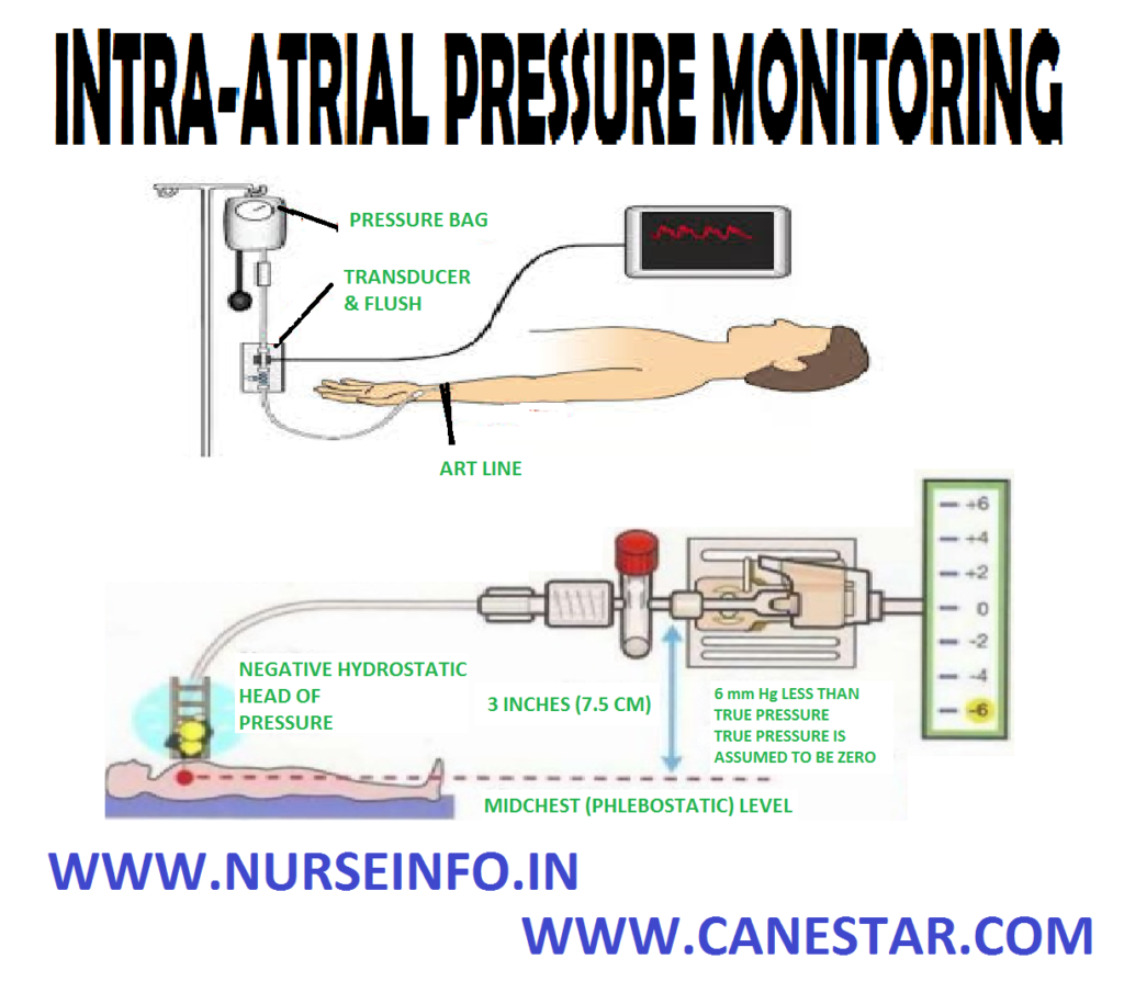
Intra-arterial pressure monitoring is a continuous and direct method of monitoring blood pressure by passing a catheter into an artery and connecting it to an electronic pressure sensor called transducer

FORMULA

Arterial pressure equals the flow through the vessel multiplied by the resistance to that flow. SVR-systemic vascular resistance

AP = CO/SVR

Normal AP range between 90/60 mm Hg and 140/90 mm Hg

PURPOSE

  • To provide continuous and more accurate intra-arterial pressure reading
  • To detect dangerously high or low blood pressure
  • To provide continuous access to arterial blood for analysis

INDICATION

  • Clients with low cardiac output and high systemic vascular resistance
  • Clients who are receiving titrated doses of vasoactive drugs or who need frequent blood sampling
  • Clients with very high and low blood pressure
  • Pulmonary embolism and cardiac tamponade
  • Extensive burns with limited intact skin surfaces

COMPONENTS

  • Intra-arterial catheter: this is introduced into an artery, from where the pressure waves are transmitted to the manometer (transducer)
  • Transducer: this is attached to the intra-arterial catheter. The transducer converts the mechanical signal (pressure) to the electrical signal (voltage)
  • Bedside monitor: this amplifies the signal coming for the transducer and displays it on the oscilloscope
  • Fluid source: this is to flash the intra-arterial catheter to prevent clotting of blood in it. Either normal saline or 5% dextrose is used to keep the arterial line patient
  • Heparin in a dose of 1 unit per ml is added to this solution; this may be increased to 4-5 units of heparin per ml when the client has history of thromboembolic diseases. It is delivered in small amounts using a flush device, without introducing the air

SPECIAL CONSIDERATIONS

  • Always watch for hemorrhage. Keep all the connections tight
  • Apply immediate pressure if the catheter is accidentally pulled out
  • Prevent the possibility of clot formation in the catheter
  • Provide a continuous infusion with heparin solution
  • Flush the catheter thoroughly after each blood specimen is withdrawn
  • Always inspect the extremity distal to the area of cannulation to detect any decrease in the circulation
  • Watch for cold fingers, cyanosis, and poor capillary filling
  • Prevent infection by maintaining strict aseptic technique and closed system, at all times
  • Never inject any medications though the arterial line
  • Keep the catheter site clean and dry to prevent skin maceration

CLIENT AND EQUIPMENT PREPARATION

  • Prepare the client with providing adequate explanations
  • Obtain informed consent
  • Instruct the client needs to clench his hand tightly at the time of cannula insertion
  • Before setting up and priming the monitoring system, wash hands thoroughly
  • Maintain asepsis by wearing personal protective equipment throughout preparation
  • Check the client’s history for an allergy or a hypersensitivity to iodine or the other local anesthetic agents

EQUIPMENT NEEDED

For catheterization:

  • Gloves gown and mask
  • Protective eye wear
  • Sterile gloves
  • 16G-20G catheter
  • Preassembled preparation kit
  • Sterile drapes
  • Sheet protector
  • Sterile towels
  • Prepared pressure transducer system
  • Local anesthetic agent (lignocaine 2%)
  • Sutures
  • Syringe and needle (21G-25G)
  • IV pole
  • Tubing and medication labels
  • Site care set (containing sterile dressing, antimicrobial ointment and hypoallergic agent)
  • Arm board and soft wrist restraints (formal site, ankle restraints)
  • Optional: shaving kit

FOR BLOOD SAMPLE COLLECTION

  • Gloves gown and mask
  • Sterile 4”/4” gauze pads
  • Protective eye wear
  • Sheet protector
  • 500 ml IV bag
  • 5 or 10 ml syringe for discard sample
  • Syringe of appropriate size and number for ordered laboratory tests
  • Laboratory request forms and labels
  • Vacutainers

PROCEDURE

  • Maintain asepsis by wearing personal protective equipment
  • Position the client for easy access to the catheter insertion site
  • Place the sheet protector under the site
  • Insert an arterial catheter by using preassembled preparation kit, the doctor prepares and anesthetizes the insertion site
  • The catheter is then inserted in to the artery and attached to the fluid-filled pressure tubing
  • The doctor may suture the catheter in place
  • Apply antimicrobial ointment and cover the insertion site with dressing
  • Immobilize the insertion site by using an arm board and soft wrist restraint
  • Activate monitor alarms as appropriate
  • Observe the pressure waveform on the monitor can enhance assessment of arterial pressure

AFTER CARE

  • Change the pressure tubing every 2-3 days, according to facility policy
  • Change the dressing at the catheter site at intervals specified by facility policy
  • Regularly assess the site for signs of infection, such as redness and swelling
  • Notify the doctor immediately if you note any such sign
  • Documentation

COMPLICATION

  • Arterial bleeding
  • Infection
  • Air embolism
  • Arterial spasm
  • Thrombosis
INTRA-ATRIAL PRESSURE MONITORING – Definition, Formula, Purpose, Indication, Components, Special Considerations, Client and Equipment Preparation, Equipment Needed, For Blood Sample Collection, Procedure, After Care and Complication (NURSING PROCEDURE)

ERGONOVINE PROVOCATION TEST

ERGONOVINE PROVOCATION TEST – Purpose, Finding, Interfering Factors, Client Preparation, Procedure, After Care and Complications (NURSING PROCEDURE)

Ergonovine provocation testing is used to diagnose coronary artery spasm and vasospastic angina accurate diagnosis of coronary artery spasm is necessary, because of its risk, ergonovine provocation testing is limited to carefully selected clients

PURPOSE

Ergonovine provocation testing is indicated in clients with atypical angina in whom coronary artery spasm is suspected

FINDING

A positive response to ergonovine includes chest pain with ST segment abnormalities, spasm visible on the arteriogram, serious dysrhythmia or a combination of those responses

INTERFERING FACTORS

Ergonovine provocation testing should not be performed if severe obstruction of a coronary artery or multivessel obstructive cardiac disease exits or in the presence of severe congestive heart failure, uncontrolled hypertension, pregnancy, acute myocardial infarction or possible cerebral hemorrhage. It is contraindicated in anyone with a history of hypersensitivity to ergonovine

CLIENT PREPARATION

  • Warn the client that chest pain is expected but will be treated immediately
  • Discontinue vasoactive medications: nitrates for 4 hours before the test, calcium channel blockers for 24 hours before rest and beta blockers for 48 hours before the test
  • Continue cardiac and hemodynamic monitoring is performed

PROCEDURE

  • The ergonovine provocation test is usually performed as part of a cardiac catheterization
  • First, the cardiac catheterization must rule out severe coronary artery obstruction
  • A pacing wire is then inserted
  • Intravenous ergonovine maleate (ergotrate maleate) is given, which will stimulate a spasm within 3 to 6 minutes
  • Its effect lasts 10 to 15 minutes. If a positive response occurs, the spasm is reversed by administering nitroglycerin intravenously
  • Bedside ergonovine provocation testing can be performed in the coronary care unit in clients who have had a cardiac catheterization to verify that the coronary arteries are not severely obstructed
  • Ergonovine is given intravenously every 5 minutes up to seven times
  • The ergonovine is stopped when ST segment changes are seen on the monitor, whether the client has pain or not. Nitroglycerin is given to reverse the spasm
  • Continuous cardiac and hemodynamic monitoring is performed. If major adverse effects are noted, the ergonovine is stopped

AFTER CARE

  • Assess the client for chest pain with or without ST segment changes, as spasm may recur after the nitroglycerin is stopped
  • Maintain monitoring and bed rest for 1-2 hours if the procedure is performed in the coronary care unit

COMPLICATION

  • Atypical chest pain
  • Bronchospasm
  • Dysrhythmia
  • Hypotension or hypertension
  • Myocardial infarctions
ERGONOVINE PROVOCATION TEST – Purpose, Finding, Interfering Factors, Client Preparation, Procedure, After Care and Complications (NURSING PROCEDURE)

ENDOMYOCARDIAL BIOPSY

ENDOMYOCARDIAL BIOPSY – Purpose, Findings, Interfering Factors, Client Preparation, Procedure, After Care and Complications (NURSING PROCEDURE)

Endomyocardial biopsy is an invasive procedure requiring cardiac catheterization. It permits sampling of right or left ventricular tissue

PURPOSE

  • An endomyocardial biopsy is usually performed to determine if a transplanted heart is being rejected
  • To diagnose myocarditis or doxorubicin (adriamycin) induced cardiomyopathy
  • To determine the cause of restrictive heart disease

FINDINGS

  • Doxorubicin-induced cardiomyopathy
  • Cardiac amyloidosis
  • Cardiac fibrosis (especially radiation injury)
  • Changes cardiomyopathy
  • Myocarditis
  • Rejection of transplanted heart
  • Scleroderma
  • Toxoplasmosis
  • Tumor infiltrates
  • Vasculitis

INTERFERING FACTORS

  • Bleeding disorders
  • Severe thrombocytopenia
  • Systemic anticoagulation
  • Uncooperative client

CLIENT PREPARATION

  • Instruct the client about the purpose and procedure
  • Inform the client that the table rotates and that the physician may ask the client to change positions or cough
  • Explain the client that when the dye is given, a feeling of warmth or flushing or a metallic taste may be tensed
  • Assist the precatheterization evaluation: blood test, including a prothrombin time test and a partial thromboplastin time test; an electrocardiogram; and chest X-ray
  • Obtain client’s height and weight
  • Assess the client fear and anxiety. Correct any misconceptions and reassure the client that the nurse, physician and technicians to assist during  the procedure will be continuously present
  • If contrast dye is going to be used, check all allergies
  • Keep the client nothing per oral after midnight, except if the catheterization is planned for late in the afternoon. In that case, a clear liquid breakfast may be given
  • Withhold the cardiac drugs as per the physician order
  • Prepare catheter site according to laboratory protocols. The femoral artery is commonly used for the percutaneous of the catheter. Usually, both side of the groin are prepared
  • Premedication is given as ordered to reduce the client’s anxiety. In some catheterization laboratories, the client is premediated to decrease the risk of allergic reaction to the contrast dye
  • Instruct the client to void before going to the catheterization laboratory

PROCEDURE

  • The procedure involves a cardiac catheterization
  • A catheter with a jaw like tip id inserted under fluoroscopy, and several small tissue samples are obtained
  • A right or left ventricular sample may be taken. For clients at high risk, such as those with a history of left ventricular thrombus or infarction, a right ventricular biopsy may be performed

AFTER CARE

  • Observe the insertion site of bleeding. Palpate around the punctured site to detect bleeding into tissue
  • If bleeding is present, exert pressure just proximal to the puncture site with a gloved hand for a minimum of 15 minutes
  • Monitor vital signs and cardiac monitor according to hospital protocol
  • Check the distal pulse for artery patency
  • Report immediately if any significant changes in vital signs, rhythm and circulation or occurrence of chest pain
  • Assess post-procedure laboratory values, such as blood count, prothrombin time, electrolytes and creatine
  • Instruct the client about strict bed rest for 12-24 hours and to keep affected extremity straight for 12 hours
  • Encourage plenty of oral fluids
  • Record type of cardiac catheterization done and client’s tolerance of the procedure

COMPLICATIONS

  • Accidental biopsy of papillary muscle or chordate tendineae
  • Hemopericardium
  • Cardiac perforation
ENDOMYOCARDIAL BIOPSY – Purpose, Findings, Interfering Factors, Client Preparation, Procedure, After Care and Complications (NURSING PROCEDURE)

ELECTROMYOGRAPHY

ELECTROMYOGRAPHY – Definition, Purpose, Client Preparation, Procedure and After Care (NURSING PROCEDURE)

An electromyography (EMG) or electromyogram is obtained by introducing needle electrodes into the skeletal muscles in order to study changes in the electrical potential of the muscles and the nerves leading to them. The electrical potentials are shown on an oscilloscope and amplified by a loudspeaker so that both the sounds and the appearance of the waves can be analyzed and compared simultaneously

DEFINITION

An electromyography is the recording of muscle activity at rest, during voluntary movement and with electrical stimulation is made by inserting small needle into the muscle

PURPOSE

  • It is useful in detecting the presence of a neuromuscular disorder and myopathies
  • It helps to distinguish weakness due to neuropathy
  • To help in the diagnosis of myasthenia gravis
  • To find out peripheral nerve injury or disease
  • To find out whether primary problem is in nerve or muscle
  • To differentiate among lesions of the anterior horn cell, root, plexus and specific nerve and muscle

CLIENT PREPARATION

  • Inform the client that he will experience a sensation similar to that of an intramuscular injection as the needle is inserted into the muscles
  • Explain the client that the muscles examined may ache for a short time following the procedure
  • Explain the procedure and ensure cooperation from the client
  • The client should have breakfast half to one hour before to the procedure
  • Provide cotton dress for the client

PROCEDURE

  • Place the client on the examination table comfortably
  • Explain the client that needle electrodes are going to place on the skin
  • Insert a needle electrode into a muscle
  • Nerve conduction studies are performed by stimulating a peripheral nerve at several points along its course and reading the muscle action potential or the sensory action potential that result
  • Surface or needle electrodes are placed on the skin over the nerve to stimulate the nerve fibers
  • The analysis of motor unit potentials both at rest and in varying degrees of muscle contraction

AFTER CARE

  • Provide bed rest for sometime
  • Monitor the vital signs
  • Provide some mild analgesic to the client
ELECTROMYOGRAPHY – Definition, Purpose, Client Preparation, Procedure and After Care (NURSING PROCEDURE)

CARDIAC ENZYME TEST

CARDIAC ENZYME TEST – Definition, Purposes, Client Preparation, Procedure, After Care and Enzyme Study Interpretation (NURSING PROCEDURE)

Enzymes are special proteins that catalyze chemical reactions in living cells. Cardiac enzymes are organ specific enzymes that are present in high concentration in myocardial tissue. Tissue damage causes a reaction of enzymes from their intracellular storage areas. For example, myocardial infarction causes cellular anoxia, which alters membrane permeability and causes spillage of enzymes into the surrounding tissue. This leakage of enzymes can be detected by rising plasma levels.

DEFINITION

Cardiac enzymes are complex compounds that are found in all tissues and that speed up the biochemical reactions of the body. Damage to body tissue causes release of the enzymes from injured cells

PURPOSE

  • To detect myocardial infarction
  • It is used in clinical evaluation and electrocardiography studies to diagnosis myocardial injury

CLIENT PREPARATION

  • Explain the procedure to the client
  • Prepare the client for venipuncture
  • Reassure the client, who is usually frightened and having chest pain and may also be in denial
  • Do not give intramuscular injections or perform repeated venipunctures, if possible, until all the initial enzyme studies are completed
  • Instruct the client about the need to repeat blood sampling
  • Determine if alcohol or drugs that affect results have been ingested

DIFFERENT TYPES OF CARDIAC ENZYMES

  • Myoglobin
  • CK-MB (creatine kinase myocardial bound
  • Troponin I
  • AST (aspartate aminotransferase)
  • LDH (lactate dehydrogenase)

PROCEDURE

  • Place the client in comfortable position
  • Place the tourniquet in a proper, if the tourniquet is in place too long, inaccurate result may occur
  • A venipuncture is necessary to obtain 5-10 ml of blood in a red-topped tube
  • Results of the enzyme levels will determinate need for admission to the hospital and emergency treatment
  • Assay of CK-MB can be done in the emergency room, with results available within 20 minutes

AFTER CARE

  • Remove the tourniquet
  • Apply gentle pressure over the punctured site
  • Place the collected blood in a correct specimen tubes
  • Sent the blood to laboratory with proper labeling and request
  • Replace the articles to the treatment room
  • Record the entire procedure in a nurse’s record

ENZYME STUDY PREPARATION

  • The enzyme most commonly used to detect myocardial infarction is creatine kinase (CK) and lactic acid dehydrogenase (LDH). CK is an enzyme found in the heart, brain and skeletal muscles. The CK isoenzymes include CK-MM, CK-MB and CK-MB
  • Isoenzymes are the various forms of CK and LDH, identified only by a process known as electrophoresis. There are three isoenzymes of CK: CK-MM (skeletal muscle), CK-MB (myocardial muscle) and CK-BB (brain)
  • Elevated CK-MB indicates myocardial damage. Elevation of CK-MB may occur within 4-6 hours and peaks 18-24 hours after the acute ischemic event
  • There are five isoenzymes for LDH (numbered 1 to 5), of which only LHD1 and LDH2 are cardiac-specific
  • If the serum concentration of LDH1 is higher than the concentration of LDH2, the pattern is said to have flipped, signifying myocardial necrosis. 80% of individuals demonstrate evaluates in LDH within 48 hours after myocardial infarction
CARDIAC ENZYME TEST – Definition, Purposes, Client Preparation, Procedure, After Care and Enzyme Study Interpretation

VECTORCARDIOGRAM

VECTORCARDIOGRAM – Purpose, Client Preparation, Procedure, After Care and Findings (NURSING PROCEDURE)

A vectorcardiogram is a graphic recording of electric forces of the heart. it is a noninvasive procedure that graphically records the direction and magnitude of the heart’s electric forces by means of a continuous series of vector loops. Three planes of the heart are recorded (frontal, sagittal and horizontal)

PURPOSE

A vectorcardiogram is used to assess ischemia, conduction defects and chamber enlargement (hypertrophy or dilation)

CLIENT PREPARATION

  • Explain to the client the purpose and procedure for vector cardiogram, no risk is involved
  • No present restrictions are required
  • Because electrode is applied to the four extremities and the chest clothing should permit easy access
  • If the male client’s chest is excessively hairy, the sites may need to be shaved

PROCEDURE

  • Establish a relaxed environment
  • Place the client in a supine position
  • Conduction jelly is placed on the electrodes and the electrodes are applied and recording is made

AFTER CARE

  • Remove the conduction jelly
  • Help the patient to a comfortable position

FINDINGS

  • Axis deviation
  • Conduction disturbances
  • Dysrhythmia
  • Hypertrophy of the ventricles
  • Therapeutic drug effects or toxicity
VECTORCARDIOGRAM – Purpose, Client Preparation, Procedure, After Care and Findings (NURSING PROCEDURE)

TRANSESOPHAGEAL ECHOCARDIOGRAPHY

TRANSESOPHAGEAL ECHOCARDIOGRAPHY – Definition, Purpose, Equipment, Pre-TTE Care, During Procedure and Post-TEE Care (NURSING PROCEDURE)

DEFINITION

It involves obtaining images of heart bypassing transesophageal echocardiography (TEE) ultrasound waves through a transducer threaded via mouth and into esophagus

PURPOSE

  • To obtain precise information in guiding surgical interventions, e.g. myocardial revascularization, repair of congenital heart defects
  • To provide clearer images than regular echocardiogram because ultrasound waves pass through less tissue
  • To give clearer visibility of heart and it structures in patient with obesity and chronic obstructive pulmonary disease
  • To aid diagnosis of cardiac mass, prosthetic valve function, aneurysm and pericardial effusions
  • To assess cardiac status of patient with known cardiac disease undergoing noncardiac procedures and during cardiac surgery

EQUIPMENT

ECG machine, echo machine, skin electrodes and lead wires BP apparatus or electronic monitoring machine, oxygen prongs, endoscope and emergency equipment

Pre-TEE CARE

  • Explain procedure and get the consent
  • Ask patient to withhold food and fluids for 6-8 hours to procedure
  • Start IV line
  • Prepare skin if chest is hairy
  • Explain that procedure will be done in cardiac catheterization laboratory
  • Administer usual dose of morning drugs as ordered with small sips of water
  • Make sure that baseline investigations are done such as blood count, electrolyte levels, ECG and regular echocardiogram
  • Remove dentures/any oral prosthesis
  • Administer stat dose of IV antibiotics if patient has artificial heart valve, congenital heart disease or history of infective endocarditis
  • Administer sedative as prescribed

DURING PROCEDURE

  • Administer sedation and topical anesthesia to back of throat as ordered
  • Attach ECG electrodes to chest and to electronic monitor
  • Administer oxygen if indicated through nasal prongs
  • If patient has nasogastric tube in place, remove before esophageal scope is inserted
  • Monitor vital signs including BP and respiratory status throughout procedure
  • Transfer patient to ward when vital signs are stable

Post-TEE Care

  • Monitor vital signs till stable
  • Position patient in upright/side lying position to support ventilation
  • Advise nil oral until gag reflex is fully restored
  • Explain that mild throat discomfort will be present for a day or two
  • Instruct to report to physician immediately if any significant throat discomfort, hemoptysis, dyspnoea, chest pain, etc
  • Advise to take throat lozenges and saline gargles/rinses to alleviate discomfort
TRANSESOPHAGEAL ECHOCARDIOGRAPHY – Definition, Purpose, Equipment, Pre-TTE Care, During Procedure and Post-TEE Care (NURSING PROCEDURE)


STRESS TESTING

STRESS TESTING – Purpose, Indications, Interfering Factors, Client Preparation, Procedure, Findings, After Care and Contraindications

Stress testing is an important noninvasive procedure for evaluating the cardiovascular status of clients who are known to have cardiac disease or who are at risk for cardiac diseases. The test increases the demand placed on the heart by increasing physical activity. Through electrocardiography tracings, it is determined whether the heart is able to meet the increased oxygen demand

PURPOSE

  • To assess the at-risk population
  • Diagnosing chest pain syndromes and dysrhythmia associated with ischemia
  • Evaluating the effectiveness of therapy (surgical or pharmacological)
  • Identifying the initial level of function in cardiac rehabilitation programs and evaluating the results

INDICATIONS

Class – I (Clear Indications for Stress Testing)

  • Suspected or proven coronary artery disease
  • Male client who present with atypical chest pain
  • Evaluate functional capacity and assess prognosis of clients with coronary artery disease (CAD)
  • Clients with exercise related palpitation, dizziness or syncope
  • Evaluate of recurrent exercise-induced arrhythmias

Class – II (Stress Testing may be Indicated)

  • Evaluation of typical or atypical symptoms in women
  • Evaluation of variant angina
  • Evaluation of clients who are on digoxin preparations or who have a right bundle-branch block

Class – III (Stress Testing is Probably Necessary)

  • Young or middle-age asymptomatic clients who have no risks factors for CAD
  • Young or middle-age asymptomatic clients

INTERFERING FACTORS

  • Severe anxiety may interfere with the client’s ability to participate fully in the stress testing
  • False-positive results may due to bundle branch block, ventricular hypertrophy or digitalization
  • False-negative results may be due to the use of beta-blockers

CLIENT PREPARATION

  • Inform the client about purpose and procedure of the test
  • Instruct the client to wear comfortable cloths and rubber-soled walking shoes
  • Instruct the client not to eat, smoke or drink alcohol for 3-4 hours before the test
  • If adenosine stresses testing being done, instruct the client to avoid theophylline-based drugs, diyridamole, over-the-counter drugs and caffeine for 24 hours
  • Routine cardiac medications are usually continued
  • Assess the following contraindications: chest pain, hypertension, thrombophlebitis, second or third degree heart block, serious dysrhythmia, and severe congestive heart failure, neurological, musculoskeletal or vascular problems that would impede mobility on the bicycle or treadmill
  • Warn the client that he or she will feel his or her racing and instruct the client to report chest pain during the procedure
  • Have emergency equipment and drug available
  • The client is attached to electrodes for recording a 12 lead ECG
  • A blood pressure cuff is put in place for quick access. A baseline blood pressure reading is obtained

PROCEDURE

  • Stress testing requires the use of a bicycle ergometer or a treadmill with continuous electrocardiac recording
  • The test is performed in a series of stages in which the client exercise for 3 minutes. Verities of protocols are used in stress testing
  • The Bruce protocol involves gradual increase in speed and intervals of short duration
  • At the end of each stage, a 12 lead ECG is recorded. After each stage, the work load or graded load is increased
  • This is accomplished by increasing the speed or resistance of the bicycle or treadmill
  • The stress testing continues until the client reaches 85% of the maximum heart rate, becomes symptomatic or displays electrocardiography changes consistent with ischemia
  • The maximum heart rate is usually determined by normograms. A gross estimate of the maximum heart rate is 220 beats per minute the client’s age
  • If the client is physically unable to exercise to the point of 85% of the maximum heart rate, a dipyridamole (persantine) scan may be performed
  • Dipyridamole may be given intravenously or by mouth. It causes coronary artery dilatation similar to the response of the coronary arteries to exercise
  • As the graded exercise begins, a multichannel ECG is recorded. A 12 lead ECG is recorded and the blood pressure is checked as each workload ends (every 3 minute increment)
  • Observe for signs to stop the stress testing, for example, falling blood pressure, three consecutive premature ventricular contractions, chest pain or exhaustion

FINDINGS

A 1 mm depression of the ST-segment is positive stress test, indicating myocardial ischemia

AFTER CARE

  • Cardiac monitoring is continued for 5-10 minutes after the testing to evaluate the client’s physiologic responses
  • Blood pressure is checked
  • Remove conduction jelly and assist in robbing the client if necessary
  • Evaluate the client’s physical and emotional response to testing
  • Instruct the client to rest and not to take hot shower or baths for 2-4 hours

CONTRAINDICATIONS

  • Uncontrolled arrhythmias
  • Uncorrected valvular diseases
  • Cardiomegaly
  • Severe anemia
  • Unstable angina
  • Uncontrolled hypertension
  • Congestive heart failure
  • Pericarditis and myocarditis
STRESS TESTING – Purpose, Indications, Interfering Factors, Client Preparation, Procedure, Findings, After Care and Contraindications

ROENTGENOGRAM

ROENTGENOGRAM – Purpose, Indications, Findings, Interfering Factors, Client Preparation, Procedure and After Care (NURSING PROCEDURE)

  • A cardiac roentgenogram is a routine screening procedure in clients suspected or known cardiac disorders. It provides information regarding the size of the heart, its shape and location of the cardiac structure and great vessels
  • The X-ray may also be used to evaluate pulmonary vasculature and determine the placement of evasive catheter and pacemaker wires

PURPOSE

  • Various views of the chest are usually obtained in a cardiac X-ray series. The four views commonly obtained are anterioposterior, lateral, right anterior oblique
  • During acute cardiac status, only portable chest X-ray studies are available to evaluate the client’s progress. Because the plate is positioned under the client, an anteroposterior view is obtained
  • This film is obtained routinely for hospitalized and preoperative clients to screen for tuberculosis and other serious pulmonary or cardiac diseases
  • It also provides a preoperative comparison film for the postoperative clients whom a pulmonary or cardiac complication develops, and it a basic radiological procedure for the clients with a suspected pulmonary disorder

INDICATIONS

  • The chest X-ray provides data about the heart including its size and shape
  • In congenital and acquired cardiac disease, the enlargement of the heart and its atria or ventricles provides information about the improper function of the cardiac valves, pulmonary or aortic arterial hypertension and venous pulmonary conduction that affect heart size

FINDINGS

  • Pneumothorax
  • Atelectasis
  • Pleural effusion
  • Pleurisy
  • Cystic fibrosis
  • Pulmonary fibrosis
  • Tumor or cyst
  • Silicosis

INTERFERING FACTORS

  • Excessive movement
  • Failure to remove jewelry or other metal from the X-ray field
  • Improper positioning

CLIENT PREPARATION

  • Instruct the client to remove all clothes and put on a hospital gown
  • Instruct the client to remove all jewelry and metal objects from the area that is to be imaged
  • Provide reassurance to the client. Young children often fear the equipment, strange room, isolation and separation from the parents

PROCEDURE

  • Ensure the client’s safety at all times, particularly when there is a risk of the client’s falling
  • The radiography table has no side rails. A Velcro waist restraint may be used, but sometimes the restraint interferes with the positioning and imaging needed
  • Position the client for the specific views needed
  • Instruct the client to remain motionless during the imaging
  • Sometimes the client is instructed to inhale deeply and hold the breath until the image is taken
  • The client must often wait in the imaging areas as the decision is made concerning whether to take additional X-ray films
  • Provide a blanket or extra gown for the client who is chilled in the cool room

AFTER CARE

  • Assist the client in dismounting from the radiography table and getting dressed, as needed
  • Record the entire procedure in the nurse’s record

PHONOCARDIOGRAM

PHONOCARDIOGRAM – Purpose, Findings, Interfering Factors, Client Preparation, Procedure and After Care

  • A phonocardiogram is a graphical recording of cardiac sounds. It is a noninvasive test that amplifies cardiac sounds, which are recorded simultaneously with the electrocardiograph reading
  • Phonocardiography is the graphic recording of heart sounds and pulse waves and their relation to time. It helps to identify, accurately time and differentiate various sounds and murmurs. It provides a permanent record for future comparison

PURPOSE

The phonocardiogram if performed to determine the exact timing of heart sounds, differentiate the varied sounds such as murmurs, splits and clicks and evaluate valvular function

FINDINGS

  • Valve disorders (stenosis or incompetence)
  • Estimate of ventricular function
  • Hypertrophic cardiomyopathies

INTERFERING FACTORS

  • Improper placement
  • Muscle tremors
  • Obesity
  • Valsalva maneuver

CLIENT PREPARATION

  • Inform the client as to the purpose and procedure of the study
  • Explain to the client that the test is painless and that no risk is involved
  • Instruct the client to remain quiet and still during the procedure

PROCEDURE

  • Electrocardiograph electrodes are applied and attached to the electrocardiogram recorder
  • Conduction jelly is applied to the chest wall
  • As the phonogram microphone is positioned at various sites over the chest wall, the client may be asked to change position, perform muscle tightening or change breathing patterns
  • Phonocardiogram may be carried out at the bedside, in the physician’s office, inside the clinic or in a cardiac laboratory

AFTER CARE

  • Remove conduction jelly from the chest and extremities
  • Record the entire procedure in the nurse’s record
PHONOCARDIOGRAM – Purpose, Findings, Interfering Factors, Client Preparation, Procedure and After Care
Nurse Info