Home Blog Page 63

Types of Headache

TYPES OF HEADACHE – Chronic Tension Headache, Migraine Headache and Cluster Headache

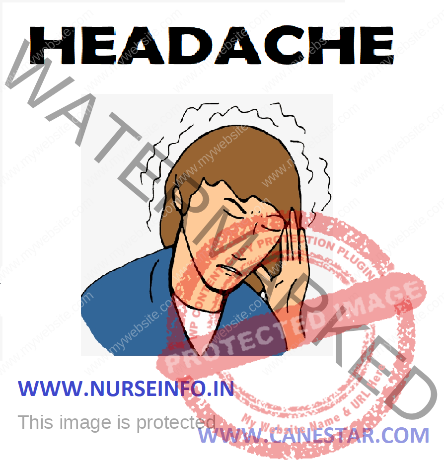
Headache is pain in any region of the head. Headaches may occur on one or both sides of the head, isolated to a certain location, radiate across the head from one point, or have a vise-like quality. A headache is a sharp pain, throbbing sensation or dull ache. Headaches may appear gradually or suddenly, and they may last less than an hour or for several days.

TYPES OF HEADACHE

  1. Chronic Tension Headache
  2. Migraine Headache
  3. Cluster Headache

CHRONIC TENSION HEADACHE

Chronic tension-type headaches maybe the result of stress or fatigue, but more than likely, they can be attributed to physical problems, psychological issues, or depression.  A pattern of chronic tension-type headaches generally begins between the ages of 20 and 40, and every personality type can experience them.

Symptoms

  • The muscles between head and neck contract for hours and days
  • A tightness around neck or even feel as if head and neck were in a cast and only certain positions seem to provide relief
  • Feeling of soreness, a tightening band around head (a ‘vice-like’ ache), a pulling, or pressure sensations
  • The pain is continuous, annoying, but not throbbing
  • Headache primarily occurs in forehead, temples or the back of head and neck
  • Changes in sleep patterns if headaches are related to anxiety, then you may have trouble falling asleep or may suffer from insomnia. If headaches are associated with depression, then you may awaken frequently during the night, awaken before you wanted to in the morning, or you may be sleeping excessively (hypersomnia).
  • Shortness of breath
  • Constipation
  • Nausea
  • Weight loss
  • Ongoing fatigue
  • Decreased sexual drive
  • Palpitations
  • Dizziness
  • Unexpected crying
  • Menstrual changes

Etiology and Risk Factors

  • Poor posture, close work under poor lighting conditions, or cramps from assuming an unnatural head or neck position for long periods of time
  • Arthritis, particularly cervical arthritis
  • Abnormalities in neck muscles, bones or disks
  • Eye strain caused when one eye is compensating for another eye’s weakness
  • Misalignment of teeth or jaws
  • Noise or lighting
  • Job conflicts and family relationships
  • Grief
  • Depression

Management

  • There are two goals when treating any type of headache: prevent future attacks, abort or relieve current plan
  • Prevention includes taking prescribed medications, avoiding or minimizing the causes, and learning self-help measures, such as biofeedback or relaxation exercises
  • NSAIDs (Nonsteroidal Anti-inflammatory Agents), fenoprofen, flurbiprofen, ketoprofen
  • Antidepressants – Tricyclics (nonsedating), protriptyline, desipramine
  • Antidepressants – tricylics (sedating), amitriptyline, doxepin

MIGRAINE HEADACHE

Migraines deserve the attention they receive, one headache can put the life ‘on hold’ for a few hours or several days. Migraine is responsible for more job absenteeism and disrupted family life than any other headache types.

Symptoms

Migraine often begins as a dull ache and then develops into a constant, throbbing and pulsating pain at the temples, as well as the front or back of one side of the head. The pain is usually accompanied by nausea and vomiting, and sensitivity to light and noise

The two most prevalent types of migraine are migraine with aura (formerly referred to as classic migraine) and migraine without aura (formerly referred to as common migraine).

Etiology

Physical and Environmental Causes

  • Stress
  • Fatigue
  • Oversleeping or lack of sleep
  • Fasting or missing a meal
  • Food or medication that affects the diameter of blood vessels
  • Caffeine
  • Chocolate
  • Alcohol
  • Menses
  • Hormonal changes
  • Changes in barometric pressure
  • Changes in altitude

Food and Diet

Specific foods are suspected of triggering at least 30 percent of the migraine headaches. Foods that contain: additives such as nitrates and nitrites (usually in processed meats), yellow (annatto) food coloring, and MSG (monosodium glutamate). Canned or processed foods, Chinese foods, tenderizer, and seasonings such as soy sauce may contain MSG.

Tyramine

Red wines and most alcoholic beverages, aged cheeses and processed meats (including pizza and hot dogs), peanuts, chicken livers, pickled foods, sourdough bread, bread and crackers containing cheese, broad beans, peas, lentils. Foods to eat in moderation include: avocados, bananas, citrus fruits, figs, raisins, red plums, raspberries, and chocolates

Management

  • There are two goals when treating migraine, or any other headache: to relieve the pain and prevent future attacks
  • Once migraine has been diagnosed, treatment will begin by identifying those circumstances or factors that trigger it
  • Keep a daily calendar of activities, foods, beverages, prescription and over-the-counter medications, physical and environmental factors, stressful situations, sleep patterns, and characteristics of the headache itself.
  • Beta-blockers, propranolol, timolol
  • Calcium channel blockers, verapamil, diltiazem
  • Antiepilepsy medication, divalproex sodium, neurontin
  • NSAIDs, fenoprofen, flurbiprofen
  • Antidepressants – tricyclics (nonsedating), protriptyline, desipramine

Self-Help Treatments for Migraine and Tension-Type Headaches

  • Counseling and psychotherapy
  • Relaxation training
  • Progressive muscle relaxation
  • Guided imagery
  • Biofeedback
  • Acupuncture
  • Physical and massage therapy

CLUSTER HEADACHE

In this the attacks come in groups. The pain arrives with little, if any, warning, and it has been described as the most severe and intense of any headache type. It generally lasts from 30 to 45 minutes, although it might persist for several hours before it disappears.

Cluster headaches frequently surface during the morning or late at night, the cluster cycle can last weeks or months and then can disappear for months or years.

Symptoms

  • The headache is usually unilateral and rarely switches sides from one attack to another
  • One might feel the pain begin around one eye, ‘like a nail or knife stabbing or piercing’ the eye, or as if someone ‘were pulling out’ eye, it may be accompanied by a tearing or bloodshot eye and a runny nose on the side of the headache.
  • It can radiate from the eye to the forehead, temple and cheek on the same side
  • The pain of a cluster headache has been described as piercing, burning, throbbing, and pulsating

Etiology

Unlike migraine headaches, cluster headaches are not the result of heredity. Sufferers, however, usually do have a history of chronic smoking, and alcohol frequently triggers a cluster headache.

Because the level of histamine increases in a person’s blood and urine during a cluster headache, which dilate or expand blood vessels, influence a cluster headache.

Management

Verapamil, prednisone, ergotamine tartrate, lithium carbonate, divalproex sodium, histamine acid phosphate

TYPES OF HEADACHE – Chronic Tension Headache, Migraine Headache and Cluster Headache

TYPES OF HEADACHE – Chronic Tension Headache, Migraine Headache and Cluster Headache

Tuberculosis

TUBERCULOSIS – Etiology, Risk Factors, Types, Pathophysiology, Clinical Manifestations, Diagnostic Evaluation, Management (Medical and Nursing)

TUBERCULOSIS

Tuberculosis is an infectious bacterial disease called by Mycobacterium tuberculosis, which most commonly affects the lungs. It is transmitted from person to person via droplets from the throat and lungs of people with the active respiratory disease.

ETIOLOGY

  • Mycobacterium tuberculosis
  • Immunocompromised person
  • Chemical industries

RISK FACTORS

  • Aging
  • Alcoholism
  • Crowded living conditions
  • Diseases that weaken the immune system
  • Health care workers
  • HIV infection
  • Homelessness
  • Low socioeconomic status
  • Malnutrition, migration from a country with a high number of cases
  • Nursing homes
  • Unhealthy immune system

TYPES

  • Pulmonary tuberculosis: if a tuberculosis infection does become active, it most commonly involves the lungs (in about 90% of cases). Symptoms may include chest pain and prolonged cough-producing sputum.
  • Extrapulmonary tuberculosis: in 15-20% of active cases, the infection spreads outside the lungs, causing other kinds of TB. These are collectively denoted as ‘extrapulmonary tuberculosis’. Extrapulmonary TB occurs more commonly in immunosuppressed persons and young children.
  • Active tuberculosis: active TB means the bacteria are active in the body. The immune system is unable to stop these bacteria from causing illness. People with active TB in their lungs can pass the bacteria on to anyone they come into close contact with. When a person with active TB coughs, sneezes or spits, people nearby may breathe in the tuberculosis bacteria and become infected.
  • Inactive tuberculosis: inactive TB infection is also called latent TB. If a person has latent TB, it means their body has been able to successfully fight the bacteria and stop them from causing illness. People who have latent TB do not feel sick, do not have symptoms and cannot spread tuberculosis.

PATHOPHYSIOLOGY AND CLINICAL MANIFESTATIONS

  • A cough lasting for more than 2-3 weeks
  • Chest pain
  • Chills
  • Discolored or bloody sputum
  • Fatigue
  • Loss of appetite
  • Night sweats
  • Pain with breathing
  • Severe headache
  • Shortness of breath
  • Slight fever
  • Tiredness or weakness
  • Weight loss

DIAGNOSTIC EVALUATION

  • Injection of protein: by injecting a protein found in TB bacteria into the skin of an arm. If the skin reacts by swelling, then the person is probably infected with tuberculosis
  • X-ray: Diagnosis of tuberculosis in the lungs may be made using an X-ray
  • Sputum test: sample of sputum is tested in laboratory to diagnose the TB
  • Bacteria: a culture of TB bacteria can also be grown in a laboratory. However, this requires specialized and costly equipment and can take six to eight weeks to produce a result.

MANAGEMENT

Medical Management

The five basic or ‘first-line’ TB drugs are:

  • Isoniazid
  • Rifampicin
  • Pyrazinamide
  • Ethambutol
  • Streptomycin

Surgical Management

If medications are ineffective, there are three surgical treatments for pulmonary TB.

  • Pneumothorax, in which air is introduced into the chest to collapse the lung
  • Thoracoplasty, in which one or more ribs are removed
  • Removal of a diseased lung

Nursing Management

Goals

The goals of management are:

  • To control the inflammatory process
  • To relieve symptoms
  • To correct metabolic and nutritional problems and promote healing
  • To achieve the previous health status

Nursing Assessment

  • Promoting airway clearance
  • Advocating adherence to treatment regimen
  • Promoting activity and adequate nutrition
  • Preventing spreading of tuberculosis infection

Nursing Diagnosis

  • Ineffective airway clearance related to increased sputum
  • Risk of infection related to lower resistance of others who are around people
  • Ineffective breathing pattern related to inflammation
  • Hyperthermia related to the infection process
  • Fluid volume deficit related to fatigue due to lack of fluid intake
  • Activity intolerance related to fatigue
  • Imbalanced nutrition, less than body requirements related to decreased appetite
  • Ineffective management, therapeutic regimen related to lack of  knowledge about the disease process
  • Impaired gas exchange related to alveoli function decline

Intervention

  • Ineffective airway clearance:

Auscultate lungs for wheezing, decreased breath sounds, coarse sounds

Use universal precautions if secretions are purulent even before culture reports

Assess cough for effectiveness and productivity

Note sputum amount, color, odor, consistency

Send sputum specimens for culture as prescribed

Institute appropriate isolation precaution if cultures are positive

Use humidity to help loosen sputum

Administer medications, noting effectiveness and side effects

Teach effective deep breathing and coughing techniques

  • Risk for infection

Monitor sputum for changes indicating infection

Monitor vital signs

Teach patient and family the purpose and techniques for infection control, such as hand washing, patient covering mouth when coughing, and maintaining isolation if necessary

Teach patient the purpose, importance and how to take medications as prescribed consistently over the long-term therapy

  • Deficient knowledge

Determine who will be the learner-patient or family

Assess ability to learn

Identify any existing misconceptions about the material to learn

Assist the learner to integrate the information into daily life

Give clear thorough explanations and demonstrations

  • Activity intolerance

Assess patient’s level of mobility

Observe and document response to activity

Assess emotional response to change in physical status

Anticipate patient’s needs to accommodate

Teach energy conservation techniques

Refer to community resources as needed

  • Ineffective therapeutic regimen management

Assess prior efforts to follow regimen

Assess patient’s perceptions of their health problem

Assess other factors that may affect success in a negative way

Inform patient of the benefits of conforming with the regimen

Concentrate on the behaviors that will make the most difference to the therapeutic effect

Include family, support system in teachings and explanations

Health Education

  • Explain about the disease condition’s causes, risk factors
  • Use universal precautions if secretions are purulent even before culture reports
  • Assess cough for effectiveness and productivity
  • Note sputum amount, color, odor, consistency
  • Send sputum specimens for culture as prescribed or PRN
  • Institute appropriate isolation precautions if cultures are positive
  • Use humidity to help loosen sputum
  • Administer medications, noting effectiveness and side effects
  • Assess patient’s perceptions of their health problem
  • Assess other factors that may affect success in a negative way
  • Inform patient of the benefits of conforming with the regimen
  • Concentrate on the behaviors that will make the most difference to the therapeutic effect

Complications

  • Miliary tuberculosis
  • Pleural effusion
  • Emphysema
  • Tuberculosis pneumonia
TUBERCULOSIS – Etiology, Risk Factors, Types, Pathophysiology, Clinical Manifestations, Diagnostic Evaluation, Management (Medical and Nursing)

TUBERCULOSIS – Etiology, Risk Factors, Types, Pathophysiology, Clinical Manifestations, Diagnostic Evaluation, Management (Medical and Nursing)

Mental Health Nurse Function

FUNCTIONS OF MENTAL HEALTH NURSE IN VARIOUS SETTINGS

Practice Setting for Psychiatric Nurses

For many years, the majority of mental health care was provided in the hospital setting. Since the 1970s, the trend has changed to treat patients in less restrictive or community based settings.

        While traditional practice settings for psychiatric nurses are psychiatric hospitals, community mental health centers, psychiatric units in the general hospitals, residential treatment facilities and private clinics, more recently alternative treatment settings have emerged. These are partial hospitalization settings, day care centers, home care, outpatient departments or ambulatory care centers. Community based treatment settings have expanded to group homes, hospice, care centers, crisis intervention centers, schools and universities, hospitals for the criminally insane, jails and prisons.

FUNCTIONS OF PSYCHIATRIC NURSE IN VARIOUS SETTINGS

Inpatient Psychiatric Ward

  • Provide for environmental safety including protecting the patient and others from injury.
  • Perform psychosocial, high-risk and physical assessment.
  • Promotion of self-care activities.
  • Medication management
  • Assisting for somatic therapies
  • Accurately observing and documenting the patient’s behavior.
  • Providing opportunities for the patient to make his own decisions and to assume responsibility for his life.
  • Providing feedback to the patient based on observations of his behavior
  • Participation in various therapies, (psychotherapy, behavior therapy, group therapy, play therapy, family therapy, etc.) individual interactions, formal and informal group situations, role play, advocating on behalf of the patient and so forth.
  • Delivering psychoeducation
  • Counseling the patient and family members
  • Cooperating with other professionals in various aspects of the patient care thereby facilitating an interdisciplinary approach to care
  • Teaching social skills and stress management strategies
  • Discharge planning and community referral and follow-up care
  • Supervise the work of subordinated
  • Maintain ward cleanliness

Psychiatric Outpatient Department

  • Performing clinical assessment
  • Assisting for psychometric assessment
  • Assisting or providing psychotherapy or behavior therapy
  • Counseling the patient and family members
  • Conducting group therapy
  • Delivering psychoeducation

ECT Treatment Setting

  • Teaching the patient prior to ECT treatment
  • Preparing the patient for ECT
  • Providing care during the procedure
  • Assisting with post-treatment
  • Providing reassurance to reduce anxiety
  • Delivering psychoeducation regarding ECT.

Psychotherapy Unit

Nurses who possess a masters degree in psychiatric nursing and are certified clinical nurse specialists may conduct individual or group psychotherapy.

  • Establishing a therapeutic relationship with the patient
  • Providing an opportunity for the patient to release tension as problems are discussed
  • Assisting the patient in gaining insight about the problem
  • Providing opportunity to practice new skills
  • Reinforcing appropriate behavior as it occurs
  • Providing consistent emotional support

Day Care Centers or Day Hospitals

In day treatment programs patients return home at night.

  • Performing clinical assessment
  • Accurately observing and documenting the patient’s behavior
  • Medication management
  • Teaching social skills
  • Counseling patient and family members
  • Delivering psychoeducation
  • Providing occupational or recreational therapy and vocational assistance

Family Therapy Units

Psychiatric nurses’ work with families at all levels of functioning

  • Assessing individual and family needs and resources
  • Facilitation of a family’s use of positive coping strategies
  • Promote adaptive family functioning by teaching communication skills and problem solving skills
  • Delivering psychoeducation

Child Psychiatric Ward

  • Assessing for biological and psychological need of the child
  • Determine the child’s strengths and abilities and develop a care plan to maintain and enhance capabilities
  • Monitor the child’s developmental levels and initiate supportive interventions, such as speech, language or occupational skills as needed
  • Provide a safe therapeutic environment, including protecting the child and others from injury
  • Cooperate with other professionals in an interdisciplinary approach to care
  • Provide adequate environment stimulation
  • Teach the child adaptive skills, such as eating, dressing, grooming and toileting
  • Demonstrate and help the child to practice self care skills
  • Provide genetic counseling if necessary
  • Deliver psychoeducation
  • Medication management
  • Provide emotional support to the parents
  • Participate in various therapies (behavior therapy, play therapy, expressive therapies, bibliotherapy, etc.)

Home Setting

  • Assessment of symptoms
  • Teaching the patient and family regarding nutrition, exercise, hygiene and the relationship between physical and emotional health.
  • Stress management
  • Daily living skills (basic money management, for example, bank accounts, rent, utility bills, use of the telephone, grocery shopping etc.,)
  • Medication management – monitoring blood levels, signs and symptoms of overdose or toxicity, teaching on dosage, side effects and purposes.
  • Administration of parenteral injections
  • Venipuncture for laboratory analysis
  • Act as a case manager and coordinate an array of services that include physical therapy, occupational therapy, social work and community services
  • Appropriate referrals to community agencies
  • Provide supportive counseling and brief psychotherapy
  • Promotion of mental health and prevention of mental illnesses.

Community Mental Health Centers

  • Identification of patients in the community
  • Refer the patients to appropriate hospitals
  • Home visiting and providing direct care to the patients in the community
  • Follow up care with special emphasis on medication regimen, improvement made and side effects, patient’s occupational function
  • Conducting public awareness programs to remove misconceptions regarding mental disorders
  • Training of paraprofessional, community leaders, school teachers and other care giving professionals in the community
  • Management of resources planning and coordination
  • Direct services, like care of families at risk for violence, abuse and dysfunction, care of homeless mentally ill patients, etc.,

Various Roles of Community Psychiatric Nurse

  • Addiction counselor
  • Counselor
  • Crisis worker
  • Advocate
  • Case manager
  • Educator
  • Researcher
  • Community developer and consultant

Hospice Care Centers

  • Helping cancer patients or terminally ill individuals through the grieving process
  • Provide supportive psychotherapy
  • Provide support groups for families of terminally ill patients.

Emergency Departments

  • Crisis intervention during natural disasters, accidents, unexpected illnesses causing increased anxiety, stress or immobilization
  • Obstetric nursing centers
  • Helping the mother in labor and support person to cope with anxiety/stress during labor
  • Providing support to bereaved parents in the event of fetal demise, abortion, birth of an infant with congenital abnormalities

Medical Inpatient Wards

Psychosocial intervention for chronic illnesses with major psychological effects, e.g. Alzheimer’s disease, multiple sclerosis, hemophilia, colostomy, amputation, etc.

Industrial Medical Centers

  • Implementing or participating in industrial substance abuse programs for employees
  • Providing crisis intervention during accidents or the acute onset of a physical or mental illness (for example, heart attack)
  • Teaching stress management

Hospitals for Criminal Insane, Jails and Prisons

  • Forensic psychiatric nurses assist patients with self-care, administration of medications and monitor the effectiveness of the treatment
  • Promote coping skills
  • Advanced nurses are able to diagnose and treat individuals with psychiatric disorders and are allowed to prescribe medications
  • Provide psychotherapy and act as consultants
  • Forensic evaluation for legal sanity
  • Assessment of potential for violence
  • Parole/probation considerations
  • Assessments of racial/cultural factors during crime
  • Sexual predator screening and assessment
  • Competency therapy
  • Formal written reports to court
  • Review of police reports
  • On scene consultation to law enforcement.
FUNCTIONS OF MENTAL HEALTH NURSE IN VARIOUS SETTINGS

NARCOLEPSY

NARCOLEPSY – Etiology, Signs and Symptoms, Diagnostic Evaluation and Management (Medical and Nursing)

Narcolepsy is a neurological disorder that affects the control of sleep and wakefulness. People with narcolepsy experience excessive daytime sleepiness and intermittent, uncontrollable episodes of falling asleep during the daytime. These sudden sleep attacks may occur during any type of activity at any time of the day

ETIOLOGY

  • The cause of narcolepsy is not known; however, scientists have made progress toward identifying genes strongly associated with the disorder. These genes control the production of chemicals in the brain that may signal sleep and awake cycles
  • Some experts think narcolepsy maybe due to a deficiency in the production of a chemical called hypocretin by the brain

SIGNS AND SYMPTOMS

  • Excessive daytime sleepiness (EDS): in general, EDS interferes with normal activities on a daily basis, whether or not a person with narcolepsy has sufficient sleep at night. People with EDS report mental cloudiness, a lack of energy and concentration, memory lapses, a depressed mood, and extreme exhaustion
  • Cataplexy: this symptom consists of a sudden loss of muscle tone that leads to feelings of weakness and a loss of voluntary muscle control. It can cause symptoms ranging from slurred speech to total body collapse, depending on the muscles involved, and is often triggered by intense emotions such as surprise, laughter, or anger
  • Usually, these delusional experiences are vivid and frequently frightening. The content is primarily visual, but any of the other senses can be involved. These are called hypnagogic hallucinations when accompanying sleep onset and hypnopompic hallucinations when they occur during awakening
  • This symptom involves the temporary inability to move or speak while falling asleep or waking up. These episodes are generally brief, lasting a few seconds to several minutes. After episodes end, people rapidly recover their full capacity to move and speak
  • Microsleep is a very brief sleep episode during which the patient continue to function (talk, put things away, etc.) and then awaken with no memory of the activities

DIAGNOSTIC EVALUATION

A physical exam and exhaustive medical history are essential for proper diagnosis of narcolepsy. Two tests that are considered essential in confirming a diagnosis of narcolepsy are the polysomnogram (PSG) and the multiple sleep latency test (MSLT).

  • Noctural polysomnogram: this overnight test measures the electrical activity of brain and heart, and the movement of muscles and eyes
  • Multiple sleep latency test (MSLT): this test measures how long it takes to fall asleep during the day
  • Spinal fluid analysis: the lack of hypocretin in the cerebrospinal fluid may be a marker for narcolepsy. Examining spinal fluid is a new diagnostic test for narcolepsy

The Epworth Sleepiness Scale measures daytime sleepiness. Use the following scale to choose the most appropriate number for each situation:

0 = would never sleep

1 = slight chance of dozing or sleeping

2 = moderate chance of dozing or sleeping

3 = high chance of dozing or sleeping

Situation            Chance of dozing or sleeping

Sitting and reading                —

Watching TV                           —

Sitting inactive in a public place   —

Being a passenger in a motor vehicle for an hour or more —

Lying down in the afternoon    —

Sitting and talking to someone    —

Sitting quietly after lunch (no alcohol)     —

Stopped for a few minutes in traffic while driving   —

Total score (add the scores up)    —

A total score of 10 or more is considered sleepy. A score of 18 or more is very sleepy

MANAGEMENT

  • Schedule sleep periods: take a few brief, scheduled naps during the daytime (10-15 minutes each). Try to get a good night’s sleep during the same hours each night. Planned naps can prevent lapses into sleep
  • Avoid caffeine, alcohol, and nicotine: these substances interfere with sleep
  • Avoid over-the-counter drugs that cause drowsiness: some allergy and cold medications can cause drowsiness, so should be avoided
  • Involve employers, coworkers and friends: alert others so that they can help when needed
  • Carry a tape recorder: record important conservations and meetings, in case you fall asleep
  • Break up larger tasks into small pieces: focus on one small thing at a time
  • Exercise on a regular basis: exercise can make you feel more awake during the day and stimulate sleep at night. For example, take severe short walks during the day
  • Avoid activities that would be dangerous if you had a sudden sleep attack: if possible, do not drive, climb ladders, or use dangerous machinery. Taking a nap before driving may help you to manage any possible sleepiness
  • Wear a medical alert bracelet or necklace: a bracelet or necklace will alert others if you suddenly fall asleep or become unable to move or sleep
  • Eat a healthy diet: aim for a diet rich in whole grains, vegetables, fruits, low fat dairy, and lean sources of protein. Eat light or vegetarian meals during the day and avoid heavy meals before important activities
  • Relax and manage emotions: narcolepsy symptoms can be triggered by intense emotions, so you may benefit from practicing relaxation techniques, such as breathing exercises, yoga or massage

Medical Management

Common medications used to treat narcolepsy symptoms include:

  • Stimulants: stimulants are the mainstay of drug treatment for narcolepsy. These include modafinil, a stimulant used during the day to promote wakefulness and alertness
  • Sodium oxybate: this strong drug maybe prescribed if one has severe cataplexy. Sodium oxybate is also known as GHB, or the ‘date rape drug’,  but is considered safe for treating narcolepsy when used responsibly to promote sound sleep, diminish daytime sleepiness, and reduce incidences of cataplexy
  • Antidepressants: selective serotonin reuptake inhibitors (SSRIs) used to treat depression may also be used to help suppress REM sleep, and alleviate symptoms of cataplexy, hallucinations and sleep paralysis

NURSING MANAGEMENT

  • Instruct patient to follow as consistent a daily schedule for retiring and arising as possible
  • This promotes regulation of the circadian rhythm, and reduces the energy required for adaptation to changes
  • Instruct to avoid heavy meals, alcohol, caffeine, or smoking before retiring
  • Though hunger can also keep one awake, gastric digestion and stimulation from caffeine and nicotine can disturb sleep
  • Instruct to avoid large fluid intake before bedtime. For patients may need to void during the night
  • Increase daytime physical activities as indicated, to reduce stress and promote sleep
  • Instruct to avoid strenuous activity before bedtime. Over fatigue may cause insomnia
  • Discourage pattern of daytime naps deemed necessary to meet sleep requirements or if part of one’s usual pattern. Napping can disrupt normal sleep patterns. However, the elderly do better with frequent naps during the day to counter their shorter night-time sleep schedule
  • Suggest use of soporifics such as milk. Which contains L-tryptophan that facilitates sleep
  • Recommend an environment conductive to sleep or rest (e.g. quiet, comfortable temperature, ventilation, darkness, closed door). Suggest use of earplugs or eye shades as appropriate
  • Suggest engaging in a relaxing activity before retiring, such as warm bath, calm music, reading an enjoyable book, relaxation exercises
  • Explain the need to avoid concentrating on the next day’s activities or on one’s problems at bedtime
  • Suggest using hypnotics or sedatives as ordered
  • If unable to fall asleep after about 30 to 45 minutes, suggest getting out of bed and engaging in a relaxing activity. Provide nursing aids (e.g. back rub, bedtime care, pain relief, comfortable position, relaxation techniques)
  • Organize nursing care: eliminate nonessential nursing activities. Prepare patient for necessary anticipated interruptions/disruptions
  • Attempt to allow for sleep cycles of at least 90 minutes
  • Move patient to room farther from the nursing station if noise is a contributing factor.
  • Post a “do not disturb” sign on the door.
NARCOLEPSY – Etiology, Signs and Symptoms, Diagnostic Evaluation and Management (Medical and Nursing)

NARCOLEPSY – Etiology, Signs and Symptoms, Diagnostic Evaluation and Management (Medical and Nursing)

MULTIPLE SCLEROSIS (MS)

MULTIPLE SCLEROSIS (MS) – Etiology and Risk Factors, Pathophysiology, Signs and Symptoms, Complications, Diagnostic Evaluation and Management

DEFINITION

Multiple sclerosis (MS) is a disease in which immune system attacks the protective sheath (myelin) that covers nerves. Myelin damage disrupts communication between brain and the rest of body. Ultimately, the nerves themselves may deteriorate a process that is currently irreversible.

ETIOLOGY AND RISK FACTORS

These factors may increase risk of developing multiple sclerosis

  • Age: Multiple sclerosis can occur at any age, but most commonly affects people between the ages of 15 and 60
  • Sex: Women are about twice as likely as men are to develop MS
  • Family History: If one of parents or siblings has had MS, you are at higher risk of developing the disease
  • Certain Infections: a variety of viruses have been linked to MS, including Epstein-Barr, the virus that causes infectious mononucleosis
  • Race: white people are at highest risk of developing MS
  • Climate: MS is far more common in countries with temperate climates
  • Certain autoimmune diseases: like thyroid diseases, type 1 diabetes or inflammatory bowel disease
  • Smoking: smokers who experience an initial event of symptoms that may signal MS are more likely that nonsmokers to develop a second event that confirms relapsing-remitting MS

PATHOPHYSIOLOGY

Early in the diseases course, MS involves recurrent bouts of CNS inflammation —- results in damage to both the myelin sheath surrounding axons as well as the axons themselves —- severe demyelination, decreased axonal and oligodendrocyte numbers, and gliotic scarring —- an autoimmune response directed against CNS antigens is suspected —- activation of T-cell mediated or T-cell-plus-antibody-mediated autoimmune responses — significant axonal injury is occurs in cortical demyelinating lesions.

SIGNS AND SYMPTOMS

Signs and symptoms of multiple sclerosis vary, depending on the location of affected nerve fibers. Multiple sclerosis signs and symptoms may include:

  • Numbness or weakness in one or more limbs that typically occurs on one side of body at a time, or the legs and trunk
  • Partial or complete loss of vision, usually in one eye at a time, often with pain during eye movement
  • Double vision or blurring of vision
  • Tingling or pain in parts of body
  • Electric-shock sensations that occur with certain neck movements, especially bending the neck forward
  • Tremor, lack of coordination or unsteady gait
  • Slurred speech
  • Fatigue
  • Dizziness
  • Problems with bowel and bladder function

COMPLICATIONS

People with multiple sclerosis also may develop:

  • Muscle stiffness or spasms
  • Paralysis, typically in the legs
  • Problems with bladder, bowel or sexual function
  • Mental changes, such as forgetfulness or mood swings
  • Depression
  • Epilepsy

DIAGNOSTIC EVALUATION

  • Blood tests: it helps to rule out infectious or inflammatory diseases with  symptoms similar to MS
  • Spinal tap (lumbar puncture): in which a small sample of fluid is removed from spinal canal for laboratory analysis. This sample can show abnormalities in white blood cells or antibodies that are associated with MS. Spinal tap can also rule out viral infections and other conditions with symptoms similar to MS
  • MRI: which can reveal areas of MS (lesions) on brain and spinal cord

MANAGEMENT

  • Corticosteroids: such as oral prednisone and intravenous methylprednisolone, are prescribed to reduce nerve inflammation. Side effects may include insomnia, increased blood pressure, mood swings, and fluid retention
  • Plasma exchange (plasmapheresis): the liquid portion of part of blood (plasma) is removed and separated from blood cells. The blood cells are then mixed with a protein solution (albumin) and put back into body
  • Beta interferons: these medications, which are injected under the skin or into muscle, can reduce the frequency and severity of relapses. Beta interferons can cause side effects such as flu-like symptoms and injection-site reactions.
  • Glatiramer acetate: this medication may help block immune system’s attack on myelin. The medication must be injected beneath the skin. Side effects may include skin irritation at the injection site
  • Dimethyl fumarate: this twice-daily oral medication can reduce relapses. Side effects may include flushing, diarrhea, nausea and lowered white blood cell count
  • Fingolimod: this once-daily oral medication reduces relapse rate. Heart rate must be monitored for six hours after the first dose because heartbeat may be slowed. Other side effects include high blood pressure and blurred vision
  • Teriflunomide: this once-daily medication can reduce relapse rate. Teriflunomide can cause liver damage, hair loss and other side effects. It is also known to be harmful to a developing fetus
  • Natalixumab: this medication is designed to block the movement of potentially damaging immune cells from bloodstream to brain and spinal cord. The medication increases the risk of a viral infection of the brain called progressive multifocal leukoencephalopathy. It is generally given to people who have more severe or active MS, or who do not respond to or cannot tolerate other treatments
  • Mitoxantrone: this immunosuppressant drug can be harmful to the heart and is associated with development of blood cancers. Mitoxantrone is usually used only to treat severe, advanced M.
  • Physical therapy: a physical or occupational therapist can teach like stretching and strengthening exercises
  • Muscle relaxants: muscle relaxants such as baclofen and tizanidine may help
  • Medications to reduce fatigue
  • Other medications: medications may also be prescribed for depression, pain and bladder or bowel control problems that are associated with MS

NURSING MANAGEMENT

Nursing Diagnosis

  1. Fatigue related to decreased energy production, increased energy requirements to perform activities

Interventions

  • Note and accept presence of fatigue
  • Indentify and review factors affecting ability to be active: temperature extremes, inadequate food intake, insomnia, use of medications, time of day
  • Schedule ADLs in the morning if appropriate
  • Determine need for walking aids. Provide braces, walkers, or wheelchairs. Review safety considerations
  • Accept when patient is unable to do activities
  • Plan care consistent rest periods between activities. Encourage afternoon nap
  • Assist with physical therapy. Increase patient comfort with massages and relaxing baths
  • Stress need for stopping exercise or activity just short of fatigue
  • Investigate appropriateness of obtaining a service dog
  • Recommend participation in groups involved in fitness or exercise
  • Self-care deficit related to neuromuscular, perceptual impairment

Interventions

  • Determine current activity level and physical condition. Assess degree of functional impairment using 0-4 scale
  • Encourage patient to perform self-care to the maximum of ability as defined by patients. Do not rush patient
  • Assist according to degree of disability allow as much autonomy as possible
  • Encourage patient input in planning schedule
  • Allot sufficient time to perform tasks, and display patience when movements are slow
  • Encourage scheduling activities early in the day or during the time when energy level is best
  • Note presence of fatigue
  • Anticipate hygienic needs and calmly assist as necessary with care of nails, skin, and hair; mouth care; shaving
  • Provide assistive devices and aids as indicated: shower  chair, elevated toilet seat with arm supports
  • Provide massage and active or passive ROM exercises on a regular schedule. Encourage use of splints or footboards as indicated
  • Reposition frequently when patient is immobile. Provide skin care to pressure points, such as sacrum, ankles and elbows. Position properly and encourage to sleep prone as tolerated
  • Consult with physical and occupational therapist
  • Problem-solve ways to meet nutritional and fluid needs
  • Encourage stretching and toning exercises and use of medications, cold packs, and splints and maintenance of proper body alignment, when indicated
  • Low self-esteem related to change in structure and function

Interventions

  • Establish and maintain a therapeutic nurse-patient relationship, discussing fears and concerns
  • Acknowledge reality of grieving process related to actual or perceived changes. Help patient deal realistically with feelings of anger and sadness
  • Support use of defense mechanisms, allowing patient to deal with information in own time and way
  • Note withdrawn behaviors and use of denial or over concern with body and disease process
  • Review information about course of disease, possibility of remissions, prognosis.
  • Provide accurate verbal and written information about what is happening and discuss with patient
  • Explain that labile emotions are not unusual. Problem-solve ways to deal with these feelings
  • Assess interaction between patients. Note changes in relationship
  • Note presence of depression and impaired thought processes, expressions of suicidal ideation
  • Discuss use of medications and adjuncts to improve sexual function
  • Provide open environment for patient to discuss concerns about sexuality, including management of fatigue, spasticity, arousal and changes in sensation
  • Powerlessness and hopelessness related to illness-related regimen, unpredictability of disease

Intervention

  • Note behaviors indicative of powerlessness or hopelessness. Patient may say statements of despair
  • Discuss plans for the future. Suggest visiting alternative care facilities, taking a look at the possibilities for care as condition changes
  • Encourage and assist patient to identify activities he or she would like to be involved in within the limits of his or her abilities
  • Acknowledge reality of situation, at the same time expressing hope for patient
  • Assist patient to identify factors that are under own control. List things that can or cannot be controlled
  • Encourage patient to assume control over as much of own care as possible
  • Discuss needs openly with patient, setting up agreed-on routines for meeting identified needs
  • Incorporative patient’s daily routine into home care schedule or hospital stay, as possible.
  • Refer to vocational rehabilitation as indicated.
  • Risk for ineffective coping related to physiological changes, psychological conflict and impaired judgment

Intervention

  • Assist current functional capacity and limitations; note presence of distorted thinking processes, labile emotions, cognitive dissonance
  • Determine patient’s understanding of current  situation and previous methods of dealing with life’s problems
  • Discuss ability to make decisions, care for children or dependent adults, handle finances
  • Maintain an honest, reality-oriented relationship
  • Encourage verbalization of feelings and fears, accepting what patient says in a nonjudgmental manner
  • Encourage patient to tape-record important information and listen to the recording periodically
  • Provide clues for orientation: calendars, clocks, notecard, and organizers
  • Observe nonverbal communication: posture, eye contact, movements, gestures and use of touch. Compare with verbal content and verify meaning with patient as appropriate
 MULTIPLE SCLEROSIS (MS) – Etiology and Risk Factors, Pathophysiology, Signs and Symptoms, Complications, Diagnostic Evaluation and Management

MULTIPLE SCLEROSIS (MS) – Etiology and Risk Factors, Pathophysiology, Signs and Symptoms, Complications, Diagnostic Evaluation and Management

Colorectal Cancer

COLORECTAL CANCER – Causes, Stages, Pathophysiology, Signs and Symptoms, Diagnostic Tests and Medical Management

DEFINITION

Colorectal cancer, commonly known as colon cancer or bowel cancer, is a cancer from uncontrolled cell growth in the colon or rectum (parts of the large intestine), or in the appendix. Colorectal cancer refers to the malignancies of colon and rectum.

CAUSE

  • High intake of fat
  • Alcohol
  • Red meat
  • Obesity
  • Smoking
  • Lack of physical exercise
  • Older age
  • Male gender
  • Family history of colorectal cancer and polyps
  • Presence of polyps in the large intestine
  • Inflammation bowel diseases
  • Chronic ulcerative colitis

STAGES OF COLORECTAL CANCER

Colon and rectal cancer are staged according to how far they have spread through the walls of the colon and rectum and whether they have spread to other parts of the body.

Staging Colon Cancer

Stage O: Stage O cancer of the colon is very early cancer. The cancer is found only in the innermost lining of the colon.

Stage I: the cancer has spread beyond the innermost lining of the colon to the second and third layers and involves the inside wall of the colon. The cancer has not spread to the outer wall of the colon or outside the colon.

Stage II: the tumor extends through the muscular wall of the colon, but there is no cancer in the lymph nodes (small structures that are found throughout the body that produce and store cells that fight infection

Stage III: the cancer has spread outside the colon to one or more lymph nodes (small structures that are found throughout the body that produce and store cells that fight infection)

Stage IV: the cancer has spread outside the colon to other parts of the body, such as the liver or the lungs. The tumor can be any size and may or may not include affected lymph nodes.

Staging Rectal Cancer

Rectal cancer is staged much the same way as colon cancer, but because the tumor is much lower down in the colon, the treatment options may vary.

Stage O: in stage O rectal cancer, the tumor is located only on the inner lining of the rectum. To treat this early stage cancer, surgery can be performed to remove the tumor or a small section of the rectum where the cancer can be removed.

Stage I: this is an early form or limited form of cancer. The tumor has broken through the inner lining of the rectum but has not made it past the muscular wall

Stage II: this cancer is a little more advanced. The tumor has penetrated all the way through the bowel wall and may have invaded other organs, such as the bladder, uterus, or prostate gland

Stage III: the tumor has spread to the lymph nodes (small structures that are found throughout the body that produce and store cells that fight infection)

Stage IV: the tumor has spread to distant parts of the body (metastasized). The tumor can be any size and sometimes is not that large. The liver and lung are two favored places for rectal cancer to spread.

PATHOPHYSIOLOGY

Cancers of the colon and rectum (colorectal cancer) start when the process of the normal replacement of lining cells goes away —- mistakes in mucosal cell division occur frequently —- these cells begin to divide independently of the normal checks and balances that control growth —- lead to growths within the colon called polyps —- polyps are precancerous tumors that grow slowly over the course of years and do not spread — as polyps grow, additional genetic mutations further destabilize the cells —- make the cells more bizarre and invade other layers of the large intestine (such as the submucosa or muscular layer), the precancerous polyp has become cancerous (within at least 8 to 10 years) —- once a colorectal cancer forms, it begins to grow in two days.

First Way — first, the cancer can grow locally and extend through the wall of the intestine —- invade adjacent structures, making the mass (called the primary tumor) more of a problem and harder to remove — cause pain or fullness, or cause blockages of the colon or nearby structures.

Second Way — second, as the cancer grows it begins the process of metastasis, shedding thousands of cells a day into the blood and lymphatic system — spread first to local lymph nodes —- spread to the liver, the abdominal cavity, and the lung are the next most common destinations of metastatic spread.

SIGNS AND SYMPTOMS

The symptoms and signs of colorectal cancer depend on the location of tumor in the bowel, and whether it has spread elsewhere in the body (metastasis). The classic warning signs include:

  • Worsening constipation
  • Blood in the stool
  • Weight loss, fever
  • Loss of appetite
  • Nausea and vomiting in someone over 50 years old
  • Rectal bleeding
  • Anemia
  • Weight loss
  • Change in bowel habits

DIAGNOSTIC TESTS

  • Stool test for colon cancer: finding colon cancer early is key to beating it. That is why doctors recommend a yearly fecal occult blood test, which tests for invisible blood in the stool, an early signs of colon cancer.
  • Fecal occult blood test: fecal occult blood is tested for the presence of microscopic or invisible blood in the stool, such as a growth, or polyp, or cancer in the colon or rectum. If microscopic blood is detected, it is important for doctor to determine the source of bleeding to properly diagnose and treat the problem.

Reason for blood to appear in Stool

Blood may appear in the stool because of one or more of the following conditions:

  • Benign (noncancerous) or malignant (cancerous) growths or polyps of the colon
  • Hemorrhoids (swollen blood vessels near the anus and lower rectum that can rupture causing bleeding)
  • Anal fissures (splits or cracks in the lining of the anal opening)
  • Intestinal infections that cause inflammation
  • Ulcers
  • Ulcerative colitis
  • Crohn’s disease
  • Diverticular disease, caused by outpouchings of the colon wall
  • Abnormalities of the blood vessels in the large intestine
  • Colonoscopy for colon cancer: one of the best tools for detecting colon cancer is a colonoscopy. Colonoscopy is an outpatient procedure during which large bowel (colon and bowel) is examined from the inside. Colonoscopies are usually used to evaluate symptoms like abdominal pain, rectal bleeding, or changes in bowel habits. They are also used to screen for colorectal cancer.

Colonoscopy

The procedure is performed by a doctor experienced in colonoscopy and lasts approximately 30 to 60 minutes. Medications will be given into vein to make patient feel relaxed and drowsy. Patient will be asked to lie on his/her left side on the examining table. During a colonoscopy, the doctor uses a colonoscope, a long, flexible, tubular instrument about 1/2 inch in diameter that transmits an image of the lining of the colon so the doctor can examine it for any abnormalities. The colonoscope is inserted through the rectum and advanced to the other end of the large intestine.

The scope bends, so the doctor can move it around the curves of his/her colon. Patient may be asked to change position occasionally to help the doctor move the scope. The scope also blows air into the colon, which expands the colon and helps to visualize better.

Patient may feel mild cramping during the procedure. Patient can reduce the cramping by taking several slow, deep breaths during the procedure. When the doctor has finished, the colonoscope is slowly withdrawn while the lining of bowel is carefully examined.

  • Sigmoidoscopy for colorectal cancer screening: sigmoidoscopy enables the physician to look at the inside of the large intestine from the rectum through the last part of the colon, called the sigmoid colon
  • CT scan and MRI for colon cancer
  • Genetic testing for colon cancer

MEDICAL MANAGEMENT

Chemotherapy

Chemotherapy uses drugs to destroy cancer cells. Chemotherapy can be used to destroy cancer cells after surgery, to control tumor growth or to relieve symptoms of colon cancer.

Radiation Therapy

Radiation Therapy uses powerful energy sources, such as X-rays, to kill any cancer cells that might remain after surgery, to shrink large tumors before an operation so that they can be removed more easily, or to relieve symptoms of colon cancer and rectal cancer. Radiation therapy is rarely used in early-stage colon cancer, but is a routine part of treating rectal cancer, especially if the cancer has penetrated through the wall of the rectum or travelled to nearby lymph nodes. Radiation therapy, usually combined with chemotherapy, may be used after surgery to reduce the risk that the cancer may recur in the area of the reaction where it began.

Targeted Drug Therapy

Drugs that target specific defects that allow cancer cells to proliferate are available to people with advanced colon cancer, including bevacizumab (avastin), cetuximab (erbitux) and panitumumab (vectibix). Targeted drugs can be given along with chemotherapy or alone. Targeted drugs are typically reserved for people with advanced colon cancer.

Alternative Treatment

Alternative treatments may help you cope with a diagnosis of colon cancer. Nearly all people with cancer experience some distress. Common signs and symptoms of distress after diagnosis might include sadness, anger, difficulty concentrating, difficulty sleeping and loss of appetite. Alternative treatments may help redirect thoughts away from your fears, at least temporarily, to give some relief.

Alternative treatments that may help relieve distress include:

  • Art therapy
  • Dance or movement therapy
  • Exercise
  • Meditation
  • Music therapy
  • Relaxation exercises
COLORECTAL CANCER – Causes, Stages, Pathophysiology, Signs and Symptoms, Diagnostic Tests and Medical Management

COLORECTAL CANCER – Causes, Stages, Pathophysiology, Signs and Symptoms, Diagnostic Tests and Medical Management

ADDISON’S DISEASE (ADRENAL INSUFFICIENCY)

ADDISON’S DISEASE (ADRENAL INSUFFICIENCY) – Etiology, Pathophysiology, Signs and Symptoms, Diagnostic Evaluation and Nursing Management

Addison’s disease is a disorder that occurs when the body produces insufficient amounts of certain hormones produced by adrenal glands. In Addison’s disease, adrenal glands produce too little cortisol and often insufficient levels of aldosterone as well.

Glucocorticoids: These hormones, which include cortisol, influence our body’s ability to convert food fuels into energy, play a role in immune system’s inflammatory response and help our body respond to stress.

Mineralocorticoids: These hormones, which include aldosterone, maintain body’s balance of sodium and potassium to keep blood pressure normal.

Androgens: These male sex hormones are produced in small amounts by the adrenal glands in both men and women. They cause sexual development in men and influence muscle mass, libido and a sense of well-being in both men and women.

ETIOLOGY

  • Primary Adrenal Insufficiency: Addison’s disease occurs when the cortex is damaged and does not produce its hormones in adequate quantities. The failure of adrenal glands to produce adrenocortical hormones is most commonly the result of the body attacking itself (autoimmune disease). For unknown reasons, immune system views the adrenal cortex as foreign, something to attack and destroy.

Others causes of adrenal gland failure may include:

  1. Tuberculosis
  2. Other infections of the adrenal glands
  3. Spread of cancer to the adrenal glands
  4. Bleeding into the adrenal glands
  • Secondary Adrenal Insufficiency: Adrenal Insufficiency can also occur if pituitary gland is diseased. The pituitary gland makes a hormone called adrenocorticotropic hormones (ACTH), which stimulates the adrenal cortex to produce its hormones. Inadequate production of ACTH can lead to insufficient production of hormones.
  • Another more common cause of secondary adrenal insufficiency occurs when people who take corticosteroids for treatment of chronic conditions, such as asthma or arthritis, abruptly stop taking the corticosteroids.

PATHOPHYSIOLOGY

  • Addison’s disease is a chronic condition that results from partial or complete adrenal destruction
  • ACTH acts primarily to regulate the adrenal release of glucocorticoids, primarily cortisol, mineralocorticoids including aldosterone; and sex steroids that supplement those produced by the gonads.
  • ACTH secretion is controlled by corticotrophin releasing hormone from the hypothalamus and by negative feedback control by the glucocorticoids.
  • Cortisol deficiency causes decreased liver gluconeogenesis. Glucose levels of patients on insulin maybe dangerously low.
  • Aldosterone deficiency causes increased renal sodium loss and enhances potassium reabsorption. Sodium excretion causes a reduction in water volume that leads to hypotension.
  • Androgen deficiency may result in decreased hair growth in axillary and pubic areas, loss of erectile function, or decreased libido.

Autoimmunity is the most common cause of adrenal insufficiency —- this leads to gradual destruction and loss of cortical tissues — cortisol deficiency causes decreased liver glucogenesis (glucose level of patients on insulin may be dangerously low) — this further leads to deficiency of glucocorticoids as well as mineralocorticoids — this results adrenocortical hypofunction

SIGNS AND SYMPTOMS

Addison’s disease symptoms usually develop slowly, often over several months, and may include:

  • Muscle weakness and fatigue
  • Weight loss and decreased appetite
  • Darkening of your skin (hyperpigmentation)
  • Low blood pressure, even fainting
  • Salt craving
  • Low blood sugar (hypoglycemia)
  • Nausea, diarrhea or vomiting
  • Muscle or joint pains
  • Irritability
  • Depression
  • Body hair loss or sexual dysfunction in women

In acute adrenal failure (Addisonian crisis), the signs and symptoms may also include:

  • Pain in lower back, abdomen or legs
  • Severe vomiting and diarrhea, leading to dehydration
  • Low blood pressure
  • Loss of consciousness
  • High potassium (hyperkalemia)

DIAGNOSTIC EVALUATION

  • Blood test: measuring blood levels of sodium, potassium, cortisol and ACTH gives an initial indication of whether adrenal insufficiency may be causing signs and symptoms. A blood test can also measure antibodies associated with autoimmune Addison’s disease.
  • ACTH stimulation test: this test involves measuring the level of cortisol in blood before and after a injection of synthetic ACTH. ACTH signals adrenal glands to produce cortisol. If adrenal glands are damaged, the ACTH stimulation test shows that output of cortisol in response to synthetic.
  • Insulin-induced hypoglycemia test: occasionally, this test has to be done if pituitary disease is a possible cause of adrenal insufficiency (secondary adrenal insufficiency). The test involves checking blood sugar and cortisol levels at various intervals after an injection of insulin. In healthy people, glucose levels fall and cortisol levels increase.
  • Imaging tests: computerized tomography (CT) scan of abdomen to check the size of adrenal glands and look for other abnormalities that may give insight to the cause of the adrenal insufficiency.

MANAGEMENT

  • Oral corticosteroids, fludrocortisones to replace aldosterone. Hydrocortisone, prednisone or cortisone acetate maybe used to replace cortisol.
  • Corticosteroid injections
  • Androgen replacement therapy: to treat androgen deficiency in women, dehydroepiandrosterone can be prescribed.
  • An ample amount of sodium is recommended, especially during heavy exercise, when the weather is hot, or gastrointestinal upsets, such as diarrhea.

Management of Addisonian Crisis

Addisonian crisis is a life-threatening situation that results in low blood pressure, low blood levels of sugar and high blood levels of potassium. This situation requires immediate medical care. Treatment typically includes intravenous injections of:

  • Hydrocortisone
  • Saline solution
  • Sugar (dextrose)

Nursing Management

Assessment

Assessing the Patient

  • Assess the daily weights or intake and output to monitor fluid volume
  • Check the pulse careful, at least every 4 hours
  • Assess bone prominences for pressure ulcers in immobilized clients
  • Monitor for exposure to cold and infections
  • Assess for manifestations of sodium and potassium imbalance
  • Skin should be assessed for changes in color and turgor which could indicate chronic adrenal insufficiency

Diagnosis

  1. Risk for Injury: Addisonian crisis related to adrenal insufficiency

Interventions

  • Monitor for sudden profound weakness, severe abdominal, back and leg pain
  • Immediately on admission, 1000 ml of normal saline with water soluble glucocorticoid added is rapidly infused.
  • Hypoglycemia is controlled by a glucose infusion
  • Monitor BP, administer IV infusion and medication
  • Monitor hourly urine output and minimize exposure to emotional and physical stress
  • Observe for manifestation of glucocorticoid overdose and overhydration, such as generalized edema, hypertension, psychosis and loss of consciousness.
  • When the client can tolerate food and fluids by mouth, steroid replacement can be administered orally
  • Keep bed in lowest position, to increase ease with which patient can get into bed and decrease the possibility of failing
  • Keep side rails up to all times unless patient refuses
  • Instruct patient to call for assistance when getting into or out of bed
  • Deficient fluid volume related to inability to conserve fluid secondary to glucocorticoid deficiency.

Interventions

  • Monitor intake and output hourly
  • Monitor blood pressure and heart rate hourly until normal
  • Monitor weigh daily: to determine fluid and nutritional needs. Fluid deficit results in weight loss. Fluid restoration results in weight gain
  • Administer intravenous fluids as prescribed
  • Monitor hemoglobin, blood urea nitrogen and serum creatinine daily
  • Administer cortisol as prescribed
  • Ineffective coping related to inability to respond to stressors secondary to adrenal insufficiency

Interventions

  • Decrease environmental stressors (noise, lights, and temperature changes) patient has reduced ability to respond to any stressors. External stressors need to be controlled until patient is able to cope in his usual manner
  • Explain all procedures and interventions to the patient this will help to reduce the fair and anxiety
  • Maintain consistency of care providers for first 24 hours. Consistency of personnel increases the patient trust and reduce stress
  • Provide care in calm and unhurried manner. If the nurse is calm the patient is more likely to be calm
  • Encouraging family members to remain with patient, if they are comforting to him. The presence of family members often increases comfort and security and reduce stress.
ADDISON’S DISEASE (ADRENAL INSUFFICIENCY) – Etiology, Pathophysiology, Signs and Symptoms, Diagnostic Evaluation and Nursing Management

ADDISON’S DISEASE (ADRENAL INSUFFICIENCY) – Etiology, Pathophysiology, Signs and Symptoms, Diagnostic Evaluation and Nursing Management

Anemia

ANEMIA – Etiology, Classification, Clinical Manifestations and Management

Anemia is a reduction in red blood cells (erythrocytes) which in turn decreases the oxygen carrying capacity of the blood. Anemia reflects on abnormality in RBC number, structure and function.

ETIOLOGY

  • Loss of RBC’s: Occurs with bleeding, potentially from a major source, such as the gastrointestinal tract, the uterus, the nose, or a wound.
  • Decreased production of RBC’s: Can be caused by a deficiency in cofactors (including folic acid, vitamin B12 and iron) required for erythropoiesis, RBC production may also be reduced if the bone marrow is suppressed (e.g. by tumor, medication, toxin) or is in adequately stimulated because of a lack of erythropoietin (as occur in chronic renal disease).
  • Increased destruction of RCB’s: May occur because of an overactive RES(reticuloendothelial system) or because the bone marrow produces abnormal RCB’s that are then destroyed by the RES (sickle cell anemia)

CLASSIFICATION OF ANEMIA

  • Morphologic
  • Etiologic

Morphologic Classification:

  • Normocytic, normochromic
  • Macrocytic, normochromic
  • Microcytic, hypochromic

Etiological Classification:

Decreased erythrocyte production (hypoproliferative anemia)

  • Decreased hemoglobin production
    • Iron Deficiency
    • Thalassemias
  • Defective DNA synthesis
    • Cobalamin deficiency
    • Folic acid deficiency
  • Decreased number of erythrocyte precursors
    • Aplastic anemia
    • Chronic diseases
    • Chemotherapy
  • Blood loss
    • Acute : Trauma, blood vessel  rupture
    • Chronic: Gastritis, menstrual flow, hemorrhoids

Increased erythrocyte destruction (hemolytic anemia)

  • Intrinsic: Abnormal hemoglobin (sickle cell anemia), enzyme deficiency (G-6-PD)
  • Extrinsic: Physical trauma, medications, and toxins

IRON DEFICIENCY ANEMIA

        Iron deficiency anemia typically results when the intake of dietary iron is inadequate for hemoglobin synthesis. The body can store about one fourth to one third of its iron, and it is not until those stores are depleted that iron deficiency anemia actually begins to develop. It is defined as anemia associated with either inadequate, absorption or excessive loss of iron; it is chronic, microcytic and hypochromic anemia.

ETIOLOGY

  • In children, adolescents, and pregnant women, inadequate iron in diet; required to keep up with increased growth.
  • For adults, blood loss (from ulcers, gastritis, inflammatory bowel disease, or gastrointestinal tumors)
  • In premenopausal women, menorrhagia
  • In chronic alcoholics, chronic blood loss from gastrointestinal tract
  • Iron malabsorption after gastrectomy or with celiac disease

CLINICAL MANIFESTATION

Body System: Integumentary

Mild (10-14 g/dl): None

Moderate (6-10 g/dl): None

Severe (<6 g/dl): Pallor, jaundice, pruritis

Body System: Eyes

Mild (10-14 g/dl): None

Moderate (6-10 g/dl): None

Severe (<6 g/dl): Icteric conjunctiva and sclera, blurred vision

Body System: Mouth

Mild (10-14 g/dl):  None

Moderate (6-10 g/dl): None

Severe (<6 g/dl): Glossitis, smooth tongue

Body System: cardiovascular

Mild (10-14 g/dl): Palpitation

Moderate (6-10 g/dl): increased palpitations, bounding pulse

Severe (<6 g/dl): tachycardia, systolic mummur, angina, MI

Body System: Pulmonary

Mild (10-14 g/dl): Exertional dyspnea

Moderate (6-10 g/dl): Dyspnea

Severe (<6 g/dl): Tachypnea, orthopnea, dyspnea

Body System: neurologic

Mild (10-14 g/dl): None

Moderate (6-10 g/dl): Roaring in ears

Severe (<6 g/dl): headache, vertigo, irritability, impaired though process

Body System: Gastrointestinal

Mild (10-14 g/dl): none

Moderate (6-10 g/dl): none

Severe (<6 g/dl): anorexia, hepatomegaly, splenomegaly, difficulty in swallowing, sore mouth

Body System: musculoskeletal

Mild (10-14 g/dl): none

Moderate (6-10 g/dl): none

Severe (<6 g/dl): bone pain

Body System: general

Mild (10-14 g/dl): none

Moderate (6-10 g/dl): fatigue

Severe (<6 g/dl): sensitivity to cold, weight loss, lethargy

DIAGNOSTIC EVALUATION

  • History and physical examination:
    • History: Ask the patient for socioeconomic status, any injuries, any disease etc
      • In physical examination check the vital signs temperature, pulse, respiration, blood pressure. Assess the skin color and signs of anemia. Check the body weight.
  • Red blood cell size and color: with  iron deficiency anemia, red blood cells are smaller and paler in color than normal
  • Hematocrit: this is the percentage of blood volume made up by red blood cells. Normal levels are generally between 34.9 and 44.5 percent for adult women and 38.8 to 50 percent for adult men. These values may change depending on age.
  • Hemoglobin: lower than normal hemoglobin levels indicate anemia. The normal hemoglobin range is generally defined as 13.5 to 17.5 grams (g) of hemoglobin per deciliter (dL) of blood for men and 12.0 to 15.5 g/dL for women. The normal ranges for children vary depending on the child’s age and sex.
  • Ferritin: this protein helps store iron in body, and a low level of ferratin usually indicates a low level of stored iron
  • Endoscopy: to check for bleeding from a hiatal hernia, an ulcer or the stomach with the aid of endoscopy
  • Colonoscopy: to rule out lower intestinal sources of bleeding
  • Ultrasound: women may also have a pelvic ultrasound to look for the cause of excess menstrual bleeding, such as uterine fibroids.

MANAGEMENT

        To treat iron deficiency anemia, it is recommend taking iron supplements.

MEDICAL MANAGEMENT

  • Oral iron preparations – ferrous sulfate, ferrous gluconate, and ferrous fumarate available
  • In case, oral iron is poorly absorbed or poorly tolerated, or iron supplementation is needed in large amounts, intravenous or intra muscular administration of iron dextran needed.
  • Before parenteral administration, a small test dose administered to avoid risk of anaphylaxis
  • Emergency medications (e.g. ephinephrine) should be close at hand. If no signs of allergic reaction occurred after 30 minutes, remaining dose administered.
  • IM injection causes local pain and stain skin. Side effects minimized by using Z-track technique for administering iron dextran deep into gluteus maximus muscle
  • Because of problems with IM administration, IV route is preferred.

DIETARY MANAGEMENT

Foods rich in iron include:

  • Red meat
  • Pork
  • Poultry
  • Seafood
  • Beans
  • Dark green leafy vegetables, such as spinach
  • Dried fruit, such as raisins and apricots
  • Iron – fortified cereals, breads and pastas
  • Peas

Choose foods containing vitamin C to enhance iron absorption

To enhance the body’s absorption of iron by drinking citrus juice or eating other foods rich in vitamin C at the same time. Vitamin C in citrus juices, like orange juice helps body to better absorb dietary iron.

Vitamin C is also found in:

  • Broccoli
  • Grape fruit
  • Kiwi
  • Leafy greens
  • Melons
  • Oranges
  • Peppers
  • Strawberries
  • Tangerines
  • Tomatoes
ANEMIA – Etiology, Classification, Clinical Manifestations and Management

Seizure or Epilepsy

SEIZURE OR EPILEPSY – Types, Etiology, Diagnostic Evaluation and Nursing Management

A seizure is a sudden disruption of the brain’s normal electrical activity accompanied by altered consciousness and other neurological and behavioral manifestations. Epilepsy is a condition characterized by recurrent seizures with symptoms that vary from a momentary lapse of attention to severe convulsions.

TYPES OF SEIZURES

  • Grandmal seizures: this type of seizures presents as a generalized tonic-clonic seizures that often begins with a loud cry before the person having the seizure loses consciousness and falls to the ground. The muscles become rigid for about 30 seconds during the tonic phase of the seizure and alternately contract and relax during the clonic phase, which lasts 30-60 seconds. The skin sometimes acquires a bluish tint and the person may bite his tongue, lose bowel or bladder control, or have trouble breathing.  A grand mal seizures last between two and five minutes, and the person may be confused or have trouble talking when the regains consciousness. The period of time immediately or have trouble talking when he regains consciousness. The period of time immediately following a seizure is known as the ‘postictal’ state.
  • Primary generalized seizures: this is a primary generalized seizures that occurs when electrical discharges begin in both halves (hemispheres) of the brain at the same time. Primary generalized seizures are more likely to be major motor attacks than to be absence seizures.
  • Absence (petit mal) seizures: this type of seizure generally begins at about the age of four, and usually stops by the time the child becomes an adolescent. Petit mal seizures usually begin with a brief loss of consciousness and last between one and 10 seconds. A person having a petit mal seizure becomes very quiet and may blink, stare blankly, roll his eyes, or move his lips. A petit mal seizure lasts 15-20 seconds.  When it ends, the person who had the seizure resumes whatever he was doing before the seizure began. He will not remember the seizures and may not realize that anything unusual has happened.
  • Myoclonic seizures: this type of seizure is characterized by brief, involuntary spasms of the tongue or muscles of the face, arms, or legs. Myoclonic seizures are most likely to occur first thing in the morning.
  • Simple partial seizures: this type of seizure does not spread from the focal area where they arise. Symptoms are determined by what part of the brain is affected. The patient usually remains conscious during the seizure.
  • Complex partial seizures: this type of seizures presents with a distinctive smell, taste, or other unusual sensation (aura) may signal the start of a complex partial seizure. Complex partial seizures start as simple partial seizures, but move beyond the focal area and cause loss of consciousness. Complex partial seizures can become major motor seizures. Although a person having a complex partial seizure may not seem to be unconscious, he does not know what is happening and may behave inappropriately. He will not remember the seizure, but may seem confused or intoxicated for a few minutes after it ends.

ETIOLOGY

Most cases of epilepsy are of unknown origin. Sometimes, however, a genetic basis is indicated, and other cases maybe traceable to birth trauma, lead poisoning, congenital brain infection, head injury, alcohol or drug addiction, or the effects of organ disease. Known causes of epilepsy and other seizure disorders can include:

  • Brain tumor
  • Cerebral hypoxia
  • Cerebrovascular accident
  • Convulsive or toxic agents
  • Alcohol and drug use withdrawal
  • Eclampsia
  • Hormone changes during pregnancy and menstruation
  • Exogenous factors (sound, light, cutaneous stimulation)
  • Fever (especially in children)
  • Head injury
  • Heatstroke
  • Infection (acute or chronic)
  • Metabolic disturbances (diabetes mellitus, electrolyte imbalances)
  • Withdrawal from, or hereditary intolerance of alcohol
  • Kidney failure
  • Degenerative disorders (senile dementia)

DIAGNOSTIC EVALUATION

  • The first step in diagnosing a seizure disorder is to determine whether or not the patient ‘did’ or ‘did not’ actually have seizures. To do this the following is required:
  • Past-medical history
  • Careful history of clinical presentation and events related to alleged seizure
  • General physical and neurological examination
  • Diagnostic testing which include:
  • Computed tomography (CT Scan)
  • Magnetic Resonance Imaging (MRI)
  • Electroencephalogram (EEG)
  • Video EEG
  • Single Proton Emission Computerized Tomography

MANAGEMENT

Once a seizure disorder of epilepsy has been diagnosed the first line of treatment is usually medication therapy that focuses on reducing the frequency and severity of the seizures.

The goal is to find a medication that will control the seizures but not produce side effects. Because many people will continue on medication for many years, selection of a good first drug is extremely important.

  • Anticonvulsants and other prescription agents are usually prescribed based on the type of seizures that the patient is experiencing. The following medications are frequently prescribed:

Benzodiazepines examples include: clonazepam, clorazepate, diazepam

Phenytoin (Dilantin): A synthetic drug that is classified as a hydantoin. It is used for the treatment of simple partial, complex partial and generalized tonic-clonic seizures.

Carbamazepine: used as a first line agent for the treatment of simple partial, complex partial and generalized tonic-clonic seizures

Lamotrigine: used when seizures are focal in onset, tonic-clonic, atypical absence or myoclonic in nature

Valproate: used for the management of myoclonic, tonic, atonic, absence and generalized tonic-clonic seizures especially with patients with one or more type of generalized seizure

Phenobarbital: once a mainstay in the treatment of seizures (especially status epilepticus), Phenobarbital is now being replaced by other anticonvulsants but can still be used for the treatment of generalized seizures except for absence and partial seizures

  • Surgical Intervention:

The most common surgical areas include:

Temporal lobectomy

Frontal lobectomy

Hemispherectomy

Corpus clallostomy (splitting of the two hemispheres of the brain)

  • Placement of a Vagus Nerve Stimulator (VNS): A VNS is an implantable device that is used to decrease seizure frequency. In some cases it eliminates seizure activity altogether. It is a surgically implanted device that is placed in the chest wall (similar to a pacemaker), with a wire that is threaded to the Vagus nerve in the neck. Once in place the Vagus Nerve Stimulator is programmed (using a magnet), to stimulate the Vagus nerve at preset intervals. Patients are sent home with a magnet as well to trigger the device at the onset of seizure

Nursing Care and Management of Seizures in the Acute Setting

Before (and During) Seizure Care

  • If the patient is seated when a major seizure occurs, ease them to the floor
  • Provide privacy if possible
  • If patient experiences an aura, have them lie down to prevent injury
  • Remove eyeglasses and loosen restrictive clothing
  • Do not try to force anything into the mouth
  • Guide the movements to prevent injuries (do not restrain patient)
  • Stay with the patient throughout the seizure to ensure safety
  • Time the seizure
  • Verbalize events as they happen to assist with more accurate recall later
  • If not already available have someone retrieve O2 and suction

Postseizure Care

  • Position patient on their side to facilitate drainage of secretions
  • Provide adequate ventilation by maintaining a patent airway
  • Suction secretions if necessary to prevent aspiration
  • Allow the patient to sleep post seizure

Status Epilepticus: Seizures lasting at least 5 minutes or two or more seizures in a row without complete recovery in between are termed as status epilepticus.

Initial Nursing Management

  • ABCs of life support
  • Position patient to avoid aspiration or inadequate oxygenation
  • If possible as soft oral airway can be placed (again do not force teeth apart)
  • Suction and O2  must be available
  • Monitor respiratory function with ongoing pulse oximetry
  • IV access should be secured
  • Frequent monitoring of neurological examination and vital signs
  • Monitor ABGs
  • Monitor Glucose
  • Treat hyperthermia

Anticonvulsant Therapy for Management of Status Epilepticus

The following drug therapy regimen is used to status epilepticus

Time 0-3 minutes: Lorazepam 4-8 mg IVP (2mg/min)

Time 4-23 minutes: Phenytoin (Dilantin) 20 mg/kg (about 1 gm) in NS at (50 mg/min)

Time 22-33 minutes: Phenytoin (Dilantin) 5-10 mg/kg

Time 37-58 minutes: Phenobarbital 20 mg/kg IV

Time 58-68 minutes: Phenobarbital 5-10 mg/kg

Patient and Family Education for Seizures or Epilepsy

General Health

  • Trigger signs (patient specific if possible)
  • Regular exercise
  • Regular sleep patterns
  • Showers or bath
  • Good oral hygiene (some anticonvulsant can cause gingival hyperplasia)
  • Eat well rounded meals at routine times
  • Avoid excess sugar, caffeine or other trigger foods
  • Noisy environments should be avoided
  • Avoid bright flashing or fluorescent lights
  • Use a screen filter on the computer screen to avoid glare
  • Do not use recreational or street drugs
  • Avoid work/recreation that could cause injury if a seizure was to occur
  • Swim with a friends only and avoid be alone in pool
  • Avoid contact sports
  • Avoid emotional stress
  • Counselling for stress reduction or depression may be warranted
SEIZURE OR EPILEPSY – Types, Etiology, Diagnostic Evaluation and Nursing Management

IRRITABLE BOWEL SYNDROME (IBS)

IRRITABLE BOWEL SYNDROME (IBS) – Etiology and Risk Factors, Pathophysiology, Signs and Symptoms, Diagnostic Evaluation and Management (medical and nursing)

Irritable bowel syndrome (IBS) is a common disorder that affects large intestine, characterized by cramping, abdominal pain, bloating gas, diarrhea and constipation.

ETIOLOGY AND RISK FACTORS

  • Foods: carbonated beverages and some fruits and vegetables may lead to bloating and discomfort in some people with IBS
  • Stress: signs and symptoms are worse or more frequent during stressful events.
  • Hormones: because women are more likely to have IBS, many women find that signs and symptoms are worse during or around their menstrual periods.
  • IBS symptoms first appear before the age of 35 for about half of those with  the disorder
  • More women than men are diagnosed with this condition
  • Have a family history of IBS

PATHOPHYSIOLOGY

Lesions typically develop in several separated segments of bowel. Examination of the bowel tissue by endoscopy reveals edematous, heavy, reddish purple area ——-

Enlarged lymph nodes appear in the submucosa and Peyer’s patches are seen in the intestinal mucous membrane —–

These areas undergo small superficial ulcerations with granulomas and fissures ——

Fissures may completely penetrate the bowel wall, leading to fistulas and abscesses ——

Fistulae, in turn, release toxic substances from the intestine into the bloodstream, the abdominal cavity and other organs —–

Collection of lymphocytes throughout the mucosa, submucosa and serosa —–

The small bowel becomes congested and thickened, narrowing the lumen

SIGNS AND SYMPTOMS

  • Abdominal pain or cramping
  • A bloated feeling
  • Gas (flatulence)
  • Diarrhea or constipation
  • Mucus in the stool

DIAGNOSTIC EVALUATION

  • Flexible sigmoidoscopy: this test examines the lower part of the colon with a flexible, lighted tube (sigmoidoscope)
  • Colonoscopy: in this, a small, flexible tube is used to examine the entire length of the colon
  • Computerized tomography (CT): CT scans produce cross-sectional X-ray images of internal organs
  • Lactose intolerance tests: Lactase is an enzyme required to digest the sugar (lactose) found in dairy products
  • Blood tests: celiac disease (nontropical sprue) is sensitivity to wheat protein that also may cause signs and symptoms like those of irritable bowel syndrome

MANAGEMENT

  • Fiber supplements: Taking fiber supplements, such as psyllium or methylcellulose with fluids may help control constipation
  • Anti-diarrheal medications: over-the-counter medications, such as loperamide can help control diarrhea
  • Eliminating high-gas foods, especially cabbage, broccoli, and cauliflower
  • Anticholinergic medications: some people need medications that affect certain activities of the autonomic nervous system (anticholinergics) to relieve painful bowel spasms
  • Antidepressant medications, such as tricyclic antidepressant or a selective serotonin reuptake inhibitor (SSRI). These medications help relieve depression as well as inhibit the activity of neurons that control the intestines.
  • Alosetron: alosetron is a nerve receptor antagonist that is designed to relax the colon and slow the movement of waste through the lower bowel
  • Lubiprostone: lubiprostone is approved for adult women and men who have IBS with constipation

Nursing Management

Nursing Diagnosis

  • Activity intolerance related to fatigue and weakness
  • Constipation related to immobility, pain, medication, and decreased GI motility
  • Diarrhea related to acute infectious process
  • Disturbed body image related to presence of feeding tube.
  • Imbalanced nutrition: less than body requirements related to enteral feeding problems
  • Ineffective therapeutic regimen management related to lack of knowledge of long-term management of disease and consequences of not following treatment plan and unwillingness to modify lifestyle.

Interventions

  • Change in bowel habit
  • Lethargy, nausea, backache and bladder symptoms may be used to support diagnosis
  • Provide information about self-help covering lifestyle, physical activity, diet, and symptom-targeted medication
  • Encourage people to identify and make the most of their leisure time and to create relaxation time
  • Assess physical activity levels, ideally using the general practice physical activity
  • Assess diet nutrition and give general advice
  • Review the person’s fiber intake and adjust (usually reduce) according to symptoms. Discourage intake of insoluble fiber (bran). If more fiber is needed, recommend soluble fiber such as ispaghula powder, or foods high in soluble fiber (oats)
  • If the person wants to try probiotics, advise it to take the dose recommended by the manufacturer for at least 4 weeks while monitoring the effect
  • Discourage use of aloe vera for IBS
  • Ensuring patients are properly nourished and hydrated will be priority of nursing care and includes monitoring weight, I and O, provision for adequate fluid intake, monitoring BP, tachycardia
  • Administration and management of alternative forms of nutrition (TPN, enteral feedings)
  • Assess self-feeding abilities and assist as needed, and administer medications as ordered (appetite stimulants, antiemetic, antidiarrheals, laxatives, stool softeners, antacids, proton pump inhibitors, H2 blockers)
  • Monitoring critical lab values will be ongoing and include potassium, sodium, magnesium, albumin and CBC with differential WBCs and BUN/creatinine
  • Patient education will include instruction in appropriate fluid intake, appropriate balance of foods, diet planning
IRRITABLE BOWEL SYNDROME (IBS) – Etiology and Risk Factors, Pathophysiology, Signs and Symptoms, Diagnostic Evaluation and Management (medical and nursing)

IRRITABLE BOWEL SYNDROME (IBS) – Etiology and Risk Factors, Pathophysiology, Signs and Symptoms, Diagnostic Evaluation and Management (medical and nursing)
Nurse Info