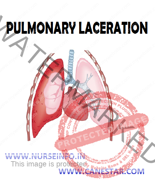

PULMONARY LACERATION – Etiology, Classification, Pathophysiology, Diagnostic Evaluation and Management
- Pulmonary laceration: in which lung tissue is torn or cut, differs from pulmonary contusion in that the former involves disruption of the macroscopic architecture of the lung
- Pulmonary hematoma: when lacerations fill with blood, the result is pulmonary hematoma, a collection of blood within the lung tissue
ETIOLOGY
Pulmonary laceration is a common result of:
- Penetrating trauma
- Blunt trauma
- Broken ribs
- Shearing forces
- Violent compression of the chest
CLASSIFICATION
- Type I lacerations: which occur in the mid-lung area, the air-filled lung bursts as a result of sudden compression of the chest. Also called compression-rupture lacerations, type 1 are the most common type and usually occur in a central location of the lung. They tend to be large, ranging in size from 2 to 8 cm.
- Type 2 lacerations: result when the lower chest is suddenly compressed and the lower lung is suddenly moved across the vertebral bodies. It is also called compression-shear, tends to occur near the spine and have an elongated shape. Type 2 lacerations usually occur in younger people with more flexible chests.
- Type 3 lacerations: which are caused by punctures from fractured ribs, occur in the area near the chest wall underlying the broken rib. Also called rib penetration lacerations, type 3 lacerations tend to be small and accompanied by pneumothorax
- Type 4 lacerations: also called adhesion tears, occur in cases where a pleuropulmonary adhesion had formed prior to the injury, in which the chest wall is suddenly fractured or pushed inward.
PATHOPHYSIOLOGY
Due to etiological factors (causes) —- a pulmonary laceration (causes) —- air to leak out of the lacerated lung and into the pleural space (results in) —- pneumothorax, hemothorax, hemopneumothorax
DIAGNOSTIC EVALUATION
- X-ray: a chest X-ray of a right-sided pulmonary contusion associated with flail chest and subcutaneous emphysema. Contusion may mask pulmonary laceration on chest X-ray
- CT scanning: on a CT scan, pulmonary lacerations show up in a contused area of the lung typically appearing as cavities filled with air or fluid that usually have a round or ovoid shape due to the lung’s elasticity
- Thoracoscopy may be used in both diagnosis and treatment of pulmonary laceration
MANAGEMENT
- As with other chest injuries, such as pulmonary contusion, hemothorax, and pneumothorax, pulmonary laceration can often be treated with just supplemental oxygen, ventilation, and drainage of fluids from the chest cavity
- A thoracostomy tube can be used to remove blood and air from the chest cavity. About 5% of cases require surgery, called thoracotomy.
Surgical Treatment
- Lobectomy: in which a lobe of the lung is removed
- Pneumonectomy: in which an entire lung is removed
COMPLICATIONS
- Pulmonary abscess
- Bronchopleural fistula (a fistula between the pleural space and the bronchial tree)
- Air embolism: in which air enters the bloodstream, is potentially fatal, especially when it occurs on the left side of the heart